A prospective study of assessment of visual outcome with rigid gas permeable contact lens in keratoconus patients.
- Guru Prasad N S , Consultant, IKSHA Eye Hospital, Madikeri.
- Dhananjaya KH , Assistant Professor, Department Of Ophthalmology, Shri Atal Bihari Vajpayee Medical College and Research Institute, Bangalore.
- Gajaraj T Naik , Assistant Professor, Department of Ophthalmology, Karwar Institute of Medical Sciences, Karwar.
- Prashanthkumar Achar , Professor, Department of Ophthalmology, Karwar Institute of Medical Sciences, Karwar.
Article Information:
Abstract:
BACKGROUND & OBJECTIVES: Keratoconus is a non-inflammatory, degenerative disease that compromises the structural integrity of the collagen matrix with in corneal stroma characterized by cone shaped ectasia and thinning of the stroma in the area of cone causing irregular astigmatism. Contact lenses are the main stay of nonsurgical management in moderate & advanced keratoconus. The primary goal of fitting different types of contact lenses is to provide optimum visual acuity with good comfort without affecting the health of cornea. This study aims at visual rehabilitation of keratoconus patients with various types of Rigid Gas Permeable lens. MATERIALS & METHODS: It is a hospital based prospective cohort study with a minimum of 50 patients diagnosed with keratoconus either unilateral or bilateral. Diagnosis and staging of keratoconus into mild moderate and severe is done by Using keratometry and using Modified Krumeich classification. Corneal topography is done using pentacam. Selection of suitable Rigid Gas permeable contact lens is on the basis of manifest refraction and degree of keratoconus. Patient fitting parameters are assessed and rigid gas permeable lens is fitted. Visual acuity is assessed post fitting rigid gas permeable lens. RESULTS: Out of 94 eyes 24 (25.53%) had mild, 34(36.17%) had moderate, 36(38.30%) had severe keratoconus. Among that 91 were not satisfied with UCVA and BCVA with spectacles. For 91 eyes RGP trial was done and there was significant improvement in visual acuity. CONCLUSION: Corneal distortion induced high corneal irregular astigmatism is the most frequent cause of increase of aberration and reduced ocular optical quality in keratoconus. Patients had reduced visual acuity and with minimum improvement with spectacles. Proper RGP contact lens provides effective method in reshaping of ocular surface combined with tear film and finally improving visual acuity significantly.
Keywords:
Article :
INTRODUCTION:
Keratoconus is a progressive ectatic disease of the cornea that typically develops during the adolescent years and results in gradual corneal thinning and steepening.(1).
The prevalence in studies can range from 1 to 2300 per 100,000 in Central India (0.0003%2.3%)(2).The prevalence of keratoconus is approximately 54 Per 100,000 in population. The average annual incidence rate is 2 per 100,000 Population.
Keratoconus is classified based on keratometry values mild if less than 48.00D Moderate if >48.00Dand <54.00D, severe if >54D.(3).
Contact lenses are the main stay of nonsurgical management in moderate & advanced keratoconus. The primary goal of fitting different types of contact lenses is to provide optimum visual acuity with good comfort without affecting the health of cornea.
While spectacles may provide acceptable visual acuity in the early stages of keratoconus, contact lens are the mainstay of vision correction.(4).
Various contact lens options such as rigid gas permeable lens soft toric lenses, piggy back. Rose k contact lenses, McGuire lens, hybrid lenses are available.
MATERIALS AND METHODS:
Source of data:
Patient diagnosed to have keratoconus attending in outpatient department.
Methods of Collection of Data:
Study design: Hospital based Prospective Cohort Study.
Study period:1.5 years
Inclusion Criteria:
1. Patients willing to give informed written consent for the study
2. Diagnosed cases of keratoconus where visual acuity is not satisfactory with spectacles
3. Diagnosed cases of keratoconus where visual acuity is not satisfactory with soft toric lens and agreeing for rigid gas permeable lens.
Exclusion Criteria:
1. Patient with corneal opacity involving visual axis.
2.Patients with ocular surface disorders and severe atopy
3.Patients with Acute corneal hydrops.
4. Patients with known contraindications for rigid gas permeable lens.
5.Patients Of keratoconus who have undergone keratoplasty.
6. Patients not willing to give written informed consent.
Methodology:
After obtaining approval and clearance from the Institutional Ethics Committee, the patients fulfilling the inclusion criteria will be enrolled for the study after obtaining informed consent. Case record form with follow up chart. It is a hospital based prospective cohort study with a minimum of 50 patients diagnosed with keratoconus either unilateral or bilateral. Demographic data (age, gender, address, occupation) and a detailed history will be taken. A complete ocular examination along with workup for keratoconus will be done. All the patients in the study will undergo BCVA, retinoscopy, Cycloplegic refraction (when necessary), torch light examinations, slit lamp examination, direct ophthalmoscopy and documentation of signs. Diagnosis and staging of keratoconus into mild moderate and severe is done by using keratometry and using Modified Krumeich classification.
Corneal topography is done using Pentacam.
Selection of suitable Rigid Gas permeable contact lens is on the basis of manifest refraction and degree of keratoconus. Patient fitting parameters are assessed and rigid gas permeable lens is fitted. Visual acuity is assessed post fitting rigid gas permeable lens and followed up at 3 months and assessed for visual acuity, lens intolerance and any side effects are noted.
Assessment tools
1. Demographic data
2. Slit lamp Examination
3. Visual acuity by Snellen chart
4. Subjective refraction
5. Keratometry
6. Corneal topography- pentacam
7. Rigid gas permeable lens/McGuire lens/rose k/easy k lens.
Outcome measures:
Efficacy parameters (Clinical outcome parameters)
1. Best corrected visual acuity.
2. Any complications post fitting rigid gas permeable lens.
STATISTICAL ANALYSIS:
The Statistical analysis was performed by STATA 11.2 (College Station TX USA).
Descriptive Statistics performed this study. Age distribution, gender distribution, laterality, Keratoconus stage, grade of visual acuity, base curve was reported as frequency and percentage. Age, base curve, keratometry, Log MAR visual acuity and base curve reported as mean and standard deviation.
SAMPLE SIZE:
Based on previous study Joonseo Hwang et al (29)
Sample size calculation - n= (Z1)2 * (S)2 / D2
Where Z1 – Z value associated with alpha -1.64
S –Pooled Standard Deviation -5.82 D-
Absolute precision -2 n=(1.64)2*(5.82)2( 2)
n=46
Total sample size(round off)=50.
RESULTS:
During the study period a total of 50 keratoconus patients were evaluated.
A.DISTRIBUTION OF PATIENTS BASED ON GENDER
TABLE1: DISTRIBUTION OF PATIENTS BASED ON GENDER
|
SEX |
Number of Cases |
Percentage |
|
Male |
19 |
38% |
|
Female |
31 |
62% |
|
Total |
50 |
100% |
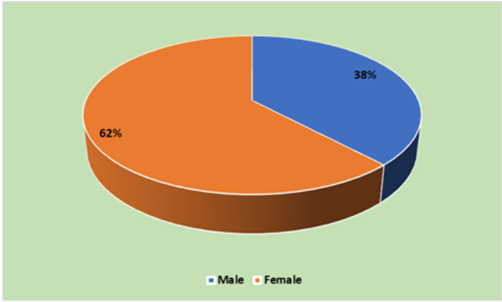
GRAPH 1: GENDER DISTRIBUTION
This table and chart depicts the gender distribution of patients in our Study, in which 38% were male and 62%were females
B.DISTRIBUTION OF PATIENTS BASED ON AGE
TABLE 2: DISTRIBUTION OF PATIENTS BASED ON AGE
|
AGE IN YEARS |
Number of Cases |
Percentage |
|
13-20 |
20 |
40% |
|
21-25 |
20 |
40% |
|
26-30 |
9 |
18% |
|
31-41 |
1 |
2% |
|
Total |
50 |
100% |
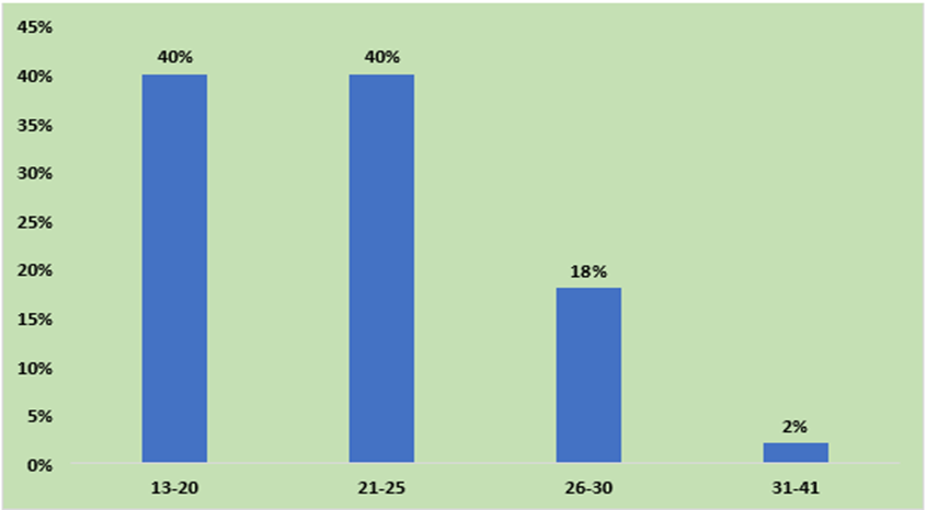
GRAPH 2: AGE WISE DISTRIBUTION
|
|
Mean |
SD |
Min- Max z |
|
Age (In Years) |
22.34 |
4.54 |
13-41 |
This table and graph shows the age wise distribution of patients included in the study. The majority of the subjects were in the age group of 13 to 25 years.Mean age of presentation is (22.34+/-4.54yrs) Minimum age being 13 and max being 41.
C.DISTRIBUTION OF PATIENTS ACCORDING TO LATERALITY
TABLE 3: DISTRIBUTION OF PATIENTS ACCORDING TO LATERALITY
|
|
Number of Cases |
Percentage |
|
Bilateral |
44 |
88% |
|
Unilateral |
6 |
12% |
|
Total |
50 |
100% |
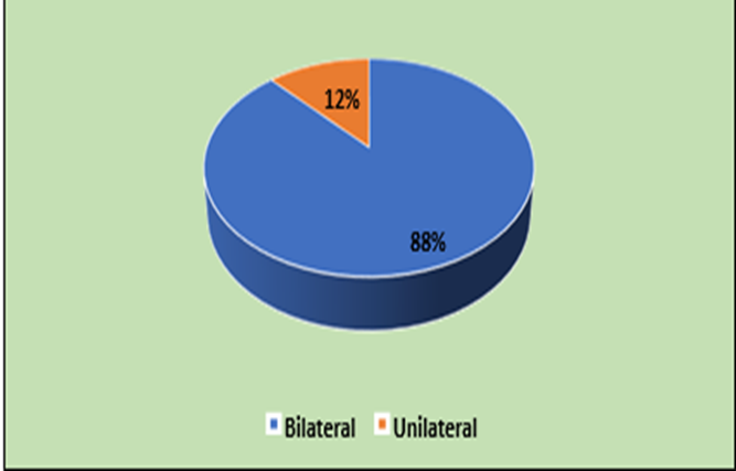
GRAPH 3: DISTRIBUTION OF PATIENTS ACCORDING TO LATERALITY
This table and graph shows the Distribution of Patients according to laterality. 88% had bilateral presentation and 12% had unilateral presentation.
D.STAGING OF KERATOCONUS BASED ON KERATOMETRY
TABLE 4: STAGING OF KERATOCONUS
|
|
Number of Cases |
Percentage |
Mild (<48D) |
24 |
25.53% |
Moderate (48-54) |
34 |
36.17% |
Severe (>54) |
36 |
38.30% |
Total |
94 |
100% |
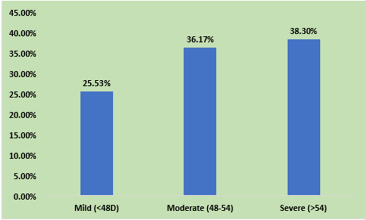
GRAPH 4: STAGING OF KERATOCONUS
The above table and graph depicts staging of keratoconus. Most common stage of keratoconus presenation is severe type 38.30%.
E.UCVA AMONG THE STUDY POPULATION
TABLE 5: DISTRIBUTION OF PATIENTS EYES BASED ON UCVA
|
|
NUMBER OF CASES |
PERCENTAGE |
|
6/6 -6/18 |
18 |
19.15% |
|
6/24 – 6/60 |
57 |
60.64% |
|
5/60-3/60 |
14 |
14.89% |
|
<3/60 |
5 |
5.32% |
|
Total |
94 |
100 |
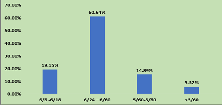
GRAPH 5: DISTRIBUTION OF PATIENTS EYES BASED ON UCVA
The above table depicts distribution of patients based on uncorrected visual acuity. Majority of study population 60.64% had an visual acuity of 6/24-6/60, 19.15% had visual acuity of 6/6-6/18 , 14.89% had an visual acuity of 5/60-3/60 and .32% had visual acuity less than 3/60 .
F.BCVA WITH SPECTACLES AMONG STUDY POPULATION
TABLE 6: BCVA WITH SPECTACLE CORRECTION
|
|
Number of Cases |
Percentage |
|
6/6 -6/18 |
52 |
55.32% |
|
6/24 – 6/60 |
38 |
40.43% |
|
5/60-3/60 |
4 |
4.26% |
|
<3/60 |
|
|
|
Total |
94 |
100% |
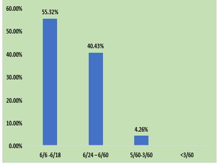
GRAPH 6: BCVA WITH SPECTACLE CORRECTION
The above table and graph depicts BCVA with spectacle correction..55.32% patients improved t0 6/6 to 6/18, 40.43% patients improved to 6/24 to 6/60, and 4.26% improved to 5/60-3/40.
G.BCVA WITH RGP CONTACT LENS
TABLE 7: BCVA WITH RGP CONTACT LENS
|
|
Number of Cases |
Percentage |
|
6/6 -6/18 |
90 |
98.90% |
|
6/24 – 6/60 |
1 |
1.10% |
|
5/60-3/60 |
|
|
|
<3/60 |
|
|
|
Total |
91 |
|
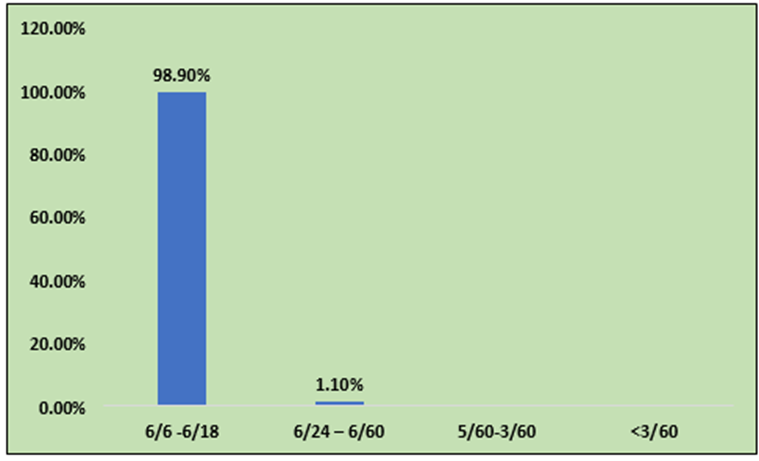
GRAPH 7: BCVA WITH RGP CONTACT LENS
The above table and graph depicts BCVA with RGP contact lens.98.90% had improved to 6/6 to 6/18. 1.10% had improved to 6/24 to 6/60.
H. BASE CURVE WITH RIGID GAS PERMEABLE LENS
TABLE 8: BASE CURVE WITH RGP LENS
|
BASE CURVE |
Number of Cases |
Percentage |
|
4-5 |
3 |
3.30% |
|
5-6 |
19 |
20.88% |
|
6-7 |
45 |
49.45% |
|
>7 |
24 |
26.37% |
|
Total |
91 |
100% |
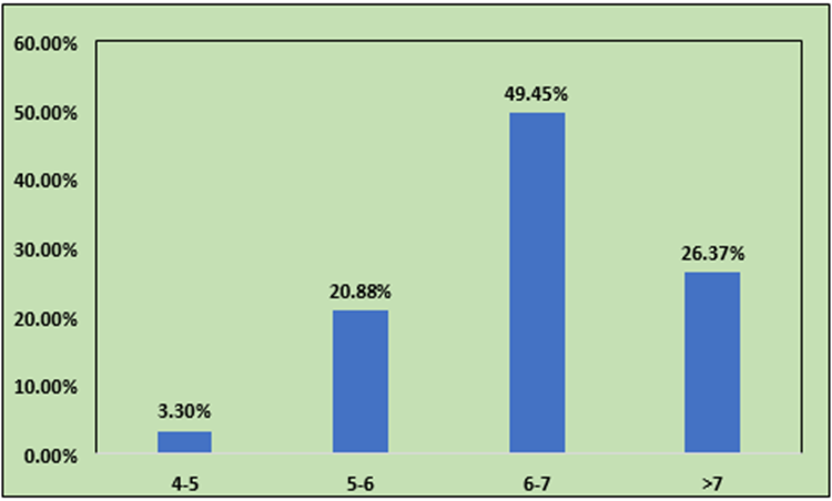
GRAPH 8: BASE CURE WITH RGP LENS
The above table and graph depicts base curve used for contact lens. The most common base curve were ranged from 6 to 7 followed by more than 7.
DISCUSSION:
Keratoconus is a progressive non inflammatory disorder of cornea that usually manifests during adolescence. Kennedy et al,6 in their clinical and epidemiologic study of keratoconus reported keratoconus prevalence rate of 54.5 per 1,00,000 in the general population. Prevalence of keratoconus as reported by Rabinowitz is between 50 and 230 per 1,00,000 in the population7. The incidence and prevalence could not be commented in our study because the study was conducted only among the patients attending our hospital. The disease has an insidious onset and a variable severity and patients may not seek ophthalmic care for a long time. As the disease is asymmetric, one eye may be affected more than the other. Some patients are able to perform their routine activities with the vision in the better eye.
Keratoconus usually affects the younger age group7. A visual impairment in this age group could hamper the quality of life of the affected individuals. Hence providing them with a good option for correcting the condition is highly beneficial. In our study the mean age of the patients was 22.34+/-4.54 years, indicating a young population.
The gender predilection may vary for keratoconus8. Most commonly is seen in females than males, Even in our study females (62%) more preponderant than males 38(%).
Although keratoconus is generally a bilateral corneal disorder, its progression is often unequal, resulting in marked asymmetry between eyes.9 Forme fruste keratoconus describes an early or subclinical stage in which characteristic topographic changes are present despite the absence of overt clinical features. A descriptive study on keratoconus conducted by N Lim at Jordan eye hospital reported a bilateral incidence of 92%. The reported incidence of unilaterality of keratoconus varies from 14.4%–41%(10). Among 50 patients included in this study population, 88% patients had bilateral keratoconus 12% had unilateral keratoconus.
Keratoconus can be divided into mild(<48d), moderate(48-54) , severe(>54). In our study population of 94 eyes ,38.38% had severe keratoconus ,36.17% of them had moderate keratoconus and 25.53% of mild keratoconus. Keratoconus, a degenerative disorder of cornea leading to thinning and ectasia of cornea, causes characteristic irregular light reflexes on distant direct ophthalmoscopy and streak retinoscopy and irregular mires on keratometry.11 The patient develops a irregular myopic astigmatism that is gradually progressive, due to which patients complain of blurred vision and reduced visual acuity.12 In Our study population of 94 eyes, Majority 60.64% had an visual acuity of 6/24-6/60, 19.15% had visual acuity of 6/6-6/18 , 14.89% had an visual acuity of 5/60-3/60 and .32% had visual acuity less than 3/60 .
When glasses were prescribed, 55.32% patients improved to 6/6 to 6/18, 40.43% patients improved to 6/24 to 6/60, and 4.26% improved to 5/60-3/40. The distortion of corneal architecture associated with keratoconus produces irregular astigmatism, leading to increased optical aberrations and compromised visual performance.13.
By providing a regular optical surface over the irregular cornea, RGP lenses help minimize the effects of corneal astigmatism and aberrations, resulting in enhanced visual quality and better visual outcomes.14. Xie PY et al, in their study on The evaluation of Visual quality in keratoconus eyes corrected by rigid gas-permeable contact lens analysed 30 eyes of mild keratoconus, 13 eyes of moderate keratoconus and 11 eyes of severe keratoconus.15 RGP contact lens corrected visual acuity was increased significantly compared with spectacles corrected VA .In our study population, RGP lens were fitted in 91 patients. Among them BCVA with RGP contact lens 98.90% had improved to 6/6 to 6/18. 1.10% had improved to 6/24 to 6/60. All our patients were comfortable on follow up.Thus, as per our study RGP lenses are a very good treatment option for patients of Keratoconus.
CONCLUSION:
In this study we concluded that keratoconus commonly presents during adolescence. Mean age being 22.34+/-4.54 years. Female preponderance was found, 62% were females and 38% were males. Bilateral presentation was found to be more common.88% had bilateral involvement and 12% had unilateral involvement. Majority of them had moderate to severe grade of keratoconus. Corneal distortion induced high corneal irregular astigmatism is the most frequent cause of increase of aberration and reduced ocular optical quality in keratoconus. Patients had reduced visual acuity and with minimum improvement with spectacles. Proper RGP contact lens provides effective method in reshaping of ocular surface combined with tear film and finally improving visual acuity significantly.
SUMMARY
A total of 50 patients with diagnosed keratoconus were included in the study. History with complete ocular examination with pentacam was done for all the patients. Symptoms and signs were noted. The severity of keratoconus was classified based on keratometry.
In our study of 50 patients (94 eyes) was diagnosed keratoconus. Females have more preponderance. Usually bilateral in presentation. Patients were classified in mild, moderate and severe keratoconus.24(25.53%) had mild keratoconus ,34(36.17%) had moderate keratoconus,36(38.30%) had severe keratoconus. UCVA, BCVA with spectacles of the patients were done. Patients whose vison was not improving with spectacles or not satisfactory was taken in to RGP trial. Out of 94 eyes, 3 eyes had 6/6 vison with spectacles. Remaining for 91 eyes, based on mean keratometery and manifest refraction proper RGP lens was selected and fitted. BCVA was done post fitting.
In our study, we conclude that rigid gas permeable lens with proper base curve, diameter and power with respect to severity of keratoconus can significantly improve the visual acuity of the patients than using spectacles.
REFERENCES:
1. AMichael K Smolek.What is Keratoconus.In:Brian S Boxer Wachler.Modern
2. Management Of Keratoconus,1st ed.India :Jaypee;2008.P:3.
3. Gokhale NS. Epidemiology of keratoconus. Indian J Ophthalmol. 2013 Aug;61(8):382–3.
4. Kanski JJ,Bowling Brad, Clinical ophthalmology A systemic approach,8th ed.China:Elseviersaunders;2016, p.213.
5. Bilgin LK, Yilmaz S, Araz B, Yüksel SB, Sezen T. 30 years of contact lens prescribing for keratoconic patients in Turkey. Contact Lens Anterior Eye J Br Contact Lens Assoc. 2009 Feb;32(1):16–21.
6. Espandar L, Meyer J. Keratoconus: overview and update on treatment. Middle East Afr J
7. Ophthalmol. 2010 Jan;17(1):15–20.
8. Kennedy RH, Bourne WM, Dyer JA. A 48-year clinical and epidemiologic study of keratoconus. Am J Ophthalmol. 1986 Mar 15;101(3):267–73.
9. Rabinowitz YS. Keratoconus. Surv Ophthalmol. 1998;42(4):297–319. doi:10.1016/s0039-6257(97)00119-7 Ophthalmol. 2000 Aug;84(8):834–6.
10. McMonnies CW. Mechanisms of rubbing-related corneal trauma in keratoconus. Cornea.
11. 2009 Jul;28(6):607–15.
12. Davidson AE, Hayes S, Hardcastle AJ, Tuft SJ. The pathogenesis of keratoconus. Eye.
13. 2014 Feb;28(2):189–95.
14. Lim N, Vogt U. Characteristics and functional outcomes of 130 patients with keratoconus attending a specialist contact lens clinic. Eye (Lond). 2002 Jan;16(1):54-9. doi:
15. 10.1038/sj.eye.6700061. PMID: 11913889.
16. Nowak DM, Gajecka M. The Genetics of Keratoconus. Middle East Afr J Ophthalmol. 2011;18(1):2–6.
17. Genetic epidemiological study of keratoconus: evidence for major gene determination -
18. PubMed [Internet]. [cited 2021 Jul 16]. Available from:
19. https://pubmed.ncbi.nlm.nih.gov/10951465/
20. Zadnik K, Mannis MJ, Johnson CA, Rich D. Rapid contrast sensitivity assessment in keratoconus. Am J Optom Physiol Opt. 1987 Sep;64(9):693–7.
21. Leung KK. RGP fitting philosophies for keratoconus. Clin Exp Optom. 1999;82(6):230–5.
22. Xie PY, Wang D, Yang LN, Zhou WJ. The evaluation of visual quality in keratoconus eyes corrected by rigid gas-permeable contact lens. [Zhonghuayankezazhi] Chinese journal of ophthalmology. 2005 Dec;41(12):1086-91.