A PROSPECTIVE OBSERVATIONAL STUDY TO EVALUATE THE COMPLICATIONS OF TRANSVERSUS ABDOMINIS RELEASE FOR VENTRAL HERNIA AT 1, 2, 6 AND 12 MONTHS POST-OPERATIVELY.
- Pranab Kumar Mandal , Associate Professor, Master Of Surgery (MS), General Surgery, IPGMER & SSKM HOSPITAL, 244, AJC Bose Rd, Kolkata 700020, West Bengal.
- Srinjay Maji , Senior Resident, Master of Surgery (MS), General Surgery, Tufanganj Sub Divisional Hospital, 8M69+H9F, Tufanganj, 736159, West Bengal.
Article Information:
Abstract:
Introduction: Complex ventral hernia repair remains surgically challenging because of high post-operative morbidity and recurrence rates. Transversus Abdominis Release (TAR) has emerged as an effective posterior component separation technique for abdominal wall reconstruction. Aims: To evaluate postoperative complications and outcomes following ventral hernia repair at 1, 2, 6, and 12 months follow-up. Materials and methods: The present study was Institution-based Clinicopathological Prospective Observational Study. This Study was conducted over 18 months. Department of General Surgery, Male and Female Surgery wards, Main OT Complex, TCCOT, IPGMER and SSKM Hospital. 33 consecutive patients. Result: The majority of patients had midline hernias, followed by subcostal, iliac, and lower abdominal defects. Postoperative complications such as seroma, wound-related complications, and intestinal obstruction were observed in the early follow-up period but showed complete resolution in most cases by 6 months. Pain scores demonstrated a progressive decline over time, with most patients reporting minimal or no pain by 2 months. Surgical site occurrences were seen in a limited number of patients and resolved with conservative management. Importantly, no recurrence or hematoma was observed at 1-year follow-up. Overall mortality was low. Conclusion: Ventral hernia repair, when performed with appropriate technique and patient selection, yields favourable outcomes with low long-term complication and recurrence rates. Early postoperative complications may occur but are generally self-limiting. Long-term follow-up confirms the durability and safety of ventral hernia repair, supporting its continued use as a standard surgical approach.
Keywords:
Article :
INTRODUCTION:
Ventral hernia repair is among the most commonly performed surgical procedures worldwide and represents a significant healthcare burden, particularly in populous countries such as India. In the United States alone, nearly 300,000 ventral hernia repairs are performed annually, highlighting the substantial clinical importance of this condition [1]. Despite considerable advances in surgical techniques and prosthetic mesh materials, recurrence rates following ventral hernia repair remain high, ranging from 15% to 21%, with even higher rates reported after incisional hernia repair. Recurrence adversely affects patient quality of life and presents ongoing challenges in achieving durable and functional abdominal wall reconstruction.
To improve surgical outcomes and reduce recurrence rates, several clinical guidelines have been established. Current recommendations favor sublay mesh placement over onlay techniques, while posterior component separation is generally preferred to anterior approaches in complex repairs. Registry-based evidence, including data from the HerniaMed registry, demonstrates increasing adoption of open sublay and preperitoneal repairs, along with a gradual rise in endoscopic sublay techniques for incisional hernias between 2010 and 2019. However, substantial international variability in ventral hernia management persists due to differences in healthcare resources, surgeon expertise, and patient demographics, particularly obesity prevalence. For instance, obesity rates are significantly higher in the United States compared to India, influencing both hernia pathogenesis and operative strategy. Furthermore, anatomical variations such as comparatively smaller abdominal girth among Asian populations suggest that procedures like component separation may become necessary at lower defect widths than in Caucasian populations.
Despite the availability of evidence-based guidelines, multicentric studies and registry analyses continue to reveal discrepancies between recommended practices and real-world management. The HerniaMed registry reported that although mesh reinforcement is recommended for small umbilical hernias (<2 cm), approximately 76.5% of such cases were still repaired using suture techniques alone. This highlights a persistent gap between established recommendations and actual clinical practice, emphasizing the need for institution-specific and region-specific outcome data.
A thorough understanding of anterior abdominal wall anatomy is fundamental for successful ventral hernia repair. Anatomically, the abdominal wall is a hexagonal structure bounded superiorly by the xiphoid process, inferiorly by the pubic symphysis, laterally by the inguinal ligaments, and posteriorly by muscles including the erector spinae and quadratus lumborum. Its musculature consists of a midline group formed primarily by the paired rectus abdominis muscles separated by the linea alba, and an anterolateral group comprising the external oblique, internal oblique, and transversus abdominis muscles enclosed within layered fascial sheaths. The linea alba is a vital fibrous midline structure formed by fusion of the aponeuroses of the lateral abdominal muscles. Restoration of the linea alba is central to functional abdominal wall reconstruction and has been associated with improved structural integrity and enhanced patient quality of life. Preservation of the neurovascular supply, particularly the superior and inferior epigastric vessels and segmental innervation, is essential to maintain postoperative muscle function and prevent atrophy.
Preoperative patient optimization plays a critical role in improving surgical outcomes and minimizing recurrence. Patient-related factors such as obesity, smoking, diabetes, and poor nutritional status significantly affect wound healing and postoperative complications. Obesity, in particular, is strongly associated with increased recurrence rates, and current guidelines recommend postponing elective repair in patients with a body mass index (BMI) ≥50 kg/m² until substantial weight reduction is achieved [2]. Smoking impairs tissue oxygenation and immune function, thereby increasing the risk of wound infection; cessation for at least 3–4 weeks prior to surgery has been shown to reduce these complications. Similarly, adequate glycemic control with HbA1c levels ≤7% is associated with lower postoperative infection rates. Nutritional optimization is equally important, as hypoalbuminemia (serum albumin <3.0 g/dL) independently predicts poor surgical outcomes [3].
Among the various operative techniques available, sublay mesh placement within the retrorectus plane, as described in the Rives–Stoppa technique, is widely considered the gold standard because of its lower recurrence rates and superior wound outcomes compared with onlay or intraperitoneal mesh placement [4]. More recently, minimally invasive approaches such as laparoscopic repair and enhanced-view totally extraperitoneal (eTEP) repair have gained popularity due to their association with reduced wound complications, shorter hospital stay, and comparable efficacy [5]. These evolving surgical strategies reflect the ongoing effort to balance optimal anatomical reconstruction with reduced morbidity and improved postoperative recovery.
The present study aims to evaluate the outcomes of contemporary ventral hernia repair practices at our institution and to identify factors influencing surgical success and recurrence. The findings of this study may contribute to improved clinical decision-making and enhanced patient outcomes in ventral hernia management.
MATERIALS AND METHODS:
Study design: Institution-based Clinicopathological Prospective Observational Study
Place of study: Department of General Surgery, Male and Female Surgery wards, Main OT Complex, TCCOT, IPGMER and SSKM Hospital
Period of study: 18 Months
Study Population: All patients undergoing surgery for ventral hernia at Dept. of General Surgery, IPGME&R and SSKM Hospital, Kolkata.
Sample size: 33 consecutive patients who underwent surgery for ventral hernia at Dept. of General Surgery
Inclusion Criteria:
• All ventral hernia surgery performed in IPGMER & SSKM Hospital in adult population (18 years or older) from 1st January, 2023 to 1st January, 2024
• Included incisional, umbilical/ paraumbilical, Spigelian, and lumbar hernia
• Elective and emergency surgery
• Recurrent ventral hernia
• Seropositive patients
Exclusion criteria:
• Hernias with loss of domain/ above EHS-W3
Study Variable:
• Patient Characteristics (Pre-operative)
• Hernia Characteristics (Pre-operative)
• Pre-operative Investigations
• Operative Details
• Post-operative Outcomes (Follow-up data)
• Post-operative Complications
Statistical Analysis: For statistical analysis, data were initially entered into a Microsoft Excel spreadsheet and then analyzed using SPSS (version 27.0; SPSS Inc., Chicago, IL, USA) and GraphPad Prism (version 5). Numerical variables were summarized using means and standard deviations, while Data were entered into Excel and analyzed using SPSS and GraphPad Prism. Numerical variables were summarized using means and standard deviations, while categorical variables were described with counts and percentages. Two-sample t-tests were used to compare independent groups, while paired t-tests accounted for correlations in paired data. Chi-square tests (including Fisher’s exact test for small sample sizes) were used for categorical data comparisons. P-values ≤ 0.05 were considered statistically significant.
RESULTS:
Table 1: List of the Location and EHS Grades of Ventral Hernia Requiring Transversus Abdominis Release
|
LOCATION OF HERNIA |
PERCENTAGE |
EHS GRADE |
NO. OF PATIENTS |
|
RIGHT SUBCOSTAL |
24%
|
L1W2 |
3 |
|
L1W3 |
1 |
||
|
L1L2W3 |
1 |
||
|
L1L2W3R1 |
1 |
||
|
M2L1W2 |
1 |
||
|
M2L1W3 |
1 |
||
|
MIDLINE |
45% |
M2M3M4W2 |
3 |
|
M2M3M4W3 |
3 |
||
|
M3M4W2 |
3 |
||
|
M2M3M2W2 |
1 |
||
|
M3M4M5W2 |
1 |
||
|
M4M5W2 |
1 |
||
|
M2M3M4M5W3 |
1 |
||
|
M1M2M3M4W3 |
1 |
||
|
M2M3M4W2R2 |
1 |
||
|
RIGHT ILIAC |
15% |
L3W2 |
3 |
|
L3W2R1 |
2 |
||
|
LOWER ABDOMINAL |
12%
|
M4L3W2 |
1 |
|
M4M5L3W2R1 |
1 |
||
|
M3M4M5L2L3W3 |
1 |
||
|
M3M4M5L3W3R3 |
1 |
||
|
LEFT LUMBAR |
3% |
L4W2 |
1 |
|
TOTAL |
33 |
||
Table 2: Distribution of Mesh Area (cm²)
|
Mesh Area (Cm2) |
|
|
Mean |
1204.12 |
|
Std. Deviation |
559.66 |
|
Minimum |
616 |
|
Maximum |
2500 |
Table 3: Distribution of OT duration in the study population undergoing Transversus Abdominis Release.
|
OT Duration |
NO. Of Patients |
Percentage |
|
<3 Hours |
1 |
3% |
|
3-5 Hours |
14 |
42% |
|
5-7 Hours |
12 |
36% |
|
>7 Hours |
6 |
18% |
|
Total |
33 |
100% |
|
Mean ± Std. |
309.7 ± 106.93 |
|
Table 4: Distribution of time taken to resume daily activities post operatively after undergoing Transversus Abdominis Release
|
Time Taken For Daily Activity |
NO. Of Patients |
Percentage |
|
<1 Week |
20 |
61% |
|
> 1 Week |
11 |
33% |
|
NA |
2 |
6% |
|
TOTAL |
33 |
100% |
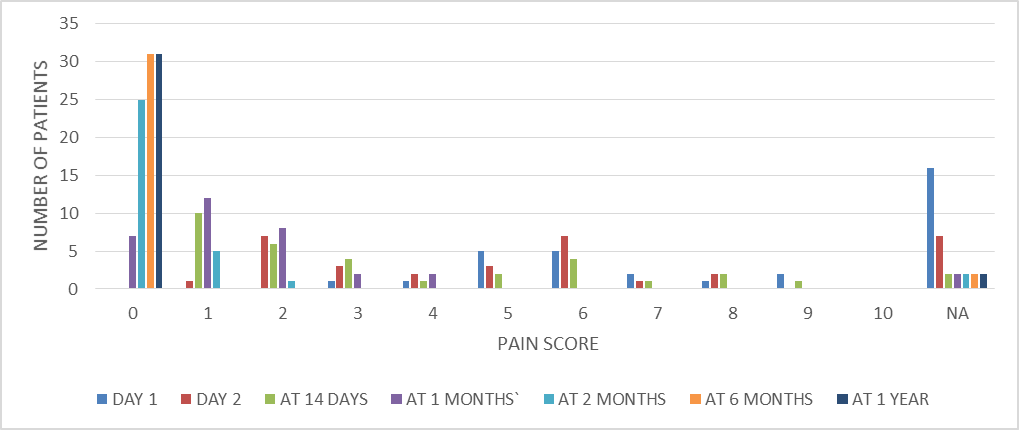
Fig. 1: Distribution of Post operative Pain score over the follow-up duration in the study population undergoing Transversus Abdominis Release
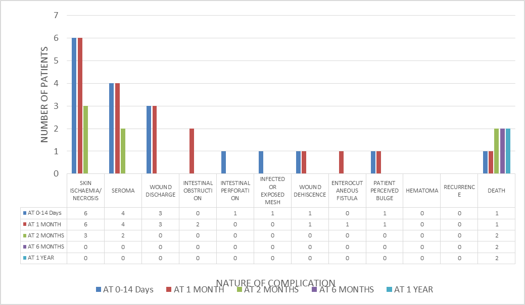
Fig.2: Distribution Of Complications In The Study Population Over The Study Time Period Following Transversus Abdominis Release.
The study population demonstrated a varied distribution of hernia defect locations. The majority of patients had a midline hernia defect (45%), followed by right subcostal defects (24%), right iliac (15%), lower abdominal (12%), and left lumbar hernias (3%). All patients underwent TAR procedures using medium-weight, macroporous prolene mesh.
Regarding mesh characteristics, out of the 33 TAR procedures, the most commonly used mesh size was 30 × 30 cm (900 cm²), which was used in 22 patients (67%). Larger meshes of 50 × 50 cm (2500 cm²) were used in 4 patients, while 40 × 40 cm (1600 cm²) mesh was used in 3 patients. Additionally, 4 patients received tailored mesh sizes including 45 × 40 cm, 40 × 38 cm, 40 × 30 cm, and 28 × 22 cm. The mean mesh surface area used in the study was 1204.12 ± 559.66 cm².
The mean operative time for TAR procedures was 309.7 ± 106.93 minutes, with a range from 155 minutes to 580 minutes. Most procedures (42%) were completed within 3–5 hours, followed by 36% taking 5–7 hours. A smaller proportion of surgeries (18%) required more than 7 hours, while only 3% were completed in less than 3 hours. Postoperatively, 61% of patients resumed daily activities within one week, whereas 33% experienced a delay beyond one week. “Daily activity” referred to basic self-care functions such as bathing, dressing, ambulation, toileting, and feeding.
Postoperative pain assessment using a numeric rating scale (0–10) showed that patients extubated on table commonly reported pain scores of 5–6 on POD-1. On POD-2, the most frequent scores were 2 and 6. A gradual reduction in pain was observed over time, with most patients reporting a score of 1 by POD-14. By one month, several patients had a score of 0, and by 2 months, the majority (25 patients) reported no pain. At 6 months, 31 patients had no pain, and this trend persisted at 1-year follow-up. Overall, pain showed a sharp immediate postoperative rise followed by a steady decline to minimal or no pain in most patients.
Postoperative complications included surgical site occurrences (SSO) such as skin ischemia/necrosis, seroma, wound dehiscence, and discharge in 8 patients, all of which resolved completely by 6 months. One patient with SSI expired, while no SSI was observed in the remaining cohort. Intestinal obstruction occurred in 2 patients at 1-month follow-up and resolved by 2 months. One patient reported a transient bulge at the previous hernia site, which also resolved by 2 months. Importantly, there were no cases of hematoma or hernia recurrence observed during the 1-year follow-up period. The total mortality in the study population was 2 patients over the study duration.
DISCUSSION:
In the present study, midline hernias were the most common defect (45%), followed by right subcostal, right iliac, lower abdominal, and left lumbar hernias. This pattern is consistent with the anatomical predisposition of midline defects due to previous laparotomies and fascial weakness, as also reported in large abdominal wall reconstruction series where midline incisional hernias predominate [5,6]. All patients underwent TAR with medium-weight macroporous polypropylene mesh, which aligns with current evidence supporting its strength and low infection profile in complex abdominal wall reconstruction [7].
The mean mesh size used in our study was 1204.12 ± 559.66 cm², with the majority of patients requiring a standard 30 × 30 cm mesh. Similar variability in mesh sizing has been described by Novitsky et al., who emphasized that TAR often requires large sublay mesh placement tailored to defect size and lateral dissection extent [7]. Larger defects requiring 40 × 40 cm or 50 × 50 cm meshes reflect the complexity of abdominal wall loss, which is comparable to outcomes reported in high-volume hernia centers [8]. The use of tailored mesh in select patients further highlights individualized reconstruction strategies, as recommended in modern ventral hernia guidelines [9].
The mean operative time in the present study was 309.7 ± 106.93 minutes, which is comparable to other TAR series reporting prolonged operative durations ranging from 3 to 8 hours depending on complexity [10]. Similarly, Petroccia et al. observed that operative time in posterior component separation is significantly influenced by defect size, adhesiolysis, and surgeon experience. Despite prolonged operative time, early functional recovery was favorable in our study, with 61% of patients resuming daily activities within one week, which is consistent with enhanced recovery protocols described in abdominal wall reconstruction literature [11].
Postoperative pain showed a predictable decline from early high scores (POD-1: 5–6) to near-complete resolution by 2 months in most patients. This trend correlates with findings by Saulis and Dumanian, who demonstrated that component separation techniques initially produce moderate pain but allow early recovery due to reduced tension repair [12]. Importantly, in our cohort, long-term pain was minimal, supporting the role of posterior component separation in improving patient comfort over time.
Surgical site occurrences were seen in 8 patients but resolved completely by 6 months, with no long-term morbidity. Similar SSO rates ranging from 10% to 25% have been reported in TAR series, particularly seroma and superficial wound complications [13]. Importantly, there was no recurrence or hematoma at 1-year follow-up in the present study, which is comparable to recurrence rates of less than 5–10% reported in contemporary TAR studies [14]. The low recurrence rate further supports TAR as a durable reconstructive option for complex abdominal wall defects.
CONCLUSION:
This prospective observational study evaluated the outcomes and complications following repair of ventral hernia over a 12-month follow-up period. The study demonstrated that Transversus Abdominis Release (TAR) with mesh reinforcement is an effective technique for complex ventral hernia repair with acceptable morbidity. Postoperative complications such as seroma, wound infection, and intestinal obstruction were observed in the early follow-up period but showed complete resolution in most patients by 6 months. Pain scores significantly decreased over time, with most patients achieving minimal or no pain by 2 months. Importantly, no recurrence or hematoma was observed at 1 year. The study highlights that although early postoperative complications may occur, long-term outcomes are favorable with low recurrence rates and good functional recovery, supporting TAR as a reliable surgical option for ventral hernia repair.
REFERENCES:
1. Henriksen NA, Bougard H, Gonçalves MR, Hope W, Khare R, Shao J, et al. Primary ventral and incisional hernias: comprehensive review. BJS Open. 2024 Dec 30;9(1).
2. Jensen KK, East B, Jisova B, Cano ML, Cavallaro G, Jørgensen LN, et al. The European Hernia Society Prehabilitation Project: a systematic review of patient prehabilitation prior to ventral hernia surgery. Hernia. 2022 Feb 25;26(3):715–726.
3. Looft P, Alfarawan F, Bockhorn M, El-Sourani N. Beyond Traditional Repair: Comparing eTEP and Open Sublay for Ventral Hernia Repair. JCM. 2025 Apr 9;14(8):2586.
4. Misiakos EP, Patapis P, Zavras N, Tzanetis P, Machairas A. Current Trends in Laparoscopic Ventral Hernia Repair. JSLS. 2015 Jan 1;19(3):e2015.00048.
5. Misiakos EP, Machairas A, Patapis P, Liakakos T. Laparoscopic Ventral Hernia Repair: Pros and Cons Compared With Open Hernia Repair. JSLS : Journal of the Society of Laparoendoscopic Surgeons. 2008 Jan 1;12(2):117–125.
6. Hawn MT, Snyder CW, Graham LA, Gray SH, Finan KR, Vick CC. Long-term follow-up of technical outcomes for incisional hernia repair. J Am Coll Surg. 2010;210(6): 898–905.
7. Itani KM, Hur K, Kim LT, et al. Comparison of laparoscopic and open repair of ventral incisional hernia. Surg Endosc. 2010;24: 1119–1126.
8. Novitsky YW, Elliott HL, Orenstein SB, Rosen MJ. Transversus abdominis release: a novel approach to posterior component separation. Am J Surg. 2012;204(2): 709–716.
9. Petroccia R, Inaba K, Okoye O, et al. Outcomes of posterior component separation with TAR in complex abdominal wall reconstruction. J Trauma Acute Care Surg. 2015;79(6): 1001–1008.
10. Slater NJ, Montgomery A, Berrevoet F, et al. Criteria for definition of a complex abdominal wall hernia. Hernia. 2014;18(1): 7–17.
11. Petroccia R, et al. Operative outcomes in posterior component separation technique. J Trauma Acute Care Surg. 2015;79: 1001–1008.
12. Atema JJ, Furnée EJ, Maeda Y, et al. Guidelines for abdominal wall reconstruction and enhanced recovery. Surg Clin North Am. 2018;98(3): 507–523.
13. Saulis AS, Dumanian GA. Periumbilical rectus abdominis muscle perforator preservation in abdominal wall reconstruction. Plast Reconstr Surg. 2002;109(1): 242–250.
14. Holihan JL, Nguyen DH, Nguyen MT, et al. Mesh location in ventral hernia repair: systematic review. Surgery. 2016;159(5): 1366–1379.