ROLE OF 16 SLICE COMPUTED TOMOGRAPHY UROGRAM IN CHARACTERIZING FOCAL RENAL LESIONS
- Dr. Supriya A S , Assistant Professor, Department of Radio Diagnosis, Subbaiah institute of medical sciences Shivamogga. India
- Dr Kavya Shree , Assistant Professor, Department of Radio Diagnosis, Subbaiah institute of medical sciences Shivamogga. India
- Dr. Chetan K K , Assistant Professor, Department of Radio Diagnosis, Subbaiah institute of medical sciences Shivamogga. India
- Dr Bharat MP , Professor, Department of Radio Diagnosis, Subbaiah institute of medical sciences Shivamogga. India
Article Information:
Abstract:
Background: Accurate characterization of focal renal lesions is essential for differentiating benign from malignant masses and guiding appropriate management. Multidetector computed tomography urography (CTU) offers high spatial resolution, multiphasic imaging, and advanced post-processing capabilities that improve lesion detection and characterization. Objectives: To evaluate the role of 16-slice CT urography in characterizing focal renal lesions, assess lesion morphology and enhancement characteristics, compare attenuation values before and after intravenous contrast administration, and correlate CT findings with histopathological or surgical diagnoses. Materials and Methods: This prospective hospital-based study was conducted in the Department of Radio Diagnosis, Subbaiah Institute of Medical Sciences, Shivamogga, over a period of one year. Fifty patients aged 4–79 years with clinically suspected or ultrasonographically detected renal lesions were evaluated using a 16-slice multidetector CT scanner. Multiphasic CT urography was performed with unenhanced, corticomedullary, nephrographic, and excretory phase imaging. Lesion characteristics including size, location, attenuation values, vascularity, calcification, necrosis, local invasion, and distant metastasis were assessed. CT diagnoses were correlated with histopathological examination and surgical findings. Diagnostic performance was calculated using sensitivity, specificity, positive predictive value (PPV), negative predictive value (NPV), and accuracy. Results: Among the 50 renal lesions studied, 33 (66%) were benign and 17 (34%) were malignant. The majority of patients belonged to the 60–69 years age group (38%), with a male predominance (56%). Renal cell carcinoma (RCC) was the most common malignant lesion, accounting for 24% of all renal lesions and 71% of malignant lesions. Benign lesions included angiomyolipoma (20%), abscess (20%), complex cyst (18%), and oncocytoma (8%). Malignant lesions demonstrated significantly higher attenuation values than benign lesions in both corticomedullary and nephrographic phases (p < 0.001). Features such as renal vein invasion, inferior vena cava thrombosis, necrosis, and distant metastases were strongly associated with malignancy. CT urography achieved a sensitivity of 100%, specificity of 94%, PPV of 90%, NPV of 100%, and overall diagnostic accuracy of 96% in differentiating benign from malignant renal lesions. Conclusion: Sixteen-slice CT urography is a highly accurate and reliable imaging modality for the evaluation of focal renal lesions. Its ability to provide multiphasic enhancement patterns, detailed lesion characterization, and assessment of local and distant disease extent makes it an invaluable tool in the diagnosis, staging, and preoperative planning of renal masses.
Keywords:
Article :
INTRODUCTION:
Computed tomography (CT) plays a crucial role in the evaluation of renal lesions by differentiating solid from cystic masses and further categorizing cystic lesions according to their probability of malignancy. Owing to its high sensitivity in detecting renal abnormalities, CT is widely utilized to guide clinical decision-making regarding surgical intervention, additional imaging follow-up, or conservative management.
Renal cell carcinoma (RCC) most commonly presents between the ages of 50 and 70 years and exhibits a male predominance with a male-to-female ratio of approximately 2:1.1,2 RCC is the eighth most common adult malignancy, accounting for nearly 2% of all cancers and representing 80–90% of primary malignant renal neoplasms in adults.3,4 Transitional cell carcinoma (TCC) of the renal pelvis is also more frequently observed in males and is typically diagnosed between 60 and 70 years of age.5,6 Wilms tumour is the most common renal mass in the paediatric population, accounting for more than 85% of paediatric renal tumours7,8 and approximately 7% of all childhood malignancies.9 The peak incidence occurs between 3 and 4 years of age, although the disease can occur in children aged 1–11 years. Renal oncocytoma constitutes approximately 5% of surgically resected primary epithelial renal neoplasms in adults. These tumours generally occur during the sixth and seventh decades of life, with a peak incidence around 55 years of age, and demonstrate a male predominance of approximately 2:1.10,11
Accurate identification and characterization of renal masses are essential, particularly in distinguishing malignant lesions from their benign counterparts when lesions are small. Despite advances in oncological treatment, most renal adenocarcinomas show limited response to chemotherapy and radiotherapy. Consequently, surgical excision remains the primary treatment modality and offers the best opportunity for long-term survival and potential cure, especially in patients with localized, low-stage disease
Although conventional axial renal CT has long been considered a reliable imaging modality, several limitations can affect its diagnostic accuracy. Variations in patient respiration during image acquisition may lead to motion artefacts or scanning gaps, thereby compromising image quality and potentially obscuring portions of the kidneys and associated lesions. In addition, small renal tumours, particularly those measuring less than twice the image collimation thickness, may not be completely encompassed within a single image section. As a result, partial volume averaging with adjacent renal parenchyma or perinephric fat may occur, leading to inaccurate attenuation measurements and lesion characterization.
The assessment of subtle internal features within cystic renal masses, such as minimal wall thickening, fine septations, or small mural nodules, may also be hindered by partial volume effects. Furthermore, conventional CT examinations require relatively long acquisition times, with scan durations averaging approximately 2 seconds per image and interscan intervals ranging from 3.5 to 9 seconds. Under routine conditions, complete imaging of the kidneys may take more than one minute. Consequently, it is difficult to obtain images exclusively during the early cortical phase of contrast enhancement, when renal cortical enhancement reaches its peak.
Helical CT offers several advantages over conventional axial CT. Its rapid and continuous image acquisition enables complete scanning of the kidneys during a single breath-hold, thereby minimizing respiratory motion artefacts and eliminating scanning gaps. Using a standard pitch of 1:1 (table speed equal to image collimation), the entire kidney can be scanned within approximately 30 seconds while maintaining a thin image collimation of 5 mm. This improved temporal resolution allows optimal imaging during the cortical phase of renal enhancement and enhances the detection and characterization of renal lesions.
Breath-hold scanning significantly reduces the risk of image misregistration, thereby minimizing motion-related artefacts and improving visualization of renal parenchyma and lesions. Another major advantage of Helical CT is its ability to reconstruct images retrospectively at any desired level from the acquired raw data. Although image collimation and pitch must be determined before scan acquisition, the raw data can subsequently be reconstructed at various intervals, providing greater flexibility in image interpretation.
This capability allows the radiologic technologist to generate axial images precisely through the center of a renal mass. Accurate localization enhances lesion characterization by improving the precision of attenuation measurements and reducing partial volume averaging effects. Consequently, the definition and delineation of renal lesions are significantly improved.
The rapid acquisition time of Helical CT also facilitates imaging during any of the three major phases of renal contrast enhancement: the corticomedullary phase, the nephrographic phase, and the excretory phase. This multiphasic imaging capability improves the detection, characterization, and staging of renal masses by providing detailed information regarding lesion vascularity and enhancement patterns.
OBJECTIVES:
• To identify CT characteristics of renal-lesions on 16- slice Computed Tomography Urogram (CTU) with intravenous contrast administration in various clinical presentations (Sample size =50 patients) in different age groups (4 yr.-79 yr.).
• To identify the extent, mass effects, vascularity, components of focal renal-lesions & to detect any local or distant organ involvement after administration of intravenous contrast on 16- slice CTU.
• To compare HU values in renal-lesions before & after administration of I.V contrast.
• Based on CTU features to categorize the renal-lesions as benign /malignant and correlate with HPE/ surgical results.
MATERIALS AND METHODS:
Study Design: A prospective hospital-based study.
Study area: Department of Radio Diagnosis, Subbaiah institute of medical sciences Shivamogga. India.
Study Period: 1 year.
Study population: Data for study, has been collected among patients who visited department of Radiodiagnosis with suspicious renal-lesions.
Sample size: The study consisted of 50 subjects.
Sampling method: Simple random technique.
Inclusion criteria:
• Patients with lesions that are clinically suspected.
• Patients who had renal-lesions detected by ultrasound
• Patients with h/o previously resected renal lesion in one kidney & presentation in another kidney.
Exclusion criteria:
• Patients who are non-cooperative & morbidly sick.
• Patient having previous history of contrast reactions.
• Simple cysts & extra renal-lesions that have invaded renal cortex are excluded
Ethical consideration: Institutional Ethical Committee permission was obtained before the commencement of the study.
Study tools and Data collection procedure:
Hospital-based prospective study, conducted among 50 patients (Age: 4yr-79yr) with clinically diagnosed renal-lesions & patients who were identified with renal-lesions on ultrasonography & were forwarded for further evaluation over one year period. Fever, abdominal pain, hematuria & weight loss were their presenting symptoms. Patients were assessed with 16- slice Multidetector CT (GE – OPTIMA 660). After the CT examination, a probable diagnosis was given & these results were compared with HPE/ surgical results as applicable.
Protocol for CT Urogram (CTU) Patient Preparation:
To prevent complications with the administration of the contrast medium, patients were maintained in fasting for a minimum of 4 hours before the CT scan. The patient was informed of the risks associated with administering contrast, & permission was obtained before performing contrast scan.
CT Technique:
Initially, an AP topogram abdomen was obtained in each patient with breath held & in supine position. 5 mm thickness axial sections were obtained from lung bases to ischial tuberosities. An unenhanced scan was always followed by an intravenous contrast injection while inspiration was suspended. From upper to lower pole of both kidneys, sections were obtained in the Cortico-medullary(40-60s), Nephrographic-(80-120s), & Excretory(180s) phases. Where necessary, sagittal & coronal reconstructions were produced. At 2.5 mm slice thickness, reconstructions were done. More recent Multi slice CT techniques, such as curved planar resizing, volume rendering, Maximum & Minimum Intensity Projections, were done
where required. Images were viewed in a direct display console using a variety of window settings, including the abdominal window at 320/40, the lung window at 1400/-600, & the bone window at 2400/200.
The size, location, presence of the mass/ calcification/fat or any involvement of adjacent structures with pre & postcontrast attenuation values were all considered when evaluating the pathological lesions.
Statistical Methods:
In the present study, descriptive statistical analysis was done. Results for categorical measurements are reported in Number (%) whereas results for continuous measurements are reported as Mean ±SD(Min-Max). At a 5% level of significance, significance is evaluated. Diagnostic statistics including sensitivity, specificity, PPV, NPV, & accuracy have been used to determine the correlation between CT scan & final diagnosis. Chi square test, Anova test & Fisher Exact test have been used to determine the significance of relation of CT results & final diagnosis.
RESULTS:
Table1: Distribution of renal-lesions among various age groups
|
Age Group |
Frequency of renal-lesions |
Percentage (%) |
|
<15 yrs. |
1 |
2 |
|
30-39 yrs. |
3 |
6 |
|
40-49 yrs. |
8 |
16 |
|
50-59 yrs. |
10 |
20 |
|
60- 69 yrs. |
19 |
38 |
|
>70 yrs. |
9 |
18 |
|
Total |
50 |
100.0 |
In present study, patients with age ranging from 60 to 69 years old represented the highest percentage (38%). In this specific study, there was a significantly higher proportion of males (56%) as opposed to females (44%).
TABLE 2: Distribution of renal-lesions related to age in years
|
Lesion |
Age(yrs.) |
|
Total |
|||||
|
<15 yrs. |
30-39 yrs. |
40-49 yrs. |
50-59 yrs. |
60- 69 yrs. |
>70 yrs. |
No. |
% |
|
|
Complex Cyst |
0 |
0 |
0 |
2 |
3 |
4 |
9 |
18 |
|
|
|
|
22.2% |
33.3% |
44.4% |
|
|
|
|
Abscess |
0 |
3 |
4 |
2 |
1 |
0 |
10 |
20 |
|
|
30.0% |
40.0% |
20.0% |
10.0% |
|
|
|
|
|
Oncocytoma |
0 |
0 |
1 |
1 |
1 |
1 |
04 |
08 |
|
|
|
25.0% |
25.0% |
25.0% |
25.0% |
|
|
|
|
Angiomyolipoma |
0 |
0 |
2 |
2 |
5 |
3 |
12 |
24 |
|
|
|
10.0% |
20.0% |
60.0% |
10.0% |
|
|
|
|
RCC |
|
|
|
|
|
|
|
|
|
|
|
16.7% |
16.7% |
41.6% |
25.0% |
|
|
|
|
Renal Pelvic TCC |
0 |
0 |
0 |
0 |
3 |
1 |
04 |
08 |
|
|
|
|
|
75.0% |
25.0% |
|
|
|
|
Wilms Tumor |
1 |
0 |
0 |
0 |
0 |
0 |
01 |
02 |
|
100% |
|
|
|
|
|
|
|
|
|
|
|
50 |
100% |
|||||
• There was a total of 50 cases, with 33 cases (or 66%) being diagnosed as benign & 17 cases being diagnosed as malignant. RCC is the most-common type of malignant renal lesion, accounting for 71% of all malignant renal-lesions & accounting for 24% of all renal-masses.
• The youngest patient diagnosed with RCC was a 44-year- old male patient, & the oldest patient was a 79-year-old male patient. 05 out of 12 patients (42%) with RCC are between the ages of 60 & 69. The average age was 63.2 years old.
• 1 case of Wilms tumour of patients with were <15 years (100%).
• The youngest patient diagnosed with Angiomyolipomas (AML) was a 45-year-old female patient, & the oldest patient diagnosed with AML was a 73-year-old female patient. 06 out of 10 patients (60%) with AML are between the ages of 60 & 69. The average age was 61.7 years.
Graph 1: Distribution of renal-lesions related to gender
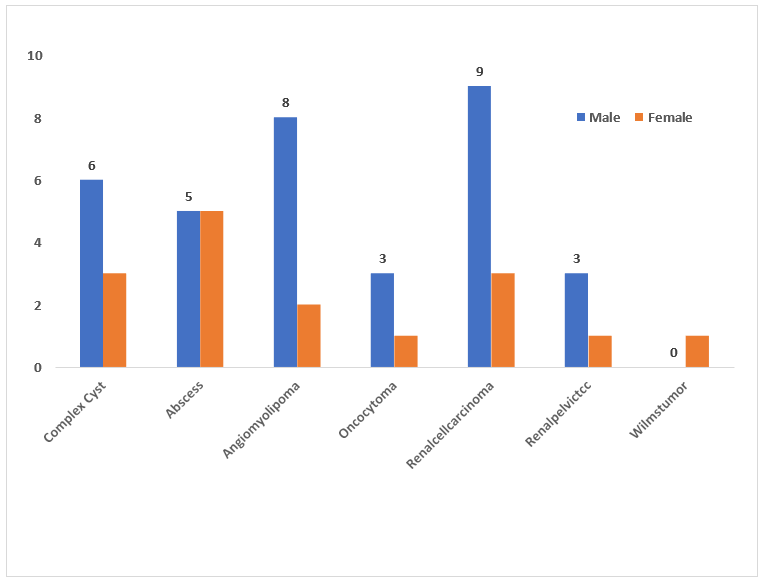
• Totally 28 (56%) males & 22 (44%) females, the M:F ratio was 1.3:1.
• Male preponderance (75.0%) in case of RCC when compared to females (25.0%), male: female ratio is 3:1.
•8 out of 10 cases of Angiomyolipomas (80.0%) were in females.
• 6 out of 9 cases of Complex cysts (66.7%) are males.
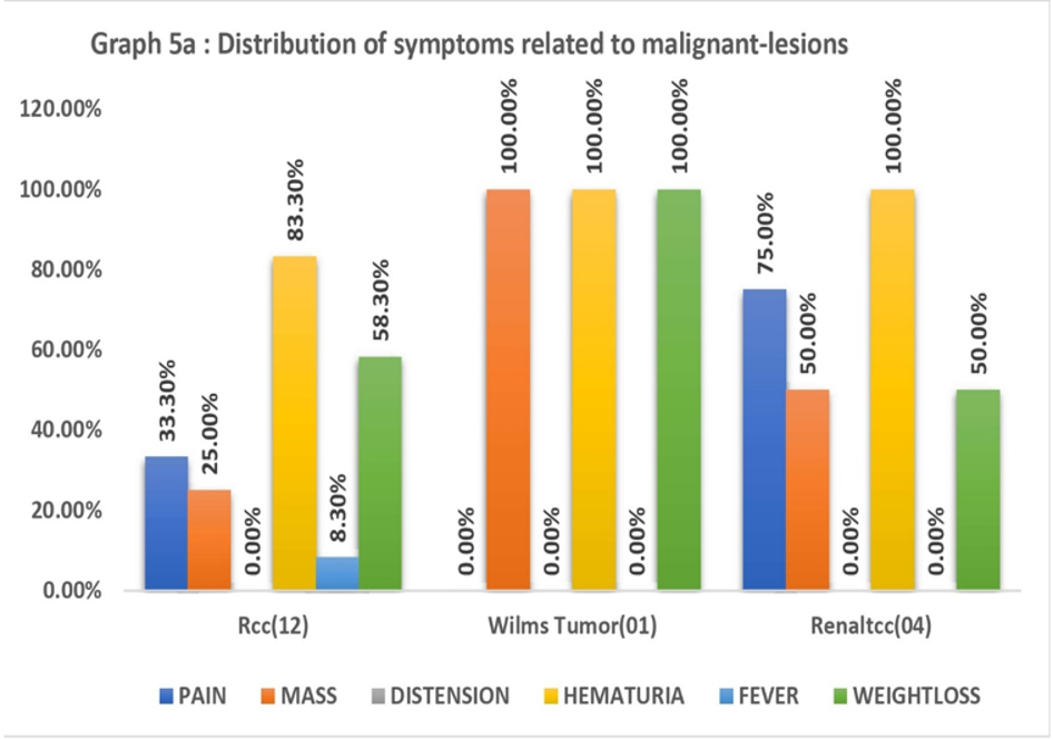
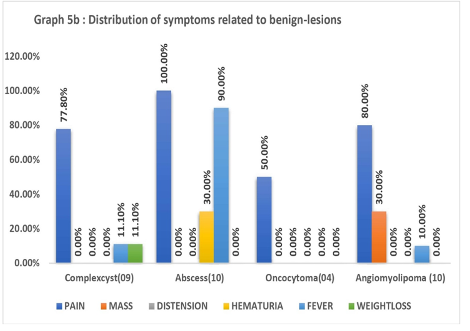
Renal lesion characteristics on CT:
• Most-common calcified renal lesion in present study is Complex cyst.
• Calcification present in 08 / 09 cases of Complex cyst (35%)
• TCC originating from renal pelvis & proximal ureter, showed an associated hydronephrosis (75%).
• In comparison to benign, malignant renal-lesions contained a significantly higher proportion of necrosis (100% Wilms tumour & 58% in RCC).
• Renal vein involvement observed among malignant renal-lesions as much as 33.3% in RCC & 1 case (100%) Wilms tumour; however, this finding was not observed in any of the benign renal-lesions.
• 3 out of 12(16.7%) RCC had IVC thrombosis.
• Lymph nodes (66.7%) & the lungs (50%) where the primary locations were RCC metastasized.
• The lymph nodes, lungs, liver, & adrenals were all affected by the metastasis by Wilms tumour.
• Renal vein; adrenals; lungs & appendicular skeleton involvement lacked among benign lesions.
• Renal-masses mostly presented in right kidney (62%).
Table 3: Comparison between HU values among renal-lesions on a 128-slice CTU during pre & post contrast
|
|
|
No. |
Mean HU |
Std. Deviation |
Minimum |
Maximum |
‘F’ value |
‘p’ value |
|
Unenhanced |
Benign |
33 |
15.71 |
10.388 |
8 |
29 |
14.696 |
<0.001 |
|
Malignant |
17 |
29.00 |
4.761 |
20 |
39 |
|||
|
Corticomedullary |
Benign |
33 |
24.29 |
16.750 |
10 |
60 |
21.190 |
<0.001 |
|
Malignant |
17 |
57.02 |
13.296 |
30 |
86 |
|||
|
Nephrographic |
Benign |
33 |
32.57 |
25.079 |
10 |
85 |
16.348 |
<0.001 |
|
Malignant |
17 |
74.70 |
22.513 |
38 |
102 |
|
|
|
No. |
Mean HU |
Std. Deviation |
Minimum |
Maximum |
‘F’ value |
‘p’ value |
|
Difference (Corticomedullary -Unenhanced) |
Benign |
33 |
8.5714 |
6.92577 |
2.00 |
22.00 |
10.766 |
<0.001 |
|
Malignant |
17 |
29.0233 |
12.52519 |
4.00 |
55.00 |
|||
|
Difference (Nephrographic-Unenhanced) |
Benign |
33 |
16.8571 |
15.02696 |
2.00 |
46.00 |
10.312 |
<0.001 |
|
Malignant |
17 |
46.6977 |
21.78852 |
9.00 |
72.00 |
|||
|
Difference (Corticomedullary -Nephrographic) |
Benign |
33 |
8.2857 |
8.38182 |
0.00 |
24.00 |
4.774 |
.011 |
|
Malignant |
17 |
18.6744 |
13.28752 |
-8.00 |
38.00 |
• On pre contrast scans, benign lesions in present study showed to have HU value: 15.71, while malignant lesions had a higher HU value: 29.
• Mean HU value of benign lesions in CMP is 24.29 & for malignant lesions is 57.02.
• In NP, benign lesions had a mean value of HU: 32.57, whereas for malignant lesions mean value of HU :74.70
• In CMP, benign lesions had mean increased value of HU: 8.5, while malignant renal-lesions had significant rise of 29.02.
• In NP, benign lesions had mean increase in value of HU: 16.8 while malignant lesions had risen in value of HU :46.69.
Table 4: HU values of various renal-lesions during pre & post contrast
|
DIAGNOSIS |
UE HU |
CMP HU |
NP HU |
CMP -UE HU |
NP – UE HU |
CMP – NP HU |
Total No. of patients |
|
COMPLEX CYST |
16.5 |
23.5 |
32.5 |
07 |
09 |
02 |
10 |
|
ABSCESS |
25.33 |
33 |
43.66 |
7.67 |
18.32 |
10.66 |
10 |
|
ONCOCYTOMA |
28 |
59 |
84 |
32 |
5 |
24 |
04 |
|
ANGIOMYOLIPOMA |
08 |
10 |
10 |
02 |
02 |
0 |
10 |
|
RENAL CELL CARCINOMA |
28.96 |
63.87 |
83.71 |
31.8 |
54.74 |
22.87 |
12 |
|
RENAL PELVIC TCC |
11 |
15 |
17.5 |
04 |
6.5 |
2.5 |
04 |
|
WILMS TUMOR |
25 |
48 |
54 |
23 |
28 |
06 |
01 |
Table 5: Sensitivity & Specificity of 128- slice CTU for renal-lesions
|
DIAGNOSIS |
True Positive |
False Positive |
False Negative |
True negative |
Total |
|
COMPLEX CYST |
7 |
2 |
0 |
41 |
50 |
|
ABSCESS |
10 |
0 |
0 |
40 |
50 |
|
ONCOCYTOMA |
3 |
1 |
0 |
46 |
50 |
|
ANGIOMYOLIPOMA |
10 |
0 |
0 |
40 |
50 |
|
RENAL CELL CARCINOMA |
12 |
0 |
0 |
38 |
50 |
|
RENAL PELVIC TCC |
4 |
0 |
0 |
46 |
50 |
|
WILMS TUMOR |
1 |
0 |
0 |
49 |
50 |
|
DIAGNOSIS |
Sensitivity |
Specificity |
PPV |
NPV |
Accuracy |
P value |
|
COMPLEX CYST |
100.0 |
97. 6 |
88.8 |
100.0 |
98 |
<0.001** |
|
ABSCESS |
100.0 |
100.0 |
100.0 |
100.0 |
100.0 |
<0.001** |
|
ONCOCYTOMA |
100.0 |
97.8 |
75.0 |
100.0 |
98 |
<0.001** |
|
ANGIOMYOLIPOMA |
100.0 |
100.0 |
100.0 |
100.0 |
100.0 |
<0.001** |
|
RCC |
100.0 |
100.0 |
100.0 |
100.0 |
100.0 |
<0.001** |
|
RENAL PELVIC TCC |
100.0 |
100.0 |
100.0 |
100.0 |
100.0 |
<0.001** |
|
WILMS TUMOR |
100.0 |
100.0 |
100.0 |
100.0 |
100.0 |
<0.001** |
Table 6: Sensitivity & Specificity of 128- slice CTU for renal-lesions
|
Final diagnosis |
Radiological diagnosis |
Total |
|
|
Malignant |
Benign |
||
|
Malignant |
17(TP) |
00(FN) |
17 |
|
Benign |
02(FP) |
31(TN) |
33 |
|
Total |
19 |
31 |
50 |
Ø Sensitivity :100 %
Ø Specificity: 94 %
Ø PPV: 90 %
Ø NPV :100 %
Ø Accuracy: 96 %
Fig 1. BOSNIAK TYPE IV CYST
Coronal reformatted and axial CT Urogram images showing partially exophytic large well-defined lobulated cystic lesion with enhancing thick wall, calcifications, and minimally enhancing hyperdense solid component within, arising from mid and lower pole of right kidney.
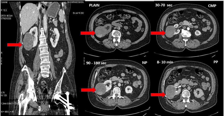
Fig 2. ONCOCYTOMA
Coronal reformatted and axial CT Urogram images showing a well-defined hypodense lesion with thick enhancing septa, few non-enhancing necrotic areas, and perilesional oedema in upper pole of right kidney
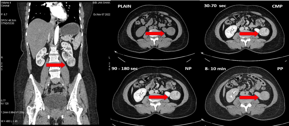
Fig 3. ANGIOMYOLIPOMA
Coronal reformatted and axial CT Urogram images showing a large well-defined heterogeneously hypodense lesion with few calcifications and irregular iso-dense component within, arising from left kidney, causing compression and displacement of adjacent bowel loops.
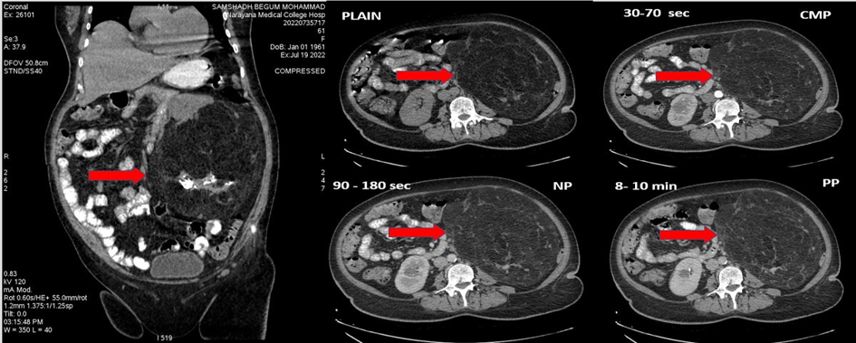
Fig 4. WILMS TUMOR
Axial CT Urogram images showing a large heterogeneously enhancing hypodense lesion replacing entire right kidney with discrete areas of calcification.
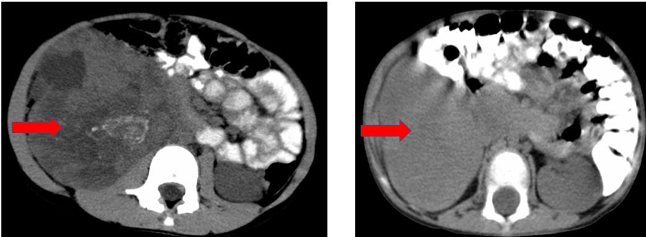
Fig 5. RCC
Sagittal reformatted and axial images showing a large well-defined heterogeneously enhancing hyperdense lesion with multiple non-enhancing necrotic areas arising from lower pole of right kidney, with mild hydronephrosis and causing mild compression with displacement of IVC.
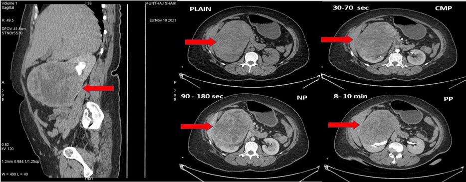
DISCUSSION:
In present study, out of a total of 50 cases studied, 33 (66%) benign & 17 (34%) malignant renal-lesions were diagnosed. The ages of the patients ranged between 4 to 79 years, & a total of 28 males, 22 females were identified in present study. RCC (n = 12) accounted for 24% of renal-lesions & 71% of malignant lesions. Other malignant lesions are TCC (n = 04) & Wilms tumor (n = 01). Benign lesions were complex cysts (n = 09), angiomyolipoma (n = 10), abscess (n = 10) & oncocytoma (n = 04).
Smith et al.12 successfully imaged 17 patients ranging in age from 38 to 78 years old who were suspected to have renal-lesions. There were 10 male patients & 7 female patients. Patients who had renal-lesions further underwent surgeries, renal biopsies, or follow-up examinations. There was a total of 10 cases of RCC, 1 case of TCC, 1 case of angiomyolipoma, 2 cases of complicated renal cysts, & 1 case of pyelonephritis.
There were a total of 70 patients, ranging in age from 4 to 84 years old, who participated in the research that was conducted by Bajwa et al.13 79. In 39 (55.7%) of the cases, neoplastic lesions were found, & in 23 (32.9%) of the cases, inflammatory lesions were found. The remainder 8 patients were diagnosed with renal injury in two, urinoma in three, & complicated cysts in three. It was determined that 32 patients had RCC, 3 patients had Wilms tumor, 2 patients had TCC, & 1 patient each had lymphoma & angiomyolipoma.
In this specific study, in the age-related distribution of renal-lesions, the highest proportion of patients (38%) belonged to the age category of 60 to 69 years. Out of a total of 50 cases, 33 were found to be benign lesions, while 17 were found to be malignant lesions. The RCC was the most-common type of renal lesion, accounting for 71% of all malignant renal-masses & 24% of all renal-masses. 05 out of the 12 patients who were diagnosed with RCC belonged to age group of 60–69 years old (71%). The patient who was diagnosed with RCC at the youngest age was 41-year-old male patient, & the patient who was diagnosed at the oldest age was 69-year-old male patient. The average age was 60.7 years old.
The results of present study are consistent with the results of Gudbjarston et al.14, who described the incidence & distribution of renal-cell cancer on a large population & found that incidence of RCC peaks in the 6th to 8th decade of life with a male to female ratio of 1.3:1. The results of this study & the results of Gudbjarston et al.14 is consistent with one another.
According to Norman Breslow et al.15, there is substantial evidence that there is a female dominance among multicentric & bilateral Wilms tumor cases (P = 0.01). There is evidence to indicate that the female percentage of unicentric cases is lower than that of multi centric & bi - lateral cases. This is despite the fact that the female percentage of unicentric cases is still greater than 50percent of total cases (P = 0.06). This corresponds well with the results of present study, which showed a slight leaning toward female preponderance in cases of Wilms tumor.
In this study, all of the malignant renal-masses (n = 17) have shown soft tissue attenuation on the pre contrast scans. They also showed an average attenuation value of57.02±13.2 in the cortico-medullary phase & 74.7±22.5 in the nephrographic- phase. On the other hand, the benign lesions (n = 33) showed a pre contrast average attenuation of 15.7±10.3, & a post contrast attenuation 24.29±16.7and 32.5±25.1in CMP & NP respectively. Important is the variation in attenuation between the CMP & the NP phases; malignant lesions showed an average increase of 29.02H U, whereas benign lesions showed an average increase of 8.5H U. Present study results correlate well with the study results of the Zagoria et al.16 study, in which the author found that vascular solid renal neoplasms showed average attenuation value of 104.46 & 90.37 in the CMP They had only included the simple cysts in study, whereas the range of benign cases included in present study are (abscess, complex cyst, angiomyolipoma & oncocytoma). The average HU of benign lesions in the study by Zagoria et al.16 was low because they had only included the simple cysts in their study. Therefore, by maintaining a check value of 20 H U as the marked increase in enhancement to differentiate between vascular neoplasms & benign lesions, a sensitivity of 100%, a specificity of 94 %, & an accuracy of 96% could be achieved. Compared to Zagoria et al.16, who achieved a sensitivity of 95.2%, a specificity of 100%, & an accuracy of 97.2%. Because of comparison between CMP & NP phase to the UE phase in this study, a higher level of sensitivity was achieved. An increase of 20 HU was considered to be indicative of malignancy. The specificity of present study was lower when compared to Zagoria et al.16 study because only lesions with high suspicion of malignancy were included, excluding the renal pathologies of certainty of benignity.
The results of this study are remarkably comparable to those obtained by Kopka et al.17, who investigated the effectiveness of UE, CMP, & NP in detecting & characterizing renal-masses. This study misinterpreted two malignant renal-lesions as benign lesions, whereas Kopka et al.17 had misdiagnosed four cases as benign. The lesser specificity in this study is primarily because of the smaller number of subjects included in our study, which is 50, compared to that of Kopka et al. study (n=173).
The high volume of contrast that was utilized in the CMP study conducted by Garant et al.18 is the basis of the elevated attenuation value that was observed. Both these studies have demonstrated a diagnostic sensitivity of one hundred percent when it comes to RCC. On MDCT scans, solid vascular lesions can be detected with a sensitivity of one hundred percent if a maximum value of 20 HU is used as the cutoff point.
In present study, all of the RCCs exhibited soft tissue attenuation on the precontract scan, & their HU ranged between 60.84±11.9 & 83.59±17.15, respectively, on CMP & NP phases. Jinzaki et al.19 have also outlined similar results in their article, in which they demonstrated that RCC, being a very vascular tumor, shows significant enhancement (>20HU) in CMP & NP. The higher attenuation value in CMP of Jinzaki et al.19 study was because of rapid infusion rate of greater contrast volume (120 ml at 3- 5 ml/s), whereas the manual injection method in this study required only 80 ml.
CONCLUSION:
In conclusion, 16-slice CT urography (CTU) demonstrated excellent diagnostic performance in the evaluation of renal masses, achieving a sensitivity of 100%, specificity of 94%, and overall accuracy of 96% in differentiating benign from malignant lesions. Renal cell carcinoma was the most common malignant renal neoplasm, accounting for the majority of malignant cases, with a higher prevalence observed among males. Malignant renal masses exhibited significant contrast enhancement, particularly during the corticomedullary and nephrographic phases, while findings such as renal vein and inferior vena cava invasion were highly suggestive of malignancy. The advantages of multidetector CT, including rapid single-breath-hold acquisition, thin-section imaging, multiphasic evaluation, and advanced three-dimensional reconstruction techniques, substantially improved lesion detection and characterization. Although the study was limited by a relatively small sample size and non-standardized contrast administration, the findings clearly indicate that 16-slice CTU, combined with appropriate post-processing and reformatting techniques, is a highly accurate and reliable imaging modality for the detection, characterization, and preoperative assessment of renal masses.
REFERENCE:
1. Ng CS, Wood CG, Silverman PM, Tannir NM, Tamboli P, Sandler CM. Renal Cell Carcinoma: Diagnosis, Staging, and Surveillance. American Journal of Roentgenology. 2008 Oct;191(4):1220–32.
2. Sheth, S., Scatarige, J.C., Horton, K.M., Corl, F.M. and Fishman, E.K. (2001). Current Concepts in the Diagnosis and Management of Renal Cell Carcinoma: Role of Multidetector CT and Three-dimensional CT. RadioGraphics, 21(suppl_1),pp.S237–S254.
3. Browne RFJ, Meehan CP, Colville J, Power R, Torreggiani WC. Transitional Cell Carcinoma of the Upper Urinary Tract: Spectrum of Imaging Findings. RadioGraphics [Internet]. 2005 Nov ;25(6):1609–27.
4. Carrim ZI, Murchison JT. The prevalence of simple renal and hepatic cysts detected by spiral computed tomography. Clinical radiology [Internet]. 2003 ;58(8):626–9.
5. Uejima Y, Niimi H, Kato R, Furuichi M, Sato S, Kitajima I, et al. Infected simple renal cyst due to Streptococcus pneumoniae rapidly diagnosed by the melting temperature mapping method: a case report. BMC Pediatrics [Internet]. 2021 Jun 5 ;21(1).
6. Lowe LH, Isuani BH, Heller RM, Stein SM, Johnson JE, Navarro OM, et al. Pediatric Renal Masses: Wilms Tumor and Beyond. RadioGraphics. 2000 Nov;20(6):1585–603.
7.
8. Choyke PL. Imaging of hereditary renal cancer. Radiologic Clinics of North America. 2003 Sep;41(5):1037–51.
9. World Journal of Clinical Cases - Baishideng Publishing Group [Internet]. Wjgnet.com. 2022.
10. Lai H-Y, Chen C-K, Lee Y-H, Tsai P-P, Chen J-H, Shen W-C. Multicentric aggressive angiomyolipomas: a rare form of PEComas. AJR American journal of roentgenology [Internet]. 2006 Mar 1;186(3):837–40.
11. Taveira-DaSilva A, Moss J. Management of lymphangioleiomyomatosis. F1000Prime Reports. 2014 Dec 1;6.
12. Multidetector row CT of the kidneys Atadan Tunaci, Ensar Yekeler European Journal of Radiology - October 2004 (Vol. 52, Issue 1, Pages 56-66.
13. Smith AD, Remer EM, Cox KL, Lieber ML, Allen BC, Shah SN, et al. Small (≤3 cm) hyperechoic renal masses: comparison of helical and conventional CT for diagnosing angiomyolipoma. AJR Am J Roentgenol. 1996;167(3):721-724.
14. Bajwa RPS, Sandhu P, Aulakh BS, Sandhu JS, Saggar K, Ahluwalia A. Helical CT evaluation of renal mass lesions: a prospective study. J Indian Acad Clin Med. 2007;8(3):262-264.
15. Gudbjartsson T, Hardarson S, Petursdottir V, Thoroddsen A, Magnusson J, Einarsson GV. Renal cell carcinoma in Iceland: clinicopathological features and epidemiology. Scand J Urol Nephrol. 2005;39(4):283-287.
16. Breslow N, Olshan A, Beckwith JB, Green DM. Epidemiology of Wilms tumor. Med Pediatr Oncol. 1993;21(3):172-181.
17. Zagoria RJ, Wolfman NT, Karstaedt N, Chen YM, Dyer RB. CT features of renal cell carcinoma and benign renal masses: value of contrast enhancement in differentiation. Radiology. 1990;176(3):709-713.
18. Kopka L, Fischer U, Zoeller G, Funke M, Oestmann JW, Grabbe E. Dual-phase helical CT of renal masses: value in differentiation and characterization of renal lesions. Radiology. 1997;205(2):419-425.
19. Garant M, Bonaldi VM, Taourel P, Pujol J, Michel F. Helical CT evaluation of renal masses using corticomedullary and nephrographic phase imaging. Eur Radiol. 1998;8(4):617-623.
20. Jinzaki M, Tanimoto A, Mukai M, Ikeda E, Kobayashi S, Yuasa Y, et al. Double-phase helical CT of small renal parenchymal neoplasms: correlation with pathologic findings and tumor angiogenesis. J Comput Assist Tomogr. 2000;24(6):835-842.