STUDY OF FROZEN SECTION IN ORAL SQUAMOUS CELL CARCINOMA IN CONTRAST TO METACHROMATIC STAIN, AT TMC & DR BRAM TEACHING HOSPITAL
- Dr Sayandeep Nath , Associate Professor, Department of Pathology, TMC & Dr. BRAM teaching hospital, Agartala, West Tripura
- Dr Pragyamita Datta , Assistant Professor, Department of Pathology, TMC & Dr. BRAM teaching hospital, Agartala, West Tripura
- Dr Sutanuka Khasnabish , Associate Professor, Department of Pathology, TMC & Dr. BRAM teaching hospital, Agartala, West Tripura
Article Information:
Abstract:
Aim: To determine whether or not toluidine blue is useful for intraoperative assessment of tumour margins following removal of a primary oral cavity tumour.Material and Methods: The present prospective investigation was conducted over the course of two years on 45 cases of oral cancer at the Histopathology Department of Pathology at Tripura medical college and DR BRAM. In this study, Toluidine Blue Dye and Rapid Hematoxylin (eosin method) were used for staining slides for frozen section.Results: The mean age in this study was 50.76±13.06 years. Most of the cases were reported as TNM Stage I followed by TNM Stage II & minimum cases belonged to TNM Stage IV. Out of 19 positive margins on histopathology; 16 were found to be true positive on frozen section while 6 were found to be false positive, 3 were found to false negative and 155 were found to be true negative. Out of 19 positive margins on histopathology; 15 were found to be true positive on toluidine blue while 8 were found to be false positive, 4 were found to false negative and 153 were found to be true negative.Conclusion: It can be concluded from the results that that toluidine blue, due to its capacity to properly highlight cancer cells in contrast to normal epithelium, could provide a low-cost facility in developing nations where frozen section is not readily available.
Keywords:
Article :
INTRODUCTION:
The 11th most prevalent cancer in the world is oral cancer1. Oral squamous cell carcinomas (OSCC) are the most common kind of oral cancer, constituting about 90% of all cases2. The standard method of treating OSCC is surgical resection3. Performing a complete removal of a tumour at the main site with a negative margin is a crucial goal of surgical oncology. Positive surgical margins are connected with local recurrence & poor patient outcomes when leftover malignant tissue is not detected4,5. Oral cancer is the most common cancer in the world, with an estimated 350,000 to 400,000 new cases per year. Developed nations seem to be the most common location for these occurrences & events, however they can be found in any nation. Cancer of the mouth & throat accounts for 2% of all malignant tumours & malignancies. Early detection & treatment for oral cancer will decrease the likelihood of dying from the disease & guarantee long-term survival.6
As soon as the diagnosis is made, the lesion is surgically removed with wider margins to reduce the probability of recurrence to almost none. In order to successfully remove a tumour, it is necessary to precisely delineate its soft tissue borders during surgery. Frozen section biopsy is the gold standard method, with a sensitivity of between 88.8 & 98.9 percent.7
Cryosection, also known as frozen section analysis, is a pathological method used for quick microscopic inspection & diagnosis of a specimen or disease. It is commonly employed in oncologic surgery & intraoperative patient diagnosis.8 The frozen section's layout is typically crucial for making a prompt pathology diagnosis during surgery. It is important for the surgeon to know whether or not the margins of his resection for a malignant neoplasm are clear before closing, whether or not there is an unusual disease process which required identification & to determine what to do next, & whether or not enough tissue was collected to further improve the disease process. Frozen sections can be used for this purpose; however, it should be emphasised that, while the technique is useful, it is currently only used to assess tissue morphology with updated H&E stains in order to form a conclusion & does not submit any other supporting techniques.8,9
These techniques have limitations & challenges in terms of efficiency, effectiveness, & scalability. Even in modern oncology centres, frozen sections remain the gold standard for intraoperative margin assessment (FS). Physical inspection reveals that only a tiny percentage of the margins are investigated, despite the fact that this is a reliable technique for detecting residual tumour in the collected tissue. As a result, the likelihood of sampling error cannot be discounted. Toluidine blue is a low-priced, readily available metachromatic stain that has a high affinity for DNA & RNA (TB). Normal mucosa does not readily take up the dye, whereas cancerous & premalignant cells do. In a number of studies, TB was found to be effective at detecting oral squamous cell carcinoma (OSCC) & oral premalignant lesions, ranging in age from 17-20 years. To date, only three studies have focused on TB during surgery. One study intraorally gave TB to measure the severity of the lesions before excision.9 Based on this information, we decided to use toluidine blue for intraoperative tumour margin assessment following removal of a primary oral cavity tumour. The objectives of the study are as follows:
1. To estimate the accuracy of frozen section in oral cancer in context to metachromatic stain.
2. To compare the accuracy of frozen section in context to metachromatic stain by correlating with histopathological findings.
MATERIALS AND METHODS:
The study was carried out for a period of two years (January 2024-2026) in Histopathology Department of Pathology, Tripura medical college and DR BRAM teaching hospital.
Type of Study
It’s an analytical and prospective study
Sample Size
Sample size formula with desired error of margin:
n = (Z α/2)2 X p X (1-p) / d2
where,
Zα/2 is the level of significance at 5% i.e. 95% confidence interval = 1.96
p = prevalence of suspected cases of malignancy = 5.4% = 0.116
d = desired error of margin (0.01)
n=44.38
45 patients were required for the present study.
Source of Data
Minimum 45 patients of suspected oral malignancy undergoing intraoperative surgery in OT complex of Frozen section were effectively used as the diagnostic step in suspected cases of malignancy of any organ. It was a simple, cost effective, and appropriate method that provides accurate diagnostic yield.
Inclusion criteria
1. All the diagnosed cases of oral carcinoma which will undergo surgery, will be considered as sample.
2. The patients will be recruited regardless of the age, gender, ethnicity, and tumor stage.
Exclusion criteria
1. Patients with prior history of head and neck malignancy,
2. Tumors of the oro-pharynx or having previously undergone treatment (surgery and/or radio-/chemotherapy) for the current oral cancer
3. Patients allergic to dye will be excluded from the study.
Materials required for frozen section: The frozen section specimens, as per laboratory routine,
1. Cryostat (leica CM)
2. Materials for Rapid Hand E staining (Harris hematoxylin, distilled water, scott’s tap water, eosin, absolute alcohol, glacial acetic acid, xylene and dystyrene plasticizer xylene-DPX)
3. Metachromatic Stain (Toluidine Blue Dye Staining)
4. Specimens
5. Tissue freezing medium as optimum cutting temperature compound (OCT)
6. Compound light microscope
7. Glass slide and slide marker
8. Scalpel and blade
9. Normal saline.
10. Filter paper
11. Forceps and gloves
Materials required for paraffin embedded sections
1. Automated tissue processor (leica TP)
2. Rotatory microscope (leica RM)
3. Compound light microscope
4. Leukhart’s moulds
5. Paraffin
6. Tissue float bath
7. Hot plate
8. Scalpel and blade
9. Glass blade
10. Cover slips
11. Glass marker
12. Distyrene plasticizer in (DPX) mountant
13. Filter paper
14. Gauze piece
Methods
In this study, Toluidine Blue Dye and Rapid Hematoxylin (eosin method) were used for staining slides for frozen section.
Toluidine Blue Dye staining for frozen section10
1. One per cent toluidine blue solution was kept. Once the primary tumor was excised a designated senior resident irrigated the tumor margins with normal saline, followed by 1% acetic acid.
2. The margins were then gently dried with gauze and painted with the 1% toluidine blue solution with a cotton swab.
3. The stain was left in place for 30 s after which the tissue was once again irrigated with normal saline followed by 1% acetic acid. The staining patterns were observed and noted by the senior resident independently.
4. Margins which stained dark blue was considered positive, whereas those which stained light blue or do not take the stain at all will be considered negative.
Rapid hematoxylin and eosin staining for frozen section11
1. All the resected specimen submitted for intraoperative frozen section has to be grossly examined and the representative area should be embedded in optimum cutting temperature compound (OCTC) and its frozen in cryostat at -20 degree Celsius. The sections were cut and slides were made.
2. The sections are cut via cryostat and taken on glass slide
3. Sections are stained with hematoxylin for 1.5 minutes and washed with tap water shortly
4. After being immersed in acid alcohol for 1-2 second they were then rinsed with lithium carbonate, alkaline tap water or scott’s tap water
5. stain with eosin for 10 seconds
6. Rinsed in tap water
7. Dehydrate, clear and mount in DPX
Statistical analysis
Statistical analysis was done by using descriptive and inferential statistics with the help of SPSS 24 version.
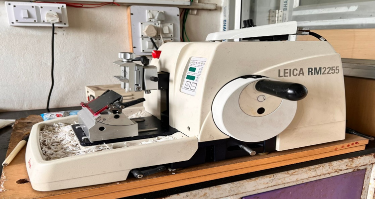
Figure 1: Rotatory microtome (Leica rm2255) use for sectioning of paraffin embedded specimens
RESULTS:
Out of 45 subjects, 34 (75.6%) were males and 11 (24.4%) were females. The mean age in this study was 50.76±13.06 years with maximum subjects from age group of 41-50 years followed by 51-60 years.
Table 1: Baseline data among the study subjects
|
Gender |
N=45 |
% |
|
Male |
34 |
75.6 |
|
Female |
11 |
24.4 |
|
Age Group (in years) |
|
|
|
30-40 |
9 |
20 |
|
41-50 |
14 |
31.1 |
|
51-60 |
13 |
28.9 |
|
>60 |
9 |
20 |
|
Mean±SD |
50.76±13.06 |
|
|
Laterality |
|
|
|
Left |
28 |
62.3 |
|
Right |
17 |
37.7 |
Most common site for the cancer in this study was buccal mucosa (31.1%) followed by Gingivobuccal sulcus (26%) & tongue (17.8%) as shown in graph 1.
Graph 1: Site distribution among the study subjects
According to histopathological diagnosis; well differentiated, moderately differentiated and poorly differentiated squamous cell carcinoma was reported among 46.7%, 42.2% and 11.1% of the subjects respectively. Most of the cases were reported as TNM Stage I followed by TNM Stage II & minimum cases belonged to TNM Stage IV (table 2).
Table 2: Histopathological diagnosis and TNM staging
|
Histopathological Diagnosis |
N=45 |
% |
|
Well Differentiated Squamous Cell Carcinoma |
21 |
46.7 |
|
Moderately Differentiated Squamous Cell Carcinoma |
19 |
42.2 |
|
Poorly Differentiated Squamous Cell Carcinoma |
5 |
11.1 |
|
TNM Staging |
|
|
|
I |
16 |
35.6 |
|
II |
14 |
31.1 |
|
III |
10 |
22.2 |
|
IVA |
2 |
4.4 |
|
IVB |
1 |
2.2 |
|
IVC |
2 |
4.4 |
About 180 margins were sampled from this pool of patients. On histopathology, 19 margins were found to be positive for cancer (graph 2).
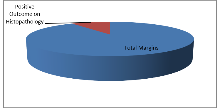
GRAPH 2: Margin analysed for carcinoma and outcome on histopathology
Out of 19 positive margins on histopathology; 16 were found to be true positive on frozen section while 6 were found to be false positive, 3 were found to false negative and 155 were found to be true negative. According to kappa analysis, there was good correlation between margin outcome of frozen section considering histopathology as gold standard (table 3).
Table 3: Comparison of margin outcome of frozen section with histopathology
|
|
Margin Status |
Final Histopathology |
Total |
|
|
|
|
+ve |
-ve |
|
|
Frozen Section |
+ve |
16 |
6 |
22 |
|
-ve |
3 |
155 |
158 |
|
|
Total |
|
19 |
161 |
180 |
|
p value |
|
0.005* |
|
|
*: statistically significant
Out of 19 positive margins on histopathology; 15 were found to be true positive on toluidine blue while 8 were found to be false positive, 4 were found to false negative and 153 were found to be true negative. According to kappa analysis, there was good correlation between margin outcome of toluidine blue considering histopathology as gold standard (table 4).
Table 4: Comparison of margin outcome of toluidine blue with histopathology
|
|
Margin Status |
Final Histopathology |
Total |
|
|
|
|
+ve |
-ve |
|
|
Toluidine Blue |
+ve |
15 |
8 |
23 |
|
-ve |
4 |
153 |
157 |
|
|
Total |
|
19 |
161 |
180 |
|
p value |
|
0.007 |
|
|
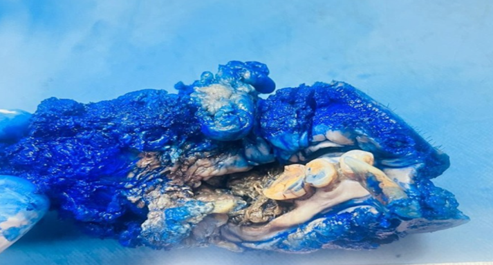
Figure 2: Resected specimen of toludine blue positive surgical margin
Sensitivity, specificity, PPV, NPV and accuracy of frozen section considering histopathology as gold standard was 84.21%, 96.27%, 72.73%, 98.10% and 95.00% respectively. Sensitivity, specificity, PPV, NPV and accuracy of toluidine blue considering histopathology as gold standard was 78.95%, 95.03%, 65.22%, 97.45% and 93.33% respectively (table 5).
Table 5: Yield (sensitivity and specificity) and reliability (PPV and NPV) of frozen section and toluidine blue as compared to histopathology
|
Parameters |
Frozen Section |
Toluidine Blue |
|
Sensitivity |
84.21% |
78.95% |
|
Specificity |
96.27% |
95.03% |
|
PPV |
72.73% |
65.22% |
|
NPV |
98.10% |
97.45% |
|
Accuracy |
95.00% |
93.33% |
DISCUSSION:
Recently, toluidine blue has been reported as a complementary method to clinical examination for the early diagnosis of oral cavity cancers & premalignant lesions. Epstein et al. discovered that the sensitivity of detecting a malignant tumour increased from 26.6% to 96.7% when using toluidine blue in conjunction with an unaided clinical exam when screening for recurrence in individuals who had previously been treated for upper aero-digestive tract malignancies.12 To be fair, it has been clinically demonstrated to be effective at detecting malignant lesions in the oral cavity.13 Although intraoperative tumour margin identification using this method has not been extensively characterised, it has shown promise. Therefore, 45 patients of oral cancer were included in the present prospective study that was conducted over the course of 2 years in the Histopathology Dept of Pathology at institute. The purpose of this research was to compare the results of intraoperative tumour margin assessment using toluidine blue & frozen sections to those obtained following the surgical removal of a primary tumour from the oral cavity.
There was male dominancy in the present study. Similar male dominancy was stated by Montasir Junaid et al14 and Sheikh AH et al15 in their study. Cancers of the mouth & throat affect men at a rate two times that of women. It's possible that men's historical propensity to consume cigarettes & alcohol contributes to this.
The mean age in this study was 50.60±11.93 years. 20% of the subjects belonged to 30-40 years of age. In 41-50 years of age, 31.1% of the subjects reported. 28.89% & 20% of the subjects were having age of 51-60 & >60 years respectively. In a study by Montasir Junaid et al14, mean age of the patients was 50.07 ± 15.73 years. Hana’a Hezam Algadi et al16 in their study revealed mean age of 54.4 ± 12.3 years. Sheikh AH et al15 in their study reported mean age of 39.56 ±7.514 years. This early presentation in their study might be due to difference in study area & design. Oral & pharyngeal cancers tend to take a long time to manifest, hence they are uncommon in younger people. The average age of these individuals when their cancer is initially diagnosed is over 55.
According to histopathological diagnosis; well differentiated, moderately differentiated & poorly differentiated squamous cell carcinoma was reported among 46.7%, 42.2% & 11.1% of the subjects respectively. Agrawal et al17 found 10 cases (55.5%) of well-differentiated SQCC & 4 cases (22.2%) of moderately differentiated SQCC in his analysis. Similarly, M. Selvamani et al18 in their study described that well-differentiated SQCC accounted for 23 cases (44.2%).
Most common site for the cancer in this study was buccal mucosa (31.1%) followed by Gingivobuccal sulcus (26%) & tongue (17.8%). 4.4% each of the subjects were having cancer at Gingivobuccal sulcus involving buccal mucosa & Lateral border of tongue. 2.2% each of the subjects were having cancer at Alveolus, Buccal mucosa involving GB sulcus & alveolus, Labiogingival sulcus, Lower Alveolus, Lower labial mucosa, Lower labial sulcus involving alveolus & Upper gingivosulcus involving alveolus site in this study. Similar Montasir Junaid et al14 in their study reported that buccal mucosa tumors was the most common site (41%) of the lesions followed by tongue (23.2%). Sheikh AH et al15 in their study stated that buccal mucosa was the most common site (48%). Hana’a Hezam Algadi et al16 in their study found that the oral cavity subsites were tongue (n = 13; 43.3%) followed by buccal mucosa (n = 4; 13.3%).
According to kappa analysis, there was good correlation between margin outcome of frozen section considering histopathology as gold standard. Sensitivity, specificity, PPV, NPV & accuracy of frozen section considering histopathology as gold standard was 84.21%, 96.27%, 72.73%, 98.10% & 95.00% respectively. Results from frozen section biopsies were consistent with those from the final histopathological study, as reported by Montasir Junaid et al. Six (4%) of the final histopathologically analysed mucosal surgical margins were positive for invasive cancer in the study by Hana'a Hezam Algadi et al.16 As validated by final histology of the same margins, the FS yielded 3 positives, 134 negatives, 3 false-negatives, & 0 false positives. The test was 100% specific (95% CI 97.3-100.0%) & 50% sensitive (11.8-88.2%). The positive present value was 100%, & the negative present value was 97.8% (95% CI, 95.2% to 99.0%). FS was 97.9 percent precise while making diagnoses. The results of the current investigation are consistent with these hypotheses.
The specificity of FS was 100%, which is quite close to the values we found (by Junaid et al)14 Gupta C et al19 in their study stated that sensitivity, specificity, PPV, NPV & accuracy of frozen section considering histopathology as gold standard was 90%, 98.32%, 69.23%, 99.57% & 97.98% respectively. These findings are in accordance to the present study. Sharma et al20 in their study mentioned that accuracy of FS was 96.74%. Chaturvedi et al21 in their study mentioned that accuracy of FS was 100%.
According to kappa analysis, there was good correlation between margin outcome of toluidine blue considering histopathology as gold standard. Sensitivity, specificity, PPV, NPV & accuracy of toluidine blue considering histopathology as gold standard was 78.95%, 95.03%, 65.22%, 97.45% & 93.33% respectively in this study. Montasir Junaid et al14 found that, out of 280 samples, 11 margins showed positive toluidine blue staining. However, only three of these 11 margins were confirmed to be positive by histology. That is, all positive margins were detected by toluidine blue, without any false negative results. After being stained with toluidine blue, all positive cases were detected, demonstrating a sensitivity of 100%; however, 8/280 (2.8%) false-positives were also detected, indicating a specificity of 97%. With a PPV of 27.2% & an NPV of 100%, toluidine was shown to have a high degree of diagnostic accuracy (97.1%). The results of the current investigation are consistent with these hypotheses.
Careful management of the tissue during excision may boost the PPV, since it has been established that inflammation related to greater mucosal stress during excision is associated with false positive results [Mashberg A et al].22 In any case, we'd like to call attention to the fact that the test's NPV was 97.45, meaning that it almost never missed a positive margin. Staining with toluidine blue was shown by Portugal et al23 to detect all positive tumour margins with few false positives & no false negatives. The study also noted that three cases of second primary lesions were discovered thanks to staining.
Screening for oral premalignant & malignant lesions with TB has a sensitivity of 77.0–100% & a specificity of 65.5–100%, respectively.19,20,24 Both Onofre et al25 & Warnakulasuriya et al26 found that TB had a false-negative rate of 0% when used to identify oral cancer. Sheikh AH et al investigation found that the dye had a sensitivity of 74% (20/27 cases were positive) & a specificity of 100% (all 23 cases were negative). We were able to get an accuracy of 86%.
Limitations
1. Large no of cases is required to carry out the study.
For correlation, cryostat facility is mandatory wherein majority of centers its is not available
CONCLUSION:
In resource-limited environments where facilities for frozen section biopsies are not readily available surgeons often rely on either their clinical judgment or on the final postoperative histopathology for assessing microscopic extension of the disease. This leads to a major disadvantage for patients and may account for high recurrence rates of OCSCC in developing countries. In our study there is high correlation between Toluidine blue and frozen section biopsies to detect malignancies.
REFERENCES:
1. Ghantous Y, Abu EI. Global incidence & risk factors of Oral Cancer. Harefuah. 2017;156(10):645–9.
2. Warnakulasuriya S. Global epidemiology of oral & oropharyngeal cancer. Oral Oncol. 2009;45(4–5):309–16.
3. Fujiwara RJT, Burtness B, Husain ZA, Judson BL, Bhatia A, Sasaki CT, et al. Treatment guidelines & patterns of care in oral cavity squamous cell carcinoma: primary surgical resection vs. nonsurgical treatment. Oral Oncol.2017;71:129–37.
4. Sutton DN, Brown JS, Rogers SN, Vaughan ED, Woolgar JA. The prognostic implications of the surgical margin in oral squamous cell carcinoma. Int J Oral Maxillofac Surg. 2003;32(1):30–4.
5. McMahon J, O'Brien CJ, Pathak I, Hamill R, McNeil E, Hammersley N, et al. Influence of condition of surgical margins on local recurrence & disease specific survival in oral & oropharyngeal cancer. Br J Oral Maxillofac Surg.2003;41(4):224–31
6. Scully C MD. International Encyclopedia of Public Health. 2008; 668-77.
7. DiNardo LJ, Lin J, Karageorge LS, Powers CN. Accuracy, utility, & cost of frozen section margins in head & neck cancer surgery. Laryngoscope 2000; 110: 1773-6.
8. Sheikh AH, Farooqui WA, Fatima S. Toluidine Staining as a Screening Tool for Soft Tissue Margins of Cancerous Oral Lesions. Research Square; 2020.
9. Kerawala CJ, Beale V, Reed M, Martin IC. The role of vital tissue staining in the marginal control of oral squamous cell carcinoma. Int J Oral Maxillofac Surg. 2000;29(1):32–5.
10. Sridharan, G, Shankar AA. Toluidine Blue: A review of its chemistry and clinical utility. J Oral Maxillofac Pathol. 2012;16:251–5.
11. Bancroft JD, Gamble M, editors. Theory and Practice of Histological Techniques. 6th ed. New York: Churchill Livingstone;2008.
12. Epstein JB, Sciubba J, Silverman S Jr, Sroussi HY: Utility of toluidine blue in oral premalignant lesions & squamous cell carcinoma: continuing research & implications for clinical practice. Head Neck 2007,29:948-958.
13. Epstein JB, Guneri P: The adjunctive role of toluidine blue in detection of oral premalignant & malignant lesions. Curr Opin Otolaryngol Head Neck Surg 2009, 17:79- 87.
14. Junaid M, Choudhary MM, Sobani ZA, et al. A comparative analysis of toluidine blue with frozen section in oral squamous cell carcinoma. World J Surg Oncol. 2012;10(1):1-5.
15. Sheikh AH, Farooqui WA, Fatima S. Toluidine Staining as a Screening Tool for Soft Tissue Margins of Cancerous Oral Lesions. Research Square; 2020.
16. Algadi HA, Abou-Bakr AA, Jamali OM, Fathy LM. Toluidine blue versus frozen section for assessment of mucosal tumor margins in oral squamous cell carcinoma. BMC Cancer. 2020 Dec;20(1):1-8.
17. Agarwal, AK, Sethi A, Sareen D, Dhingra S. Oral & oropharyngeal squamous cell carcinoma in our population: The clinic-pathological & morphological description of 153 cases. Int. J. Morphol 2011;29:686-93.
18. Selvamani M, Yamunadevi A, Basandi PS, Madhushankari GS. Prevalence of oral squamous cell carcinoma of tongue in & around Davangere, Karnataka, India: A retrospective study over 13 years. J Pharm Bioall Sci 2015;7:S491-4.
19. Gupta C, Bhola N, Jadhav A, Mishra A, Hingnikar P, Ghavat C. Does diagnostic accuracy of surgeon's perception outweigh frozen section analysis in determining intraoperative clear mucosal surgical margins in oral squamous cell carcinoma patients? NatlJ Maxillofac Surg 2020;11:182-5.
20. Sharma SM, Prasad BR, Pushparaj S, Poojary D. Accuracy of intraoperative frozen section in assessing margins in oral cancer resection. J Maxillofac Oral Surg 2009;8:357 61.
21. Chaturvedi P, Datta S, Nair S, Nair D, Pawar P, Vaishampayan S, et al. Gross examination by the surgeon as an alternative to frozen section for assessment of adequacy of surgical margin in head & neck squamous cell carcinoma. Head Neck 2014;36:557 63.
22. Mashberg A, Samit A: Early diagnosis of asymptomatic oral & oropharyngeal squamous cancers. CA Cancer J Clin 1995; 45:328-351.
23. Portugal LG, Wilson KM, Biddinger PW, Gluckman JL. The role of toluidine blue in assessing margin status after resection of squamous cell carcinomas of the upper aerodigestive tract. Arch Otolaryngol Head Neck Surg. 1996; 122(5):517–9.
24. Du E, Ow TJ, Lo YT, Gersten A, Schiff BA, Tassler AB, et al. Refining the utility and role of frozen section in head & neck squamous cell carcinoma resection. Laryngoscope. 2016;126(8):1768–75.
25. Onofre MA, Sposto MR, Navarro CM. Reliability of toluidine blue application in the detection of oral epithelial dysplasia & in situ & invasive squamous cell carcinomas. Oral Surg Oral Med Oral Pathol Oral Radiol Endod. 2001;91(5):535–40.
26. Warnakulasuriya KA, Johnson NW. Sensitivity & specificity of Ora Scan (R)toluidine blue mouth rinse in the detection of oral cancer & precancer. J Oral Pathol Med. 1996;25(3):97–103.