Echocardiographic Assessment of Valvular Regurgitation Patterns and Left Atrial Remodeling in Patients with Dilated Cardiomyopathy.
- Suhasini Atharga , Assistant Professor, Department of Cardiology, JNMC, KAHER, Belagavi, Karnataka, India.
- Shrikanth Metri , Associate Professor, Department of General Medicine, JNMC, KAHER, Belagavi, Karnataka, India.
- Sanjay Porwal , Professor, Department of Cardiology, JNMC, KAHER, Belagavi, Karnataka, India.
- Suresh Patted , Professor, Department of Cardiology, JNMC, KAHER, Belagavi, Karnataka, India.
- Sameer Ambar , Professor, Department of Cardiology, JNMC, KAHER, Belagavi, Karnataka, India.
- Prasad M. R , Associate Professor, Department of Cardiology, JNMC, KAHER, Belagavi, Karnataka, India.
- Vijayanand Metgudmath , Associate Professor, Department of Cardiology, JNMC, KAHER, Belagavi, Karnataka, India.
- Vishwanath Hesarur , Assistant Professor, Department of Cardiology, JNMC, KAHER, Belagavi, Karnataka, India.
Article Information:
Abstract:
Background: Dilated cardiomyopathy (DCM) is characterized by ventricular chamber dilatation and impaired systolic function, frequently leading to secondary valvular regurgitation and structural remodeling of the left atrium. Functional mitral and tricuspid regurgitation are common consequences of ventricular dilatation and contribute to worsening cardiac performance and adverse clinical outcomes. Echocardiography plays a pivotal role in evaluating valvular regurgitation and left atrial remodeling in patients with DCM. Methods: This hospital-based observational study was conducted at a tertiary care teaching hospital between March 2023 and April 2024. Fifty patients diagnosed with DCM and left ventricular ejection fraction (LVEF) ≤45% were enrolled. All participants underwent comprehensive two-dimensional and three-dimensional transthoracic echocardiography. Assessment included left ventricular dimensions and function, left atrial volume and left atrial volume index, mitral regurgitation (MR), tricuspid regurgitation (TR), pulmonary artery pressure (PAP) estimation, and Doppler evaluation of diastolic function. Statistical analysis was performed using SPSS version 23.0, and correlations were assessed using Spearman’s correlation coefficient. Results: Among the 50 patients studied, 54% were males and 46% were females, with the majority belonging to the 41–60-year age group. Echocardiographic evaluation demonstrated a high prevalence of functional mitral and tricuspid regurgitation associated with ventricular dilatation. Significant left atrial remodeling was observed, reflected by increased left atrial volume and left atrial volume index. An inverse correlation was noted between LVEF and left atrial volume (r = –0.336, p = 0.018) and between LVEF and left atrial volume index (r = –0.334, p = 0.017). Severity of valvular regurgitation was associated with greater left atrial enlargement and adverse ventricular remodeling. Conclusion: Patients with DCM exhibit significant valvular regurgitation and left atrial remodeling secondary to ventricular dysfunction. Echocardiographic assessment of valvular regurgitation patterns and left atrial remodeling provides valuable insights into disease severity and may serve as an important tool for risk stratification and clinical management.
Keywords:
Article :
INTRODUCTION:
Dilated cardiomyopathy (DCM) is a primary myocardial disorder characterized by left ventricular dilatation and systolic dysfunction in the absence of abnormal loading conditions or significant coronary artery disease sufficient to explain the degree of myocardial impairment. It represents one of the most common causes of heart failure, arrhythmias, sudden cardiac death, and cardiac transplantation worldwide. The disease is associated with progressive ventricular remodeling, leading to deterioration of cardiac function and adverse clinical outcomes.[1,2]
Structural remodeling of the left ventricle in DCM alters ventricular geometry and disrupts the normal relationship between the myocardium, papillary muscles, and atrioventricular valves. Consequently, functional valvular regurgitation develops despite anatomically normal valve leaflets. Functional mitral regurgitation (MR) is the most frequently encountered valvular abnormality in DCM and results from annular dilatation, papillary muscle displacement, leaflet tethering, and impaired systolic closing forces.[3,4] In addition, tricuspid regurgitation (TR) may occur secondary to right ventricular dysfunction, pulmonary hypertension, and tricuspid annular dilatation, further contributing to disease progression and symptomatic heart failure.[5]
Valvular regurgitation imposes chronic volume overload on the atria and ventricles, accelerating cardiac remodeling. Persistent elevation of left ventricular filling pressures and regurgitant flow into the left atrium promote structural and functional changes collectively termed as left atrial remodeling. These changes include atrial enlargement, fibrosis, altered compliance, and impaired reservoir function.[6] Left atrial enlargement is increasingly recognized as an integrated marker of the chronicity and severity of cardiovascular disease and has been associated with atrial fibrillation, worsening heart failure, thromboembolic events, and increased mortality.[7,8]
Echocardiography remains the cornerstone imaging modality for evaluating patients with DCM because it provides comprehensive assessment of chamber dimensions, ventricular systolic and diastolic function, valvular abnormalities, pulmonary pressures, and atrial remodeling in a non-invasive and cost-effective manner. Recent advances in two-dimensional and three-dimensional echocardiography permit accurate quantification of regurgitant lesions and left atrial volumes, thereby improving risk stratification and clinical decision-making.[9,10]
Given the prognostic significance of valvular regurgitation and left atrial remodeling in DCM, detailed echocardiographic evaluation may provide valuable insights into disease severity and progression.
AIMS AND OBJECTIVES
The present study was undertaken to evaluate valvular regurgitation patterns and left atrial remodeling in patients with dilated cardiomyopathy using echocardiography. The specific objectives were to assess the prevalence and severity of functional mitral and tricuspid regurgitation, evaluate left atrial remodeling by measuring left atrial volume (LAV) and left atrial volume index (LAVI), determine the relationship between LAV and LAVI with left ventricular systolic function as assessed by left ventricular ejection fraction (LVEF), and analyse
MATERIALS AND METHODS:
Study Design
This hospital-based observational single-centre study was conducted between March 2023 and April 2024 at KLE's Dr. Prabhakar Kore Hospital and Medical Research Centre, Belagavi, Karnataka, a tertiary care teaching hospital affiliated with KLE Academy of Higher Education and Research (Deemed-to-be-University). Patients diagnosed with dilated cardiomyopathy (DCM) with a left ventricular ejection fraction (LVEF) ≤45% were enrolled in the study. All participants underwent comprehensive transthoracic echocardiographic evaluation, including conventional two-dimensional (2D) and three-dimensional (3D) echocardiography, to assess valvular regurgitation patterns, left atrial remodeling, and left ventricular function.
Inclusion and Exclusion Criteria
The study included patients aged 25–85 years who were diagnosed with dilated cardiomyopathy (DCM) and had a left ventricular ejection fraction (LVEF) ≤45% on echocardiographic evaluation. Patients with conditions that could independently influence valvular function or cardiac chamber dimensions were excluded, including those with organic mitral, aortic, or pulmonary valve disease, previous myocardial infarction, intracardiac shunts, and prosthetic heart valves. These criteria were applied to ensure accurate assessment of valvular regurgitation patterns and left atrial remodeling attributable primarily to dilated cardiomyopathy.
Sample Size Calculation
The study consisted of total 50 patients who were diagnosed with Dilated Cardiomyopathy [DCM] with left ventricular ejection fraction of ≤ 45% in OPD or IPD patients at KLE’S Dr. Prabhakar Kore Hospital and Medical Research Centre, Belagavi were selected for this study.
Sample Size is Calculated at 95% of Confidence Interval
20% Tolerable error
5% Alteration [1.05] where,
z1- α/2 = 1.96
p = 69.2 [left atrial volume]
q = 100 – p
= 100 – 69.2
= 30.8
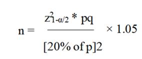
n = 50 patients
Data Collection Procedure
After obtaining institutional ethical clearance, eligible OPD and IPD patients diagnosed with dilated cardiomyopathy (DCM) and having a left ventricular ejection fraction (LVEF) ≤45% were enrolled in the study. All participants underwent comprehensive transthoracic echocardiographic evaluation using an EPIQ 7C ultrasound system equipped with an X5-1 matrix transducer. Conventional two-dimensional (2D), three-dimensional (3D), Doppler, and ECG-gated echocardiographic images were acquired in the left lateral decubitus position. Left ventricular systolic function was assessed using Simpson’s biplane method, while left atrial volume (LAV) was measured using the area-length method and indexed to body surface area to derive the left atrial volume index (LAVI). Echocardiographic assessment also included evaluation of valvular regurgitation patterns and cardiac chamber remodeling. Electrocardiography was performed in all patients, and the findings were systematically recorded in a predesigned data collection proforma for subsequent analysis.
Statistical Analysis
The collected data were coded and entered into Microsoft Excel and subsequently analysed using Statistical Package for the Social Sciences (SPSS) version 23.0. Continuous variables were expressed as mean ± standard deviation (SD), while categorical variables were presented as frequencies and percentages. Spearman’s correlation coefficient (ρ) was used to assess the relationship between left atrial remodeling parameters, including left atrial volume (LAV) and left atrial volume index (LAVI), and left ventricular function in patients with dilated cardiomyopathy. A 95% confidence interval (CI) was applied for all statistical analyses, and a p-value of ≤0.05 was considered statistically significant.
RESULTS:
Table 1. Baseline Demographic Characteristics of Study Population (n=50)
|
Variable |
Frequency (n) |
Percentage (%) |
|
Male |
27 |
54.0 |
|
Female |
23 |
46.0 |
|
Age 20–40 years |
11 |
22.0 |
|
Age 41–60 years |
20 |
40.0 |
|
Age 61–80 years |
19 |
38.0 |
Table 1 illustrates the demographic profile of the study population. Males constituted a slightly higher proportion than females. The majority of patients belonged to the middle-aged group (41–60 years), indicating that DCM predominantly affected patients in the economically productive age group.
Table 2. Clinical and Echocardiographic Characteristics of DCM Patients
|
Parameter |
Mean ± SD |
|
Age (years) |
54.2 ± 13.6 |
|
Height (cm) |
164.5 ± 8.3 |
|
Weight (kg) |
67.4 ± 11.5 |
|
Body Surface Area (m²) |
1.72 ± 0.18 |
|
LVEF (%) |
31.8 ± 7.4 |
|
LVEDD (cm) |
6.3 ± 0.9 |
|
LVESD (cm) |
5.2 ± 0.8 |
Table 2 presents the baseline echocardiographic characteristics of the study cohort. The study population demonstrated significantly impaired LV systolic function with reduced ejection fraction and enlarged LV dimensions consistent with dilated cardiomyopathy.
Table 3. Distribution of Left Atrial Remodeling Parameters
|
Parameter |
Mean ± SD |
|
LA Area (cm²) |
25.6 ± 4.8 |
|
LA Volume (2D) (mL) |
67.4 ± 18.2 |
|
LA Volume Index (mL/m²) |
39.2 ± 10.6 |
|
LA Volume (3D) (mL) |
70.8 ± 19.4 |
Table 3 depicts the left atrial remodeling characteristics among DCM patients. Increased LA volume, LAVI, and 3D LA volume indicates significant structural remodeling secondary to chronic elevation of LV filling pressures.
Table 4. Severity of Left Atrial Enlargement Based on LAVI
|
LAVI Category |
Frequency (n) |
Percentage (%) |
|
Normal (≤34 mL/m²) |
12 |
24.0 |
|
Mild Enlargement |
15 |
30.0 |
|
Moderate Enlargement |
13 |
26.0 |
|
Severe Enlargement |
10 |
20.0 |
Table 4 demonstrates the distribution of LA enlargement according to left atrial volume index. More than three-fourths of patients exhibited varying degrees of LA enlargement, reflecting chronic diastolic burden and disease progression.
Table 5. Pattern of Valvular Regurgitation in DCM Patients
|
Regurgitation Type |
Present n (%) |
|
Mitral Regurgitation |
39 (78.0) |
|
Tricuspid Regurgitation |
28 (56.0) |
|
Aortic Regurgitation |
9 (18.0) |
|
Multiple Valve Regurgitation |
22 (44.0) |
Table 5 summarizes the prevalence of valvular regurgitation among study participants. Mitral regurgitation was the most common valvular abnormality, followed by tricuspid regurgitation, reflecting ventricular and annular dilatation associated with DCM.
Table 6. Severity of Mitral Regurgitation
|
Severity |
Frequency (n) |
Percentage (%) |
|
None |
11 |
22.0 |
|
Mild |
16 |
32.0 |
|
Moderate |
14 |
28.0 |
|
Severe |
9 |
18.0 |
Table 6 illustrates the distribution of mitral regurgitation severity. Mild-to-moderate MR predominated in the study population, while a smaller proportion exhibited severe regurgitation indicative of advanced ventricular remodeling.
Table 7. Association Between Mitral Regurgitation Severity and Left Atrial Remodeling
|
MR Severity |
LA Volume (mL) Mean ± SD |
LAVI (mL/m²) Mean ± SD |
|
None |
51.2 ± 9.4 |
29.8 ± 5.2 |
|
Mild |
61.4 ± 10.8 |
35.6 ± 6.3 |
|
Moderate |
72.8 ± 12.5 |
42.1 ± 7.8 |
|
Severe |
88.6 ± 14.3 |
52.4 ± 9.5 |
|
Statistical Test |
p-value |
|
|
ANOVA/Kruskal-Wallis |
<0.001 |
|
Table 7 demonstrates the relationship between mitral regurgitation severity and left atrial remodeling. Progressive increase in LA volume and LAVI were observed with increasing MR severity, suggesting that chronic volume overload contributes significantly to atrial enlargement.
Table 8. Correlation of Left Atrial Remodeling Parameters with Left Ventricular Function
|
Variable |
Correlation Coefficient (r) |
p-value |
|
LA Volume vs LVEF |
-0.336 |
0.018 |
|
LAVI vs LVEF |
-0.334 |
0.017 |
|
3D LA Volume vs LVEF |
-0.336 |
0.010 |
Table 8 evaluates the correlation between left atrial remodeling and LV systolic function. A statistically significant inverse correlation was observed between LVEF and all LA remodeling parameters, indicating that worsening LV systolic dysfunction is associated with progressive left atrial enlargement.
DISCUSSION:
Dilated cardiomyopathy is characterized by progressive ventricular dilatation and systolic dysfunction, resulting in elevated left ventricular filling pressures, secondary valvular regurgitation, and structural remodeling of the left atrium. The present study evaluated the pattern of valvular regurgitation and the degree of left atrial remodeling in patients with dilated cardiomyopathy and demonstrated a significant inverse relationship between left ventricular systolic function and left atrial size parameters.
The demographic profile of the present study revealed a predominance of male patients (54%). Similar observations were reported by George et al.[11] who found that males constituted approximately 68% of their DCM cohort. Mancuso et al.³ reported a mean age of 52 ± 11 years among patients with non-ischemic DCM, which was comparable to the age distribution observed in the present study. These findings suggest that DCM predominantly affects middle-aged adults and is more common in males.
The present study demonstrated significant left ventricular systolic dysfunction, with a mean LVEF of approximately 31.8%. Comparable findings were reported by Mancuso et al.[12] who observed a mean LVEF of 31.5 ± 8.0% in patients with non-ischemic DCM. Likewise, Rossi et al.[8] reported that progressive decline in left ventricular systolic function was associated with increasing atrial dimensions and adverse cardiac remodeling. The reduction in ejection fraction observed in DCM leads to elevated left ventricular end-diastolic pressure and chronic atrial pressure overload, thereby promoting left atrial enlargement.
An important finding of the present study was the presence of significant left atrial remodeling, reflected by increased left atrial volume, left atrial volume index, and three-dimensional left atrial volume measurements. Similar findings were reported by Tsang et al.[13] who demonstrated that left atrial volume is a more sensitive marker of chronic diastolic burden than linear left atrial dimensions. Furthermore, the study by Lester et al.[14] established that left atrial volume index correlates more closely with cardiovascular outcomes than simple atrial diameter measurements. These observations support the concept that volumetric assessment provides a more accurate representation of atrial remodeling.
The present study demonstrated a statistically significant inverse correlation between left atrial volume and left ventricular ejection fraction (r = -0.336, p = 0.018). George et al.[11] reported an even stronger inverse correlation (r = -0.789, p < 0.01), while Rossi et al.[2] also demonstrated a significant association between increasing LA volume and worsening ventricular function. Similar observations were reported by Pritchett et al.[15] who identified LA enlargement as a marker of chronic exposure to elevated filling pressures and ventricular dysfunction. The consistency of these findings suggests that LA volume reflects the cumulative effects of long-standing hemodynamic stress.
The correlation between left atrial volume index and left ventricular systolic function observed in the present study is in agreement with previous investigations. George et al.[11] reported a strong negative correlation between LAVI and LVEF (r = -0.826, p < 0.01). Zivlas et al.[16] demonstrated that patients with severe systolic dysfunction and heart failure exhibited significantly increased LAVI values, which correlated with left ventricular end-diastolic volume, end-systolic volume, and mitral regurgitant volume. Therefore, LAVI appears to be an important marker of both structural and functional cardiac remodeling.
Another important finding of the present study was the high prevalence of mitral regurgitation among DCM patients. Functional mitral regurgitation develops secondary to left ventricular enlargement, papillary muscle displacement, and annular dilatation. Rossi et al.[8] identified mitral regurgitation as one of the strongest determinants of left atrial enlargement in DCM patients. Similarly, Mancuso et al.[12] demonstrated that mitral regurgitation independently predicted increased left atrial volume (p = 0.02). Agricola et al[17] further showed that increasing severity of functional mitral regurgitation was associated with progressive left atrial enlargement and worsening clinical outcomes.
The present study also observed tricuspid regurgitation in a substantial proportion of patients. Similar findings were reported by Topilsky et al.[18] who demonstrated that secondary tricuspid regurgitation commonly accompanies advanced left-sided heart disease and contributes to adverse prognosis. Progressive right ventricular remodeling and pulmonary hypertension resulting from chronic left-sided dysfunction are considered the major mechanisms underlying tricuspid valve incompetence in DCM.
Three-dimensional assessment of left atrial volume showed significant association with left ventricular dysfunction in the present study. Real-time three-dimensional echocardiography has been shown to provide more accurate estimation of atrial volume than conventional two-dimensional techniques. Mor-Avi et al.[19] demonstrated that three-dimensional echocardiography correlates closely with cardiac magnetic resonance imaging and reduces geometric assumptions during volume estimation. Therefore, three-dimensional LA volume assessment may improve risk stratification in DCM patients.
The prognostic significance of left atrial enlargement has been highlighted by several investigators. Rossi et al.[2] reported that patients with a left atrial volume index greater than 68.5 mL/m² had a 3.8-fold higher risk of adverse cardiovascular events. Similarly, Moller et al.[20] identified LA volume as an independent predictor of mortality and hospitalization in heart failure patients. Tsang et al.[13] further demonstrated that increasing LA volume predicts atrial fibrillation, stroke, heart failure progression, and cardiovascular mortality. These findings emphasize that LA enlargement is not merely a marker of disease severity but also an important prognostic indicator.
The findings of the present study are consistent with previous literature and reinforce the concept that progressive left ventricular dysfunction, secondary valvular regurgitation, and chronic elevation of filling pressures contribute to left atrial remodeling in DCM. The significant association between LA volume, LAVI, valvular regurgitation severity, and LVEF suggests that comprehensive echocardiographic assessment of these parameters may provide valuable prognostic information and aid in risk stratification of patients with dilated cardiomyopathy.
LIMITATIONS
The present study was limited by its single-centre design and relatively small sample size, which may restrict the generalizability of the findings. In addition, the cross-sectional nature of the study precluded assessment of longitudinal changes and clinical outcomes. Further multicentre studies with larger sample sizes and long-term follow-up are warranted to validate these findings.
CONCLUSION:
Patients with dilated cardiomyopathy commonly exhibit significant functional valvular regurgitation and left atrial remodeling. Increased left atrial volume and left atrial volume index were significantly associated with worsening left ventricular systolic dysfunction and greater mitral regurgitation severity. Comprehensive echocardiographic assessment of these parameters may serve as a valuable tool for evaluating disease severity, risk stratification, and clinical management in patients with dilated cardiomyopathy.
REFERENCES:
1. Schultheiss HP, Fairweather D, Caforio ALP, et al. Dilated cardiomyopathy. Nat Rev Dis Primers 2019;5:32.
2. Japp AG, Gulati A, Cook SA, et al. The diagnosis and evaluation of dilated cardiomyopathy. J Am Coll Cardiol 2016;67(25):2996-3010.
3. Vajapey R, Kwon D. Guide to functional mitral regurgitation: a contemporary review. Cardiovasc Diagn Ther 2021;11(3):781-92.
4. Toader DM. Echocardiographic quantification of mitral apparatus morphology and dynamics in patients with dilated cardiomyopathy. J Int Med Res 2024;52(2):3000605231209830.
5. Karahan A, Oguz E. Addressing functional mitral regurgitation in dilated cardiomyopathy: a focus on myocardial segment resynchronization through cardiac resynchronization therapy. Cureus 2024;16(11):e74374.
6. Cho IJ, Jeong H, Chang HJ. Prognostic value of left atrial volume index in patients with rheumatic mitral stenosis. Clin Cardiol 2021;44(3):364-70.
7. Lin J, Wu H, Zhang T. The correlation of left atrial diameter with preserved ejection fraction, reduced ejection fraction, and mid‐range ejection fraction. Clin Cardiol. 2023;46:1588‐1593. doi:10.1002/clc.24134
8. Rossi A, Cicoira M, Zanolla L, et al. Determinants and prognostic value of left atrial volume in patients with dilated cardiomyopathy. J Am Coll Cardiol 2002;40(8):1425.
9. Trasca L, Popescu MR, Popescu AC, et al. Echocardiography in the diagnosis of cardiomyopathies: current status and future directions. Rev Cardiovasc Med 2022;23(8):280.
10. Pender A, Lewis-Owona J, Ekiyoyo A, et al. Echocardiography and heart failure: an echocardiographic decision aid for the diagnosis and management of cardiomyopathies. Curr Cardiol Rep 2025;27(1):64.
11. George S. Left atrial volume index in patients with dilated cardiomyopathy: correlation with left ventricular function. World J Cardiovasc Dis 2016;6(9):312-9.
12. Mancuso FJN, Moisés VA, Almeida DR, et al. Left atrial volume determinants in patients with non-ischemic dilated cardiomyopathy. Arq Bras Cardiol 2015;105(1):65-70.
13. Tsang TS, Barnes ME, Gersh BJ, et al. Left atrial volume as a morphophysiologic expression of left ventricular diastolic dysfunction and relation to cardiovascular risk burden. Am J Cardiol 2002;90(12):1284-9.
14. Lester SJ, Ryan EW, Schiller NB, et al. Best method in clinical practice and research studies to determine left atrial size. Am J Cardiol 1999;84(7):829-32.
15. Pritchett AM, Mahoney DW, Jacobsen SJ, et al. Diastolic Dysfunction and Left Atrial Volume. J Am Coll Cardiol 2005;45(1):87-92.
16. Zivlas C, Triposkiadis F, Psarras S, et al. Left atrial volume index in patients with heart failure and severely impaired left ventricular systolic function: the role of established echocardiographic parameters, circulating cystatin C and galectin-3. Ther Adv Cardiovasc Dis 2017;11(11):283-95.
17. Agricola E, Oppizzi M, Maisano F, et al. Echocardiographic classification of functional mitral regurgitation. J Am Soc Echocardiogr 2004;5(5):326-34.
18. Yan Topilsky, Simon Maltais, Jose Medina Inojosa, et al., Burden of Tricuspid Regurgitation in Patients Diagnosed in the Community Setting. JACC: Cardiovascular Imaging, 2019; 12(3), March 2019:433–42.
19. Mor-Avi V, Sugeng L, Lang RM. Real-Time 3D echocardiographic quantification of left atrial volume. JACC Cardiovasc Imaging. 2012;5(8):769-77.
20. Moller JE, Hillis GS, Oh JK, et al. Left atrial volume: a powerful predictor of survival in heart failure. Circulation 2003;107(17):2207-12.