Evaluating Biventricular Strain in Chronic Obstructive Pulmonary Disease and Obstructive Sleep Apnea Using Speckle Tracking Echocardiography.
- Suhasini Atharga , Assistant Professor, Department of Cardiology, JNMC, KAHER, Belagavi, Karnataka, India.
- Shrikanth Metri , Associate Professor, Department of General Medicine, JNMC, KAHER, Belagavi, Karnataka, India.
- Sanjay Porwal , Professor, Department of Cardiology, JNMC, KAHER, Belagavi, Karnataka, India.
- Suresh Patted , Professor, Department of Cardiology, JNMC, KAHER, Belagavi, Karnataka, India.
- Sameer Ambar , Professor, Department of Cardiology, JNMC, KAHER, Belagavi, Karnataka, India.
- Prasad M. R. , Associate Professor, Department of Cardiology, JNMC, KAHER, Belagavi, Karnataka, India.
- Vijayanand Metgudmath , Associate Professor, Department of Cardiology, JNMC, KAHER, Belagavi, Karnataka, India.
- Vishwanath Hesarur , Assistant Professor, Department of Cardiology, JNMC, KAHER, Belagavi, Karnataka, India.
- Shreya Patil , Msc Echocardiographist, Belagavi, Karnataka, India.
Article Information:
Abstract:
Background: Chronic Obstructive Pulmonary Disease (COPD) and Obstructive Sleep Apnea (OSA) are common respiratory disorders associated with significant cardiovascular morbidity. Conventional echocardiography may fail to detect early myocardial dysfunction, whereas two-dimensional speckle tracking echocardiography (2D-STE) and Global Longitudinal Strain (GLS) provide sensitive assessment of subclinical left ventricular (LV) dysfunction. This study sought to compare LV deformation characteristics in patients with COPD and OSA using 2D-STE &GLS. Methods: This hospital-based comparative study was conducted in the Department of Cardiology, KLE’s Dr. Prabhakar Kore Hospital and Medical Research Centre, Belagavi, between May 2023 and March 2024. Fifty patients were enrolled, which included 30 patients with COPD and 20 patients with OSA. All participants underwent clinical evaluation, conventional echocardiography, and 2D speckle tracking echocardiography. Parameters assessed were left ventricular ejection fraction (LVEF), chamber dimensions, diastolic dysfunction, apical longitudinal strain (2-, 3-, and 4-chamber views), and GLS. Statistical analysis was performed using SPSS version 23.0, with p<0.05 considered statistically significant. Results: Among the 50 participants, 32 (64%) were males and 18 (36%) were females. Normal LVEF was observed in 45.2% of COPD patients and 54.8% of OSA patients, while severe LV dysfunction was present exclusively in the COPD group. Abnormal GLS was significantly more common in COPD than OSA patients (76.2% vs. 23.8%; p=0.047). Mean GLS was significantly reduced in COPD compared with OSA patients (-17.32±5.25% vs. -19.58±2.28%; p=0.047). Significant differences were also observed in apical 4-chamber strain (p=0.029) and apical 3-chamber strain (p=0.035). LVEF showed a significant negative correlation with COPD severity (r=-0.541, p<0.002) and GLS in both COPD (r=-0.887, p<0.001) and OSA (r=-0.452, p=0.045) groups. Conclusion: COPD is associated with significant impairment of left ventricular deformation that worsens with increasing disease severity. Although LV function is also affected in OSA, GLS measured by 2D speckle tracking echocardiography is more severely impaired in COPD, making it a valuable tool for early detection of subclinical myocardial dysfunction.
Keywords:
Article :
INTRODUCTION:
“Chronic Obstructive Pulmonary Disease (COPD) is a prevalent respiratory condition marked by enduring airflow restriction and ongoing inflammatory alterations in the airways and lung tissue. It is presently one of the foremost causes of illness and death globally and is projected to continue being a significant public health concern in the years ahead.”[1] COPD is increasingly recognized as a systemic disease with multiple extrapulmonary manifestations, among which cardiovascular complications are particularly important determinants of prognosis and survival.[2] Patients with COPD exhibit a higher prevalence of hypertension, ischemic heart disease, heart failure, and arrhythmias as compared with the general population.[3] Chronic hypoxemia, systemic inflammation, oxidative stress, and pulmonary vascular remodeling have been implicated in the development of cardiac dysfunction in COPD.[4]
“Obstructive Sleep Apnea (OSA) is characterized by recurrent episodes of upper airway obstruction during sleep, resulting in intermittent hypoxia and sleep fragmentation.”[5] OSA affects a substantial proportion of the adult population and is independently associated with increased cardiovascular morbidity and mortality.[6] Repeated nocturnal hypoxic episodes and sympathetic activation contribute to adverse cardiac remodeling and ventricular dysfunction, even in patients without clinically apparent cardiovascular disease.[7] Therefore, early detection of subclinical myocardial dysfunction in OSA patients has become an area of growing clinical interest.[7]
Conventional echocardiographic parameters, particularly left ventricular ejection fraction (LVEF), may remain normal despite the presence of early myocardial impairment.[8]“Two-dimensional speckle tracking echocardiography (2D-STE) has emerged as a sensitive, angle-independent technique for assessing myocardial deformation and ventricular mechanics.”[9] Global Longitudinal Strain (GLS) derived from speckle tracking analysis can identify subtle left ventricular systolic dysfunction before abnormalities become evident on conventional echocardiography.[9] Studies have demonstrated significantly impaired GLS values in patients with COPD despite preserved LVEF, suggesting the presence of subclinical myocardial dysfunction.[10] Similarly, reduced GLS has been reported in patients with OSA, indicating early impairment of myocardial contractile function.[11]
Although cardiac dysfunction has been extensively investigated separately in COPD and OSA, direct comparisons of left ventricular deformation characteristics between these two disorders remain limited.[10,11]
AIMS AND OBJECTIVES
The aim of this study was to assess the degree of left ventricular dysfunction in patients with chronic obstructive pulmonary disease (COPD) across different stages of disease severity and compare it with that observed in patients with obstructive sleep apnea (OSA) using Global Longitudinal Strain (GLS) measured by speckle-tracking echocardiography. This study also sought to evaluate the relationship between COPD severity and left ventricular dysfunction and to determine the extent of subclinical myocardial impairment in both the disease groups.
MATERIALS AND METHODS:
Study Design
This hospital-based comparative study was conducted in the Department of Cardiology at KLE’s Dr. Prabhakar Kore Hospital and Medical Research Centre, KAHER, Belagavi, Karnataka, from May 2023 to March 2024. The study included both male and female patients diagnosed with chronic obstructive pulmonary disease (COPD) and obstructive sleep apnea (OSA) attending the inpatient and outpatient departments.
Inclusion and Exclusion Criteria
Patients with obstructive sleep apnoea (OSA) or chronic obstructive pulmonary disease (COPD), aged between 18 to 85, were enrolled in the study. Patients who were unwilling to participate, or those who were unstable or uncooperative, or had valvular heart disease, congenital heart disease, chest deformities, asthma, tuberculosis, or COVID-19 were excluded from the study.
Sample Size Calculation
A sample size is calculated using following formula at 95% of confidence interval 20% Tolerance error, 5% Alteration(1.05).
Where,
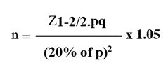
n=50.3
n=50 patients (30 patients are of COPD and 20 patients are of COPD with OSA) where,
Z1-α/2=1.96
p=number of diastolic dysfunctions=66.7%
q=100-p
=100-66.7
=33.3
Data Collection Procedure
After obtaining informed consent, all eligible participants diagnosed with either Chronic Obstructive Pulmonary Disease (COPD) (Group A) or Obstructive Sleep Apnea (OSA) (Group B) were enrolled in the study. Detailed clinical evaluation and relevant demographic data were recorded in a predesigned proforma. All participants underwent a comprehensive transthoracic echocardiographic examination. Conventional echocardiographic parameters including left ventricular end-diastolic diameter (LVEDD), left ventricular end-systolic dimension (LVESD), left ventricular end-diastolic volume (LVEDV), left ventricular end-systolic volume (LVESV), and left ventricular ejection fraction (LVEF) calculated by Simpson’s biplane method were assessed. Mitral inflow velocities, including peak early diastolic (E) and late diastolic (A) velocities as well as the E/A ratio, were used to assess diastolic function. Global longitudinal strain (GLS) measurements were also obtained using two-dimensional speckle tracking echocardiography in the apical four-chamber (A4C), apical two-chamber (A2C), and apical three-chamber (A3C) perspectives. Chest radiography (posteroanterior view), spirometry reports, and sleep study findings were also collected and documented for all study participants. The findings from all investigations were recorded and analyzed for comparison between the two study groups.
Statistical Analysis
The collected data were coded and entered into a Microsoft Excel worksheet and subsequently analyzed using Statistical Package for the Social Sciences (SPSS) software version 23.0. Continuous variables were expressed as mean ± standard deviation (SD) for data following a normal distribution, whereas data with a skewed distribution were presented as median and interquartile range (IQR). Categorical variables were summarized using frequencies and percentages. Spearman’s correlation coefficient (ρ) was used to assess the correlation between different study parameters. Statistical significance was determined at a 95% confidence interval (CI), and a p-value of ≤ 0.05 was considered statistically significant.
RESULTS:
Table 1. Baseline Demographic Characteristics of Study Participants
|
Variable |
COPD (n=30) |
OSA (n=20) |
Total (n=50) |
p-value |
|
Male |
21 (70.0%) |
11 (55.0%) |
32 (64.0%) |
|
|
Female |
9 (30.0%) |
9 (45.0%) |
18 (36.0%) |
0.279 |
|
Age 20–40 years |
1 (3.3%) |
2 (10.0%) |
3 (6.0%) |
|
|
Age 40–60 years |
7 (23.3%) |
9 (45.0%) |
16 (32.0%) |
|
|
>60 years |
22 (73.3%) |
9 (45.0%) |
31 (62.0%) |
0.122 |
Table 1 illustrates the baseline demographic characteristics of the study population. A total of 50 participants were included, comprising 30 COPD patients and 20 OSA patients. Males predominated in both groups (64%). Most participants were older than 60 years (62%). No significant differences were observed between groups regarding age or gender distribution.
Table 2. Distribution of COPD Severity According to GOLD Classification
|
GOLD Stage |
Frequency |
Percentage |
|
GOLD A |
10 |
33.3 |
|
GOLD B |
10 |
33.3 |
|
GOLD C |
4 |
13.3 |
|
GOLD D |
6 |
20.0 |
|
Total |
30 |
100 |
Table 2 shows the distribution of COPD patients according to GOLD severity stages. GOLD A and GOLD B, each accounted for one-third of the COPD population, while GOLD D represented 20% of cases, indicating a substantial proportion of patients with advanced disease.
Table 3. Cardiac Chamber Morphology in COPD and OSA Patients
|
Parameter |
COPD Normal (%) |
COPD Abnormal (%) |
OSA Normal (%) |
OSA Abnormal (%) |
|
Left Atrium |
27 (90.0) |
3 (10.0) |
20 (100) |
0 (0) |
|
LVIDd |
25 (83.3) |
5 (16.7) |
20 (100) |
0 (0) |
|
LVIDs |
25 (83.3) |
5 (16.7) |
20 (100) |
0 (0) |
Table 3 summarizes cardiac chamber morphology findings. Left atrial enlargement and left ventricular dilatation were observed predominantly among COPD patients, whereas all OSA patients demonstrated normal chamber dimensions.
Table 4. Distribution of Diastolic Dysfunction in COPD and OSA Groups
|
Grade of DDF |
COPD n (%) |
OSA n (%) |
|
Grade I |
18 (60.0) |
19 (95.0) |
|
Grade II |
4 (13.3) |
1 (5.0) |
|
Grade III |
6 (20.0) |
0 |
|
No DDF |
2 (6.7) |
0 |
|
Total |
30 |
20 |
Table 4 presents the pattern of diastolic dysfunction in both study groups. Grade I dysfunction was the most common abnormality in both groups. Advanced grades of diastolic dysfunction (Grade III) were observed exclusively among COPD patients, suggesting more pronounced cardiac involvement.
Table 5. Comparison of Conventional Echocardiographic Parameters Between COPD and OSA Groups
|
Parameter |
COPD Mean±SD |
OSA Mean±SD |
p-value |
|
LVEF (%) |
50.86 ± 14.71 |
58.81 ± 5.72 |
0.177 |
|
E velocity (m/s) |
0.72 ± 0.29 |
0.69 ± 0.17 |
0.992 |
|
A velocity (m/s) |
0.73 ± 0.33 |
0.67 ± 0.22 |
0.563 |
|
E/A ratio |
1.09 ± 0.45 |
0.85 ± 0.17 |
0.103 |
Table 5 compares conventional echocardiographic parameters between COPD and OSA patients. Although COPD patients showed lower mean LVEF and higher E/A ratio, none of these differences reached statistical significance.
Table 6. Comparison of Left Ventricular Ejection Fraction Categories Between COPD and OSA Groups
|
LVEF Category |
COPD n (%) |
OSA n (%) |
|
Severe |
5 (100) |
0 |
|
Moderate |
4 (100) |
0 |
|
Mild |
2 (50.0) |
2 (50.0) |
|
Borderline |
5 (83.3) |
1 (16.7) |
|
Normal |
14 (45.2) |
17 (54.8) |
|
Chi-square = 3.955, p = 0.047 |
||
Table 6 demonstrates the distribution of LVEF categories in both groups. Severe and moderate LV dysfunction were observed exclusively among COPD patients, while most OSA patients maintained normal ejection fraction. The association was statistically significant.
Table 7. Comparison of Speckle Tracking Echocardiographic Parameters Between COPD and OSA Groups
|
Parameter |
COPD Mean±SD |
OSA Mean±SD |
p-value |
|
Apical 4-Chamber Strain (%) |
-17.70 ± 5.43 |
-20.21 ± 1.87 |
0.029* |
|
Apical 2-Chamber Strain (%) |
-17.29 ± 5.68 |
-19.23 ± 2.91 |
0.255 |
|
Apical 3-Chamber Strain (%) |
-17.14 ± 5.82 |
-19.53 ± 2.73 |
0.035* |
|
Global Longitudinal Strain (%) |
-17.32 ± 5.25 |
-19.58 ± 2.28 |
0.047* |
|
*Statistically significant |
|||
Table 7 compares myocardial strain parameters obtained by speckle tracking echocardiography. COPD patients demonstrated significantly reduced myocardial deformation compared with OSA patients. Significant differences were observed in Apical 4-Chamber strain, Apical 3-Chamber strain, and Global Longitudinal Strain.
Table 8. Correlation Analysis of Left Ventricular Function Parameters
|
Correlation |
Spearman's r |
p-value |
|
LVEF vs COPD Severity Stage |
-0.541 |
0.002 |
|
LVEF vs GLS (COPD) |
-0.887 |
<0.001 |
|
LVEF vs GLS (OSA) |
-0.452 |
0.045 |
|
LVEF vs DDF (COPD) |
-0.433 |
0.017 |
|
LVEF vs DDF (OSA) |
0.265 |
0.258 |
Table 8 summarizes the correlation analyses. Increasing COPD severity was associated with worsening left ventricular systolic function. A strong negative correlation was observed between LVEF and GLS in COPD patients, indicating that worsening myocardial strain was associated with declining ventricular function. The relationship was weaker in OSA patients.
DISCUSSION:
This study compared LV systolic and diastolic function, conventional echocardiographic parameters, and speckle tracking-derived global longitudinal strain (GLS) in 30 COPD and 20 OSA patients. The principal finding was a significantly more impaired mean GLS in COPD (−17.32±5.25%) than OSA (−19.58±2.28%, p=0.047), with abnormal GLS in 76.2% of COPD versus 23.8% of OSA subjects (p=0.047), despite no significant difference in LVEF (50.86±14.71% vs 58.81±5.72%, p=0.177) or conventional Doppler indices (E, A, E/A; all p>0.10).
The COPD-group GLS value closely matches the pooled meta-analysis estimate of Ranjini et al. [11] (−17.055%, 95% CI −18.4 to −15.7, from 742 COPD patients), and falls outside the normal reference range of −19.7% defined by Yingchoncharoen et al.[12] in 2,597 healthy subjects — while the present OSA-group GLS (−19.58%) sits almost exactly within that normal range, confirming that subclinical systolic impairment was concentrated in the COPD arm.
Pizarro et al[8] studying 85 COPD outpatients, reported a more markedly impaired COPD GLS (−13.3±5.4% vs −17.1±1.8% in controls, p=0.04) and found no further strain decline when OSA overlapped with COPD — concluding COPD itself, not coexisting OSA, drove LV deformation abnormality. This matches the direction of the present head-to-head finding that COPD impaired strain more than isolated OSA, and the difference in absolute magnitude likely reflects differing severity-stage mix and imaging-vendor variability between cohorts.
For OSA, Altekin et al[7] showed GLS worsens gradually with apnoea severity (healthy −25.58%, mild −23.93%, moderate −21.27%, severe −16.94%). The present OSA cohort’s mean GLS (−19.58%) falls between the mild and moderate strata, consistent with its predominantly Grade 1 (95%) diastolic dysfunction and relatively preserved systolic strain.
Regional analysis showed significantly reduced COPD strain at the apical 4-chamber (p=0.029) and apical 3-chamber (p=0.035) views, paralleling Pizarro et al.’s [8] finding of severity-linked apical septal strain reduction in COPD (p=0.02).
Diastolic dysfunction was present in 93.3% of COPD subjects, closely matching the 88% reported by Caram et al.[10] who also found it independent of disease stage; their discussion cites earlier prevalence of 76% (Boussuges et al.) and >50% (Rutten et al., Funk et al.), placing the present figure at the upper end of the published range. Bhattacharjee et al.[13] studying a more severe cohort, found diastolic dysfunction rising from 41.2% in GOLD II to 92.2% in GOLD IV (p<0.001) and systolic dysfunction linked to GOLD IV (OR 1.83, p=0.014) - consistent with this study’s negative correlation between LVEF and COPD severity (ρ=−0.541, p<0.002). The 100% (mostly mild) diastolic dysfunction prevalence in the OSA group aligns with Kim SH et al.’s[14] demonstration that OSA independently impairs diastolic function early, via repetitive negative intrathoracic pressure and sympathetic surges, before systolic strain is affected.
The dissociation between a significant GLS difference (p=0.047) and a non-significant LVEF difference (p=0.177) parallels Schoos et al.[15] who found GLS - not LVEF or Doppler indices - was the only independent mortality predictor in COPD patients with preserved LVEF, reinforcing that strain detects impairment whereas conventional indices miss. The strong LVEF–GLS correlation in this study (ρ=−0.887 COPD, p<0.001; ρ=−0.452 OSA, p=0.045) supports the two measures being related but not interchangeable.
With 50 subjects, this study is smaller than several comparators - 85 in Pizarro et al.[8] 114 in Bhattacharjee et al.[13]90 in Schoos et al.[15] limiting power for sub-group and regional strain comparisons, a limitation shared across much of this literature.
LIMITATIONS
The study was conducted over a relatively short duration, which may have limited the assessment of long-term outcomes and disease progression. The sample size was relatively small, which may have reduced the statistical power of the study and limited the external validity of the findings to the broader population.
CONCLUSION:
In the present study, COPD was associated with impaired left ventricular (LV) deformation properties, with progressive worsening observed across increasing stages of disease severity. Although LV function was also affected in patients with obstructive sleep apnea (OSA), the degree of impairment was more evident in patients with COPD. Two-dimensional speckle-tracking echocardiography is used to measure Global Longitudinal Strain (GLS), was significantly reduced in the COPD group compared to the OSA group, indicating significant subclinical LV systolic dysfunction among COPD patients. These findings suggest that two-dimensional speckle-tracking echocardiography is a sensitive tool for the early detection of LV dysfunction in COPD and may aid in identifying patients at increased cardiovascular risk
REFERENCES:
1. Global Initiative for Chronic Obstructive Lung Disease (GOLD). Global Strategy for Prevention, Diagnosis and Management of COPD. 2024 Report.
2. Nussbaumer-Ochsner Y, Rabe KF. Systemic manifestations of COPD. Chest 2011;139(1):165-73.
3. Mannino DM, Thorn D, Swensen A, et al. Prevalence and outcomes of diabetes, hypertension and cardiovascular disease in COPD. Eur Respir J 2008;32(4):962-9.
4. Maclay JD, MacNee W. Cardiovascular disease in COPD: mechanisms. Chest 2013;143(3):798-807.
5. McNicholas WT. Comorbid obstructive sleep apnoea and chronic obstructive pulmonary disease and the risk of cardiovascular disease. Journal of Thoracic Disease 2018;10(Suppl 34):S4253.
6. Marin JM, Soriano JB, Carrizo SJ, et al. Outcomes in patients with chronic obstructive pulmonary disease and obstructive sleep apnea: the overlap syndrome. Am J Respir Crit Care Med 2010;182(3):325-31.
7. Altekin RE, Yanikoglu A, Baktir AO, et al. Assessment of subclinical left ventricular dysfunction in obstructive sleep apnea patients with speckle tracking echocardiography. Int J Cardiovasc Imaging 2012;28(8):1917-30.
8. Pizarro C, van Essen F, Linnhoff F, et al. Speckle tracking echocardiography in chronic obstructive pulmonary disease and overlapping obstructive sleep apnea. Int J Chron Obstruct Pulmon Dis2016;11:1823-34.
9. Cengiz Elçioğlu B, Kamat S, Yurdakul S, et al. Assessment of subclinical left ventricular systolic dysfunction and structural changes in patients with chronic obstructive pulmonary disease. Int Med J 2022;52(10):1791-8.
10. de Oliveira Caram LM, Ferrari R, Naves CR, et al. Association between left ventricular diastolic dysfunction and severity of chronic obstructive pulmonary disease. Clinics 2013;68(6):772-6.
11. Ranjini NV, Kumar SS, Desai N, et al. Speckle-tracking echocardiographic abnormalities in chronic obstructive pulmonary disease: a systematic review and meta-analysis. J Cardiovasc Imaging 2025;33(1):4.
12. Yingchoncharoen T, Agarwal S, Popović ZB, et al. Normal ranges of left ventricular strain: a meta-analysis. Journal of the American Society of Echocardiography 2013;26(2):185-91.
13. Bhattacharjee R, Deb T, Roy P, et al. Echocardiographic evidence of left ventricular dysfunction in COPD: relationship with disease severity. Medicina 2025;61(7):1260.
14. Kim SH, Cho GY, Shin C, et al. Impact of obstructive sleep apnea on left ventricular diastolic function. Am JCardiol2008;101(11):1663-8.
15. Schoos MM, Dalsgaard M, Kjærgaard J, et al. Echocardiographic predictors of exercise capacity and mortality in chronic obstructive pulmonary disease.BMC Cardiovascular Disorders 2013;13:84/.