ANAESTHETIC MANAGEMENT OF A PATIENT WITH YAMAGUCHI SYNDROME AND CLIVUS CHORDOMA UNDERGOING ENDOSCOPIC ENDONASAL DECOMPRESSION: A CASE REPORT.
- Rishi Katiyar , Consultant, Department of Critical Care Medicine, Apollo Sage Hospital, Bhopal, Madhya Pradesh, India.
- Sarika Katiyar , Professor, Department of Anaesthesiology, Bhopal Memorial Hospital and Research Centre, Bhopal, Madhya Pradesh, India.
- Kanika Suhag. , Associate Professor, Department of Anaesthesiology, Bhopal Memorial Hospital and Research Centre, Bhopal, Madhya Pradesh, India.
Article Information:
Abstract:
Background: Yamaguchi syndrome, also known as apical hypertrophic cardiomyopathy, is a rare variant of hypertrophic cardiomyopathy characterized by localized hypertrophy of the left ventricular apex. Although the prognosis is generally favorable, perioperative anaesthetic management is challenging because dynamic alterations in preload, afterload, heart rate, and myocardial contractility may precipitate left ventricular outflow obstruction, myocardial ischemia, or life-threatening arrhythmias. The coexistence of Yamaguchi syndrome with a clival chordoma requiring endoscopic endonasal decompression presents unique anaesthetic concerns due to the simultaneous need for meticulous haemodynamic stability and optimal neurosurgical operating conditions. Case Presentation: A patient diagnosed with Yamaguchi syndrome was scheduled for elective endoscopic endonasal decompression of a clival chordoma. Preoperative evaluation included transthoracic echocardiography, which demonstrated characteristic apical left ventricular hypertrophy. A comprehensive anaesthetic strategy emphasizing maintenance of sinus rhythm, avoidance of tachycardia, preservation of preload, maintenance of systemic vascular resistance, and advanced haemodynamic monitoring was adopted. Continuous invasive arterial pressure monitoring and FloTrac-based cardiac output monitoring were used to assess mean arterial pressure (MAP), systemic vascular resistance (SVR), and stroke volume variation (SVV) throughout the perioperative period. Anaesthesia was successfully conducted with stable haemodynamic parameters, uneventful extubation, and satisfactory postoperative neurological recovery. Conclusion: This case highlights the importance of individualized perioperative planning, vigilant haemodynamic monitoring, and multidisciplinary collaboration in patients with Yamaguchi syndrome undergoing major neurosurgical procedures. Goal-directed haemodynamic management facilitated successful anaesthetic care without significant cardiovascular complications.
Keywords:
Article :
INTRODUCTION:
Yamaguchi syndrome, also known as apical hypertrophic cardiomyopathy (ApHCM), is a relatively uncommon morphological variant of hypertrophic cardiomyopathy characterized by predominant hypertrophy of the left ventricular apex. First described in Japan in 1976, it accounts for approximately 15–25% of hypertrophic cardiomyopathy cases in East Asian populations but is considerably less common in Western countries [1,2]. Patients typically exhibit giant negative T waves on electrocardiography and the characteristic "ace-of-spades" configuration of the left ventricular cavity on ventriculography or echocardiography. Although many individuals remain asymptomatic, others may present with chest pain, exertional dyspnoea, palpitations, syncope, atrial or ventricular arrhythmias, myocardial ischemia, heart failure, or, rarely, sudden cardiac death [1–4].
The principal anaesthetic concerns in patients with Yamaguchi syndrome include preservation of adequate preload, avoidance of tachycardia and increased myocardial contractility, maintenance of systemic vascular resistance, and prevention of hypotension to preserve diastolic filling and coronary perfusion. Sudden reductions in preload or afterload, excessive sympathetic stimulation, and inappropriate inotropic support may precipitate haemodynamic instability or myocardial ischemia. Therefore, meticulous preoperative assessment, invasive haemodynamic monitoring, and individualized anaesthetic management are essential for minimizing perioperative cardiovascular complications [1,2,5].
Clival chordoma is a rare, slow-growing malignant neoplasm arising from embryonic notochordal remnants and accounts for a small proportion of skull-base tumours. Owing to its anatomical location, patients commonly present with headache, diplopia, cranial nerve deficits, dysphagia, or long-tract neurological signs secondary to brainstem compression. Complete surgical excision remains the cornerstone of management, with the endoscopic endonasal approach emerging as the preferred technique because it offers superior visualization, reduced surgical morbidity, and improved postoperative recovery [6–8].
The coexistence of Yamaguchi syndrome and clival chordoma is exceedingly rare, and only a limited number of reports have discussed the perioperative anaesthetic management of patients presenting with this unique clinical combination. Simultaneous achievement of cardiovascular stability and optimal neuroanaesthetic conditions requires meticulous planning, maintenance of cerebral perfusion pressure, goal-directed haemodynamic optimization, and close multidisciplinary collaboration [5,8].
This report describes the perioperative anaesthetic management of a patient with Yamaguchi syndrome undergoing endoscopic endonasal decompression for clival chordoma using advanced FloTrac™-guided haemodynamic monitoring. Continuous assessment of mean arterial pressure (MAP), systemic vascular resistance (SVR), and stroke volume variation (SVV) facilitated individualized perioperative management and contributed to a favourable surgical outcome. Goal-directed haemodynamic monitoring using minimally invasive cardiac output monitoring systems has been shown to improve perioperative cardiovascular optimization in high-risk surgical patients and therefore formed an integral component of our anaesthetic strategy [9].
CASE PRESENTATION:
A patient with a known diagnosis of Yamaguchi syndrome (apical hypertrophic cardiomyopathy) presented with progressively worsening headache, intermittent diplopia, and imbalance while walking for several months. Neurological examination revealed features suggestive of raised intracranial pressure and cranial nerve involvement. Magnetic resonance imaging (MRI) of the brain demonstrated a clival chordoma causing compression of the brainstem. After multidisciplinary evaluation by the departments of Neurosurgery, Cardiology, and Anaesthesiology, the patient was scheduled for elective endoscopic endonasal decompression of the lesion.
Preoperative cardiovascular evaluation was performed in detail because of the underlying cardiomyopathy. Electrocardiography demonstrated sinus rhythm with deep T-wave inversions in the precordial leads, consistent with apical hypertrophic cardiomyopathy. Two-dimensional transthoracic echocardiography revealed marked apical left ventricular hypertrophy with preserved left ventricular systolic function, without evidence of significant left ventricular outflow tract obstruction or valvular pathology (Figure 1). Routine laboratory investigations, including complete blood count, renal and liver function tests, coagulation profile, serum electrolytes, and blood glucose levels, were within acceptable limits for surgery.
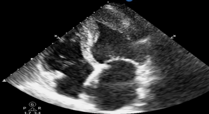
Figure 1: Preoperative transthoracic echocardiography (apical four-chamber view) demonstrating apical left ventricular hypertrophy consistent with Yamaguchi syndrome (Apical Hypertrophic Cardiomyopathy).
The patient was receiving appropriate medical therapy for hypertrophic cardiomyopathy, which was continued until the day of surgery. Adequate preoperative hydration was ensured to maintain intravascular volume, while prolonged fasting and dehydration were avoided. Premedication was kept minimal to prevent excessive sedation and respiratory depression. Blood products were cross-matched and made readily available considering the possibility of intraoperative blood loss during skull base surgery.
After arrival in the operating room, standard American Society of Anesthesiologists (ASA) monitoring was instituted, including continuous electrocardiography with ST-segment analysis, pulse oximetry, non-invasive blood pressure monitoring, capnography, and temperature monitoring. Before induction of anaesthesia, a radial arterial catheter was inserted under local anaesthesia for continuous invasive arterial pressure monitoring and repeated arterial blood gas analysis. A central venous catheter was secured under ultrasound guidance to facilitate vasoactive drug administration and haemodynamic assessment.
To optimize perioperative cardiovascular management, advanced FloTrac™ haemodynamic monitoring was connected after arterial cannulation, allowing continuous measurement of mean arterial pressure (MAP), systemic vascular resistance (SVR), stroke volume variation (SVV), cardiac output, and stroke volume. These dynamic parameters guided fluid therapy and vasopressor administration throughout surgery.
Considering the coexistence of Yamaguchi syndrome and a major neurosurgical procedure, the primary anaesthetic goals included preservation of sinus rhythm, avoidance of tachycardia, maintenance of adequate preload and systemic vascular resistance, prevention of sympathetic surges during airway manipulation, optimization of cerebral perfusion pressure, and facilitation of early postoperative neurological assessment. A multidisciplinary perioperative strategy was formulated to achieve these objectives while minimizing the risk of cardiovascular and neurological complications.
ANAESTHETIC MANAGEMENT
The patient was shifted to the operating room after confirmation of fasting status, informed high-risk consent, and availability of adequate blood products. Standard ASA monitors, including continuous electrocardiography, pulse oximetry, non-invasive blood pressure, capnography, temperature monitoring, and bispectral index (BIS) monitoring, were applied. Baseline haemodynamic parameters were recorded. A radial arterial catheter was placed under local anaesthesia for continuous invasive arterial pressure monitoring, and a central venous catheter was inserted under ultrasound guidance for administration of vasoactive drugs and perioperative fluid management.
Advanced haemodynamic monitoring using the FloTrac™ system was established immediately after arterial cannulation, providing continuous measurements of mean arterial pressure (MAP), stroke volume (SV), stroke volume variation (SVV), cardiac output (CO), cardiac index (CI), and systemic vascular resistance (SVR). These parameters were used to guide individualized fluid administration and haemodynamic optimization throughout the surgical procedure.
Induction of Anaesthesia
The primary objective during induction was to prevent sympathetic stimulation while preserving adequate preload and systemic vascular resistance. Adequate preoxygenation was performed using 100% oxygen for three minutes. Intravenous induction was achieved using carefully titrated doses of fentanyl, propofol, and a non-depolarizing neuromuscular blocking agent to facilitate smooth endotracheal intubation. Direct laryngoscopy and tracheal intubation were performed gently to minimize haemodynamic fluctuations. Additional doses of opioid were administered whenever required to attenuate sympathetic responses during airway manipulation.
Immediately after intubation, bilateral air entry was confirmed, and mechanical ventilation was initiated with oxygen-air mixture while maintaining normocapnia (end-tidal carbon dioxide 35–40 mmHg). Ventilator settings were adjusted to avoid excessive positive airway pressures that could reduce venous return and compromise preload.
Maintenance of Anaesthesia
Anaesthesia was maintained using a balanced anaesthetic technique with inhalational anaesthetic supplemented with intermittent opioid administration and neuromuscular blockade. The depth of anaesthesia was adjusted according to BIS monitoring to avoid inadequate anaesthesia or excessive myocardial depression.
Special attention was directed toward maintaining:
· Sinus rhythm throughout surgery.
· Heart rate between 60 and 70 beats/minute.
· Mean arterial pressure within 20% of baseline.
· Adequate preload while avoiding fluid overload.
· Normal systemic vascular resistance.
· Stable cerebral perfusion pressure.
Hypotension, if encountered, was treated promptly using vasopressors with predominant alpha-adrenergic activity rather than inotropic agents, thereby maintaining coronary perfusion without increasing myocardial contractility. Tachycardia and hypertension were prevented by adequate analgesia and maintenance of an appropriate depth of anaesthesia.
Goal-Directed Fluid Therapy
Fluid therapy was individualized using continuous FloTrac-derived haemodynamic variables. Stroke volume variation and cardiac output trends guided administration of balanced crystalloid solutions. Fluid boluses were administered only when indicated by dynamic parameters, thereby avoiding both hypovolaemia and excessive fluid administration, which could adversely affect cardiac function and neurosurgical exposure.
Electrolytes, arterial blood gases, blood glucose, haemoglobin concentration, and urine output were monitored periodically throughout the procedure and corrected whenever necessary.
Haemodynamic Monitoring
Continuous haemodynamic monitoring demonstrated satisfactory cardiovascular stability throughout surgery. Mean arterial pressure remained within the desired range with only minor fluctuations during induction, nasal instrumentation, tumour decompression, and emergence.
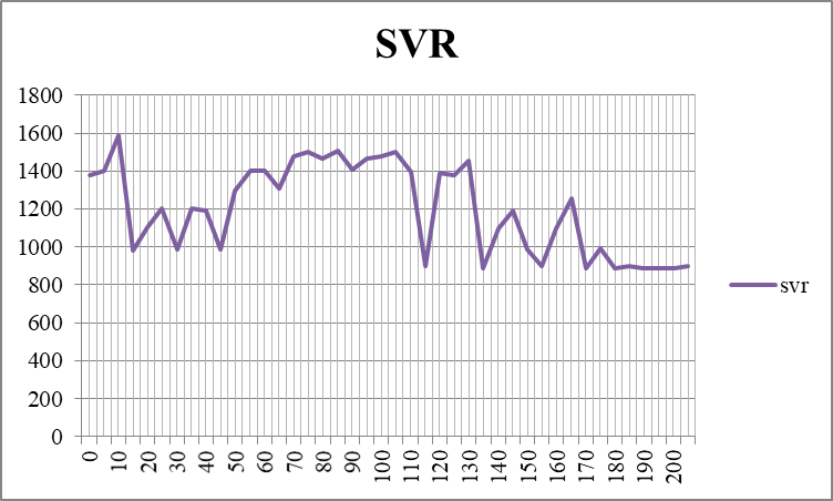
Figure 2: Perioperative variation in mean arterial pressure (MAP) demonstrating stable arterial pressure throughout the surgical procedure.
Systemic vascular resistance remained within acceptable limits throughout surgery. Transient reductions in SVR were promptly corrected with judicious vasopressor therapy and optimization of intravascular volume.
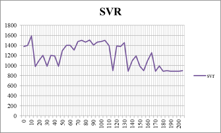
Figure 3: Perioperative variation in systemic vascular resistance (SVR) demonstrating maintenance of vascular tone using goal-directed haemodynamic management.
Stroke volume variation was continuously monitored to guide individualized fluid therapy. Dynamic assessment of preload helped avoid both hypovolaemia and excessive fluid administration during the neurosurgical procedure.
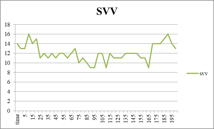
Figure 4: Perioperative variation in stroke volume variation (SVV) used to guide goal-directed fluid therapy throughout surgery.
Emergence and Postoperative Care
At completion of tumour decompression, meticulous haemostasis was confirmed. Residual neuromuscular blockade was adequately reversed after ensuring recovery of spontaneous ventilation. The trachea was extubated smoothly after confirming satisfactory respiratory effort, intact airway reflexes, haemodynamic stability, and adequate neurological responsiveness.
The patient was subsequently transferred to the neurosurgical intensive care unit for close observation. Postoperative analgesia was provided using a multimodal regimen to minimize sympathetic activation while avoiding excessive sedation. Continuous cardiovascular monitoring revealed stable haemodynamic parameters without arrhythmias, myocardial ischemia, or neurological deterioration. The postoperative period remained uneventful, and the patient demonstrated satisfactory neurological recovery.
DISCUSSION:
Yamaguchi syndrome, also known as apical hypertrophic cardiomyopathy (ApHCM), is an uncommon morphological variant of hypertrophic cardiomyopathy characterized by localized hypertrophy of the left ventricular apex. Although initially regarded as a relatively benign condition, long-term follow-up studies have demonstrated that affected patients remain at risk of atrial and ventricular arrhythmias, myocardial ischemia, thromboembolic events, progressive heart failure, and, in rare instances, sudden cardiac death [10–12]. Accordingly, careful perioperative planning and maintenance of haemodynamic stability remain central to the safe conduct of non-cardiac surgical procedures in these patients.
The principal anaesthetic objectives in patients with hypertrophic cardiomyopathy include preservation of sinus rhythm, maintenance of adequate preload and systemic vascular resistance, avoidance of tachycardia, hypotension, and excessive myocardial contractility, thereby optimizing diastolic filling and coronary perfusion [11–13]. In the present patient, these haemodynamic objectives were achieved through judicious titration of anaesthetic agents, continuous invasive monitoring, and FloTrac™-guided goal-directed haemodynamic optimization.
Preoperative transthoracic echocardiography demonstrated characteristic apical left ventricular hypertrophy without evidence of significant left ventricular outflow tract obstruction (Figure 1), facilitating perioperative risk stratification and individualized anaesthetic planning. Contemporary guidelines recommend detailed cardiovascular evaluation before major non-cardiac surgery in patients with hypertrophic cardiomyopathy to reduce perioperative morbidity and optimize clinical outcomes [12,13].
Continuous FloTrac™-based haemodynamic monitoring provided valuable real-time information during this prolonged skull-base procedure and facilitated timely therapeutic interventions whenever required. Dynamic assessment of mean arterial pressure (MAP), systemic vascular resistance (SVR), and stroke volume variation (SVV) enabled individualized fluid administration and timely correction of haemodynamic disturbances. Stable perioperative trends in MAP (Figure 2), SVR (Figure 3), and SVV (Figure 4) reflected effective maintenance of cerebral perfusion and cardiovascular stability while avoiding both hypovolaemia and fluid overload. Previous studies have shown that goal-directed haemodynamic therapy can improve tissue perfusion while reducing perioperative complications in selected high-risk surgical patients [14,15].
Endoscopic endonasal decompression for clival chordoma presents unique anaesthetic challenges because of prolonged operative duration, restricted airway access after surgical draping, potential blood loss, haemodynamic responses during skull-base drilling, and the requirement for early postoperative neurological assessment. Maintenance of adequate cerebral perfusion pressure, meticulous haemostasis, normocapnia, and smooth emergence from anaesthesia remain essential neuroanaesthetic goals [16].
Previous reports describing anaesthetic management in patients with hypertrophic cardiomyopathy undergoing non-cardiac surgery have emphasized that avoidance of sympathetic stimulation, preservation of preload and afterload, and maintenance of sinus rhythm are the key determinants of favourable perioperative outcomes [11,13,17]. The present case is consistent with these observations and further illustrates that minimally invasive haemodynamic monitoring may assist in maintaining cardiovascular stability during complex neurosurgical procedures.
The favourable perioperative outcome in this patient was likely the result of thorough preoperative cardiac assessment, multidisciplinary planning, individualized anaesthetic management, goal-directed fluid therapy, and continuous advanced haemodynamic monitoring rather than any single intervention alone. Because reports describing the coexistence of Yamaguchi syndrome and clival chordoma are exceedingly rare, this case contributes valuable clinical evidence regarding the safe anaesthetic management of such patients. Recent case reports of Yamaguchi syndrome have emphasized the importance of individualized perioperative management in preventing adverse cardiovascular outcomes [17,18]. Furthermore, our experience highlights the role of FloTrac™-guided haemodynamic optimization in achieving favourable surgical and neurological outcomes.
CONCLUSION:
Anaesthetic management of patients with Yamaguchi syndrome undergoing endoscopic endonasal decompression for clival chordoma requires a thorough understanding of both hypertrophic cardiomyopathy physiology and neuroanaesthetic principles. Maintenance of sinus rhythm, adequate preload, preserved systemic vascular resistance, avoidance of tachycardia, and vigilant goal-directed haemodynamic monitoring are essential for achieving favourable perioperative outcomes. This case suggests that meticulous perioperative planning combined with FloTrac™-guided haemodynamic optimization and multidisciplinary teamwork can contribute to safe anaesthetic management in patients with this rare clinical combination.
Learning Points
· Yamaguchi syndrome presents unique perioperative cardiovascular challenges due to impaired diastolic function and susceptibility to arrhythmias.
· Detailed preoperative cardiac assessment is essential before major neurosurgical procedures.
· Goal-directed haemodynamic monitoring using dynamic parameters such as MAP, SVR, and SVV can optimize intraoperative fluid and vasopressor therapy.
· Maintenance of preload, afterload, sinus rhythm, and controlled heart rate remains the cornerstone of anaesthetic management.
· Multidisciplinary collaboration between anaesthesiologists, cardiologists, and neurosurgeons is crucial for achieving favorable perioperative outcomes in such rare clinical scenarios.
REFERENCES:
1. Ommen SR, Ho CY, Asif IM, Balaji S, Burke MA, Day SM, et al. 2024 AHA/ACC/AMSSM/HRS/PACES/SCMR Guideline for the Management of Hypertrophic Cardiomyopathy: A Report of the American Heart Association/American College of Cardiology Joint Committee on Clinical Practice Guidelines. J Am Coll Cardiol. 2024 Jun 11;83(23):2324-2405. doi: 10.1016/j.jacc.2024.02.014. PMID: 38727647.
2. Ommen SR, Mital S, Burke MA, Day SM, Deswal A, Elliott P, et al. 2020 AHA/ACC Guideline for the Diagnosis and Treatment of Patients With Hypertrophic Cardiomyopathy: A Report of the American College of Cardiology/American Heart Association Joint Committee on Clinical Practice Guidelines. J Am Coll Cardiol. 2020 Dec 22;76(25):e159-e240. doi: 10.1016/j.jacc.2020.08.045. PMID: 33229116.
3. Maron BJ, Desai MY, Nishimura RA, Spirito P, Rakowski H, Towbin JA, et al. Diagnosis and Evaluation of Hypertrophic Cardiomyopathy: JACC State-of-the-Art Review. J Am Coll Cardiol. 2022 Feb 1;79(4):372-389. doi: 10.1016/j.jacc.2021.12.002. PMID: 35086660.
4. Eriksson MJ, Sonnenberg B, Woo A, Rakowski P, Parker TG, Wigle ED, Rakowski H. Long-term outcome in patients with apical hypertrophic cardiomyopathy. J Am Coll Cardiol. 2002 Feb 20;39(4):638-45. doi: 10.1016/s0735-1097(01)01778-8. PMID: 11849863.
5. Li J, Fang J, Liu Y, Wei X. Apical hypertrophic cardiomyopathy: pathophysiology, diagnosis and management. Clin Res Cardiol. 2024 May;113(5):680-693. doi: 10.1007/s00392-023-02328-8. PMID: 37982860.
6. Stacchiotti S, Sommer J; Chordoma Global Consensus Group. Building a global consensus approach to chordoma: a position paper from the medical and patient community. Lancet Oncol. 2015 Feb;16(2):e71-83. doi: 10.1016/S1470-2045(14)71190-8. PMID: 25638683.
7. Cavallo LM, Messina A, Cappabianca P, Esposito F, de Divitiis E, Gardner P, Tschabitscher M. Endoscopic endonasal surgery of the midline skull base: anatomical study and clinical considerations. Neurosurg Focus. 2005 Jul 15;19(1):E2. PMID: 16078816.
8. Walcott BP, Nahed BV, Mohyeldin A, Coumans JV, Kahle KT, Ferreira MJ. Chordoma: current concepts, management, and future directions. Lancet Oncol. 2012 Feb;13(2):e69-76. doi: 10.1016/S1470-2045(11)70337-0. PMID: 22300861.
9. Schnurman Z, Benjamin CG, Miceli M, Sen C. Clival Chordomas in the Endoscopic Endonasal Era: Clinical Management, Outcomes, and Complications. Neurosurgery. 2023 Apr 1;92(4):876-883. doi: 10.1227/neu.0000000000002289. PMID: 36700749.
10. Yousaf J, Afshari FT, Ahmed SK, Chavda SV, Sanghera P, Paluzzi A. Endoscopic endonasal surgery for Clival Chordomas - a single institution experience and short term outcomes. Br J Neurosurg. 2019 Aug;33(4):388-393. doi: 10.1080/02688697.2019.1567683. PMID: 30741028.
11. Ho AM, Chui PT, Lee AP, Wan S. Apical Hypertrophic Cardiomyopathy. Anesth Analg. 2015 Nov;121(5):1398-9. doi: 10.1213/ANE.0000000000000867. PMID: 26484467.
12. Maron BJ, Rowin EJ, Casey SA, Link MS, Lesser JR, Chan RH, et al. Hypertrophic Cardiomyopathy in Adulthood Associated With Low Cardiovascular Mortality With Contemporary Management Strategies. J Am Coll Cardiol. 2015 May 12;65(18):1915-28. doi: 10.1016/j.jacc.2015.02.061. PMID: 25953744.
13. Poirier H, Brusset A, Fischler M. Myocardiopathie hypertrophique apicale: un piège de l'électrocardiogramme préopératoire [Apical hypertrophic cardiomyopathy: a pitfall in preoperative electrocardiography]. Ann Fr Anesth Reanim. 2000 Oct;19(8):599-602. French. doi: 10.1016/s0750-7658(00)00265-3. PMID: 11098321.
14. Gersh BJ, Maron BJ, Bonow RO, Dearani JA, Fifer MA, Link MS, et al. 2011 ACCF/AHA Guideline for the Diagnosis and Treatment of Hypertrophic Cardiomyopathy: a report of the American College of Cardiology Foundation/American Heart Association Task Force on Practice Guidelines. Developed in collaboration with the American Association for Thoracic Surgery, American Society of Echocardiography, American Society of Nuclear Cardiology, Heart Failure Society of America, Heart Rhythm Society, Society for Cardiovascular Angiography and Interventions, and Society of Thoracic Surgeons. J Am Coll Cardiol. 2011 Dec 13;58(25):e212-60. doi: 10.1016/j.jacc.2011.06.011. PMID: 22075469.
15. Fleisher LA, Fleischmann KE, Auerbach AD, Barnason SA, Beckman JA, Bozkurt B, et al. 2014 ACC/AHA guideline on perioperative cardiovascular evaluation and management of patients undergoing noncardiac surgery: a report of the American College of Cardiology/American Heart Association Task Force on Practice Guidelines. Circulation. 2014 Dec 9;130(24):e278-333. doi: 10.1161/CIR.0000000000000106. PMID: 25085961.
16. Kristensen SD, Knuuti J, Saraste A, Anker S, Bøtker HE, Hert SD, et al. 2014 ESC/ESA Guidelines on non-cardiac surgery: cardiovascular assessment and management: The Joint Task Force on non-cardiac surgery: cardiovascular assessment and management of the European Society of Cardiology (ESC) and the European Society of Anaesthesiology (ESA). Eur Heart J. 2014 Sep 14;35(35):2383-431. doi: 10.1093/eurheartj/ehu282. PMID: 25086026.
17. Samy NMP, Thangaswamy CR, Srivats VR, Arikrishnan T. Perioperative management of apical hypertrophic cardiomyopathy (Yamaguchi Syndrome) complicating pregnancy for emergency lower segment caeserean section. Indian J Anaesth. 2020 Aug;64(8):725-727. doi: 10.4103/ija.IJA_14_20. Epub 2020 Jul 31. PMID: 32934413.
18. Giri A, Acharya S, Kamat S, Shukla S, Kumar S. Yamaguchi Syndrome: A Hidden Masquerader of Ischemic Heart Disease. Cureus. 2022 Jun 29;14(6):e26439. doi: 10.7759/cureus.26439. PMID: 35915685.