Decoding Metastatic Bone Disease through Whole-Body MRI: A Mechanistic Imaging Perspective on Tumour–Marrow Interactions.
- Prakash Asokan , MBBS, DNB (Radiodiagnosis), DMRD, FRCR, EDiR, FiOR. Assistant professor, Radiodiagnosis, Srinivasan medical college and hospital, near samayapuram Toll Plaza, Tiruchirappalli, 621112.
- Mohamed Muzaffer Ghouse , MBBS, MDRD, Associate Professor, Radiodiagnosis, Srinivasan medical college and hospital, near samayapuram Toll Plaza, Tiruchirappalli, 621112.
- Sindhuja Ramakrishnan , MBBS MDRD, Assistant professor, Radiodiagnosis, Srinivasan medical college and hospital, near samayapuram Toll Plaza, Tiruchirappalli, 621112.
Article Information:
Abstract:
Background: Bone metastases are difficult to detect at an early stage because they often begin with subtle bone marrow changes before structural bone alterations become visible on conventional imaging. Whole-body magnetic resonance imaging (WB-MRI) with diffusion-weighted imaging (DWI) has emerged as a promising technique for early detection and assessment of bone marrow involvement. Methods: This review examines the applications of WB-MRI combined with DWI in the evaluation of bone metastases. It focuses on the role of apparent diffusion coefficient (ADC) measurements, treatment response monitoring, and the integration of radiomics and artificial intelligence (AI) for quantitative image analysis and predictive modeling. Results: WB-MRI with DWI enables early identification of bone marrow metastases by detecting restricted diffusion and low ADC values, which are associated with high tumor cellularity. Treatment response can be assessed through increasing ADC values, indicating tumor reduction. Additionally, radiomics and AI enhance the extraction of imaging features, improving lesion characterization, tumor quantification, and predictive accuracy. Compared with conventional imaging and FDG-PET/CT, WB-MRI offers superior soft-tissue resolution and avoids radiation exposure, making it suitable for repeated assessments. Conclusion: WB-MRI with DWI is a valuable imaging modality for the early detection and monitoring of bone metastases. Its ability to evaluate tumor biology, quantify disease burden, and support precision medicine is further strengthened by radiomics and AI applications. The absence of ionizing radiation and superior soft-tissue contrast make WB-MRI an ideal tool for serial imaging in oncology.
Keywords:
Article :
BACKGROUND :
Bone metastasis is a significant burden of cancer, with pain, fractures and premature death as major complications. It stems from dynamic interactions between circulating tumour cells and the bone marrow, where early metastases modify the marrow prior to structural changes. Current diagnostic techniques, including computed tomography (CT) and bone scintigraphy, pick up late changes, making early diagnosis difficult [1].
WB-MRI allows direct visualisation of bone marrow involvement, whereas DWI offers functional information as it measures the diffusion of water, which relates to cellularity. Tumours with high cellularity have reduced diffusion, so DWI is a sensitive indicator of early metastatic disease and tumour burden [1,3]. WB-MRI also helps assess responses to treatment, particularly in multiple myeloma. Apparent diffusion coefficient (ADC) values enable non-invasive assessment, with rising ADC reflecting decreased tumour cellularity and treatment response, which may occur before morphological changes [2].
Recent advances have expanded WB-MRI into a quantitative tool for biological characterization. Radiomics enables extraction of features reflecting tumour heterogeneity and treatment response [4]. WB-MRI also shows comparable or superior performance to FDG-PET/CT in certain settings, offering better soft tissue contrast without radiation, making it suitable for longitudinal monitoring [5]. Overall, WB-MRI bridges imaging and tumour biology, supporting understanding of tumour–marrow interactions and data-driven cancer management.
Mechanistic Basis of Diffusion-Weighted WB-MRI in Metastatic Bone Disease
Diffusion-weighted imaging (DWI) in WB-MRI enables non-invasive assessment of tumour biology by evaluating water molecule motion influenced by cellularity and tissue structure [1,3]. Normal marrow, rich in adipocytes, allows free diffusion and high ADC values. In contrast, metastatic infiltration increases cellular density, restricting diffusion and reducing ADC values. Thus, ADC serves as a surrogate biomarker for tumour burden and proliferation [1] (Figure 1).
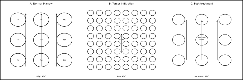
Figure 1: Mechanistic illustration of diffusion-weighted imaging (DWI) in metastatic bone disease. (A) Normal bone marrow demonstrates low cellularity with free water diffusion and high apparent diffusion coefficient (ADC) values. (B) Tumour infiltration increases cellular density, restricting diffusion and resulting in low ADC values. (C) Post-treatment changes lead to reduced cellularity and increased extracellular space, causing elevated ADC values, reflecting therapeutic response.
Restricted diffusion in metastatic lesions reflects increased cellularity, reduced extracellular space, and altered stromal architecture. Tumour-driven angiogenesis and inflammation further influence microvascular permeability, enabling WB-MRI to detect early tumour–marrow interactions before structural changes appear [3].
This effect is vital in evaluating response: chemotherapy increases extracellular space, increasing ADC, enabling DWI to predict response before size reduction. In multiple myeloma, ADC measurements have significant correlation with treatment response [2]. DWI has now evolved into a quantitative technique. Radiomics offers information about tumour heterogeneity via signal and texture features that support predictive modelling and precision medicine [4].
Compared with FDG-PET/CT, DWI reflects cellular density rather than glucose metabolism, reducing inflammatory confounding and improving specificity. WB-MRI is well suited for longitudinal monitoring [5].
Emerging Trends: Radiomics, Artificial Intelligence, and Quantitative Imaging Biomarkers
Whole-body MRI (WB-MRI) is increasingly quantitative. Radiomics produces high-dimensional imaging features from diffusion weighted imaging (DWI) that reflect tumour heterogeneity through texture and signal patterns that reflect tumour ecology [4]. These features are stable and predictive of response and enable response monitoring early on, making WB-MRI a proxy for disease biology. This is supported by artificial intelligence (AI) for feature extraction and prediction. An integration of radiomic and clinical features helps in prognostication and response prediction, linking image features to molecular disease features in personalised medicine [8].
Software such as matRadiomics increase reproducibility and translatability [9]. Techniques like diffusion-weighted imaging with background body signal suppression (DWIBS) increase lesion conspicuity and are histologically relevant [6]. WB-MRI is particularly valuable in such diseases as multiple myeloma for staging and follow-up [7]. It's more accurate than FDG-PET/CT and is radiation-free, so it's great for monitoring [5]. Radiomic features and quantitative measurements (ADC) help in diagnosis and prognostication, but need to be further validated [10].
Limitations
Whole-body MRI (WB-MRI) with diffusion-weighted imaging (DWI) is useful in clinical practice but there are drawbacks. Different imaging sequences (b-values, signal-to-noise ratio (SNR), etc.) affect the apparent diffusion coefficient (ADC) values, which are often used to compare images. False positives can occur with DWI (e.g. inflammation, bone marrow reconversion). WB-MRI has limited sensitivity for sclerotic lesions due to low water content. WB-MRI also suffers from practical limitations including length of scan, cost and lack of standardisation. Radiomics and artificial intelligence (AI) have problems with validation, reproducibility and lack of standardised processes, which limit clinical implementation [4,8,9].
Future Directions
The next steps include harmonisation, integration and validation. Standardised imaging protocols and ADC guidelines are important for reproducibility and multiparametric MRI may help lesion characterisation. Radiomics and AI allow automatic lesion detection, predictions and link imaging with molecular information, for personalised approaches to cancer management. Methods like DWIBS may increase lesion detection [6]. Large, prospective studies are needed for imaging biomarkers. WB-MRI will be a quantitative systems biology - imaging, biology and computing - approach [8-10].
CONCLUSION:
WB-MRI with diffusion-weighted imaging is a move towards imaging of tumour biology in the bone marrow. It allows mapping of the distribution of cellularity, diffusion and treatment response, as well as anatomy. WB-MRI may help to predict and diagnose cancers in precision medicine, along with radiomics and artificial intelligence. Despite these challenges, technological advances will make WB-MRI an important component in identifying and using biological knowledge for cancer diagnosis and treatment.
REFERENCES:
1. Stecco A, Trisoglio A, Soligo E, Berardo S, Sukhovei L, Carriero A. Whole-Body MRI with Diffusion-Weighted Imaging in Bone Metastases: A Narrative Review. Diagnostics. 2018; 8(3):45. https://doi.org/10.3390/diagnostics8030045
2. Wang, K., Lee, E., Kenis, S. et al. Application of diffusion-weighted whole-body MRI for response monitoring in multiple myeloma after chemotherapy: a systematic review and meta-analysis. Eur Radiol 32, 2135–2148 (2022). https://doi.org/10.1007/s00330-021-08311-z
3. Lecouvet, F.E., Chabot, C., Taihi, L. et al. Present and future of whole-body MRI in metastatic disease and myeloma: how and why you will do it. Skeletal Radiol 53, 1815–1831 (2024). https://doi.org/10.1007/s00256-024-04723-2
4. Thrussell I, Winfield JM, Orton MR, Miah AB, Zaidi SH, Arthur A, Thway K, Strauss DC, Collins DJ, Koh D-M, Oelfke U, Huang PH, O’Connor JPB, Messiou C and Blackledge MD (2022) Radiomic Features From Diffusion-Weighted MRI of Retroperitoneal Soft-Tissue Sarcomas Are Repeatable and Exhibit Change After Radiotherapy. Front. Oncol. 12:899180. doi: 10.3389/fonc.2022.899180
5. Maccioni, F., Alfieri, G., Assanto, G.M. et al. Whole body MRI with Diffusion Weighted Imaging versus 18F-fluorodeoxyglucose-PET/CT in the staging of lymphomas. Radiol med 128, 556–564 (2023). https://doi.org/10.1007/s11547-023-01622-9
6. El-Sawy Deif Allah, N.S., Abdallah, R.H., Hassan, M.S. et al. Role of diffusion weighted imaging with background body signal suppression (DWIBS) in diagnosis of breast masses and correlation with histopathological findings. Egypt J Radiol Nucl Med 55, 95 (2024). https://doi.org/10.1186/s43055-024-01260-9
7. Rodríguez-Laval V, Lumbreras-Fernández B, Aguado-Bueno B, Gómez-León N. Imaging of Multiple Myeloma: Present and Future. Journal of Clinical Medicine. 2024; 13(1):264. https://doi.org/10.3390/jcm13010264
8. Vicini, S., Bortolotto, C., Rengo, M. et al. A narrative review on current imaging applications of artificial intelligence and radiomics in oncology: focus on the three most common cancers. Radiol med 127, 819–836 (2022). https://doi.org/10.1007/s11547-022-01512-6
9. Pasini G, Bini F, Russo G, Comelli A, Marinozzi F, Stefano A. matRadiomics: A Novel and Complete Radiomics Framework, from Image Visualization to Predictive Model. Journal of Imaging. 2022; 8(8):221. https://doi.org/10.3390/jimaging8080221
10. Chiu, FY., Yen, Y. Imaging biomarkers for clinical applications in neuro-oncology: current status and future perspectives. Biomark Res 11, 35 (2023). https://doi.org/10.1186/s40364-023-00476-7