HISTOPATHOLOGICAL STUDY OF THE SPECTRUM OF SKIN LESIONS IN A TERTIARY CARE HOSPITAL- A RETROSPECTIVE STUDY.
- Syeda Iffath Tahseen , Associate Professor, Department of Pathology, GGH, Mahabubnagar.
- Jyothi Buranakunta , Associate Professor, Department of Pathology, Osmania medical college and hospital, Hyderabad.
- Syeda Aliya Rafath , Postgraduate, Department of Pathology, Government General Hospital, Mahabubnagar.
- HimaBindu Deshala , Postgraduate, Department of Pathology, Government General Hospital, Mahabubnagar.
- Lakshmi Bai Earla , Professor & Head, Department of Pathology, GGH, Mahabubnagar, Telangana, India.
Article Information:
Abstract:
Background: The skin is a complex organ with multiple vital functions. It is composed of three principal anatomical components: the epidermis with its adnexal structures, the dermis, and the subcutis, along with the melanocytic system. Skin diseases are highly prevalent in developing countries, affecting individuals across all age groups, and nearly 2000 distinct dermatological conditions have been described to date.1 These diseases exhibit significant geographical variation and encompass a wide spectrum, ranging from simple vesicular non-neoplastic lesions to potentially fatal neoplastic disorders. Histopathological examination of skin biopsies remains essential for establishing an accurate diagnosis, identifying etiological factors, and guiding clinicians in appropriate management decisions. Aim: Present study was carried out to understand prevalence and spectrum various skin lesion in at tertiary care hospital and so to educate community and physicians accordingly after study results. Materials And Methods: It was a retrospective cross-sectional study conducted over a period of 24 months from Jan 2024 to Jan 2026. All cases were retrieved from department registries and analyzed. Results: Results will be discussed during the presentation. Conclusions: An accurate diagnosis is not possible with just the clinical features. Histopathologic examination is considered the gold standard for diagnosis and categorization.
Keywords:
Article :
INTRODUCTION:
The skin, consisting of the epidermis, dermis, and subcutaneous tissue, is the body's largest organ.2 Skin lesions are prevalent worldwide and exhibit a diverse range, differing across countries and regions. Their occurrence is influenced by factors such as gender, age, systemic health conditions, socioeconomic status, education levels, cultural practices, and racial characteristics. These lesions are broadly categorized into non-neoplastic and neoplastic types .To ensure accurate diagnosis, determine the underlying cause, and support dermatologists or clinicians in choosing appropriate treatment, histopathological examination of skin biopsy specimens is essential.3 This study was undertaken to evaluate the histopathological patterns of skin lesions, analyze their distribution across different age groups and sexes, identify the most common types of lesions.
MATERIALS AND METHODS:
This retrospective cross-sectional study was conducted over 24 months from January 2024 to January 2026 in the department of pathology GGH, MBNR. All the cases were retrieved and processed via gross examination tissue processing, embedding,4-5 micrometer sections, hematoxylin and eosin staining following standard protocol further these lesions were classified as non-neoplastic and neoplastic lesions.
RESULTS:
A total of 112 cases of various skin lesions were received over a period of 24 months in a tertiary care institute, comprising 64 males and 48 females. Of these 112 cases, 94 were non-neoplastic, 10 were neoplastic, and 8 were inconclusive.
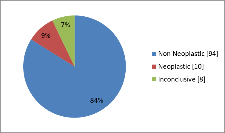
Fig:1 Type of skin lesions according to histopathology
Illustrates the overall distribution of skin lesions based on histopathology. Non-neoplastic lesions constituted the majority of cases, followed by a smaller proportion of neoplastic and inconclusive cases, emphasizing the predominance of benign conditions in the study population.
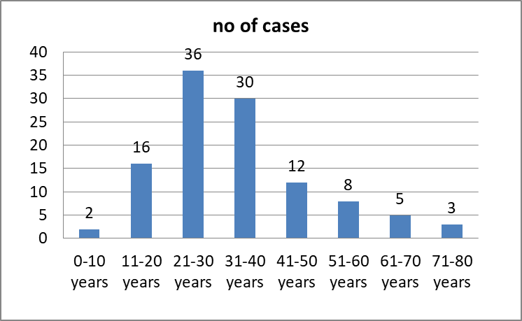
Fig:2 distribution of skin lesions according to age group
This depicts the age-wise distribution of skin lesions. The majority of cases were observed in the 21–30 years age group, indicating that skin lesions are more prevalent among young adults in this study population.
Table:1 distribution of cases according to Gender (n=112)
|
Gender |
No. of cases |
Percentage |
|
Male |
64 |
57.14% |
|
Female |
48 |
42.85% |
A male predominance was observed, with males accounting for 57.14% of cases, while females constituted 42.85%. This suggests a higher exposure of males to environmental and occupational risk factors.
Table:2 Distribution Of Non-neoplastic Skin Lesions
|
Skin lesions |
No of cases |
Percentage |
|
Leprosy |
37 |
39.36 |
|
Pityriasis Rosea |
5 |
5.31 |
|
Seborrheic Keratosis |
2 |
2.12 |
|
Contact Dermatitis |
1 |
1.06 |
|
Sarcoidosis |
2 |
2.12 |
|
Psoriasis |
2 |
2.12 |
|
Lichen Planus |
3 |
3.19 |
|
Verruca Vulgaris |
2 |
2.12 |
|
Myrmecia |
4 |
4.25 |
|
Discoid Lupus Erythematosis |
3 |
3.19 |
|
Nevus |
6 |
6.38 |
|
Chromoblastomycosis |
1 |
1.06 |
|
Morphea |
3 |
3.19 |
|
Folliculitis Decalvans |
1 |
1.06 |
|
Pityriasis Rubra Pilaris |
1 |
1.06 |
|
Erythema Annulare Centrifugum |
2 |
2.12 |
|
Chronic Lupus Erythematosus |
1 |
1.06 |
|
Verrucous Lesions |
7 |
7.44 |
|
Pemphigus |
3 |
3.19 |
|
Inflammatory Lesion |
5 |
5.31 |
|
Granulomatous Inflammation |
3 |
3.19 |
Leprosy was the most common lesion, accounting for 39.36% of cases, followed by verrucous lesions (7.44%) and nevi (6.38%). Other conditions such as lichen planus, pemphigus, and granulomatous inflammation were observed in smaller proportions, highlighting the wide spectrum of inflammatory and infectious dermatoses.
Table 3 Distribution of Neoplastic Skin Lesions(n=10)
|
Skin Lesions |
No Of Cases |
Percentage |
|
Squamous Cell Carcinoma |
5 |
50% |
|
Basal Cell Carcinoma |
4 |
40% |
|
Melanoma |
1 |
10% |
Squamous cell carcinoma was the most common malignancy (50%), followed by basal cell carcinoma (40%), while melanoma accounted for 10% of cases. This indicates a predominance of keratinocytic malignancies in the study population.
Table 4 COMPARATIVE OVERVIEW OF MY STUDY WITH OTHER STUDIES
|
Study (Year) |
Sample Size |
Common Age Group |
Gender Predominance |
Common Non-Neoplastic Lesion |
Common Neoplastic Lesion |
Overall Pattern |
|
Padvi et al. (IJPO 2024)4 |
374 |
<40 years (mean ~38 yrs) |
Male (60%) |
Dermatitis (28%), Leprosy |
Squamous cell carcinoma (41.7%), Basal cell carcinoma |
Predominantly non-neoplastic (98.4%) |
|
Dawande et al. (Cureus 2023)5 |
50 |
21–30 years |
Male (78%) |
Leprosy (24%) |
Squamous cell carcinoma (14%) |
Non-neoplastic predominant (66%) |
|
Goswami et al. (JFMPC 2022)6 |
610 |
>60 years |
Male predominance |
Vesiculobullous diseases(33.3%), Leprosy, Seborrheic keratosis |
Basal cell carcinoma (44.8%) |
Majority non-neoplastic (87.1%) |
|
Present Study |
112 |
21-30 |
Male(57.14) |
Leprosy(39.3%) |
Squamous cell carcinoma(50%) |
Non-neoplastic predominant(83.92%) |
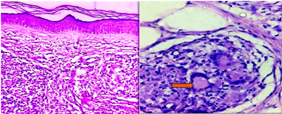
Figure3: Figure4:
Fig 3 Lepromatous Leprosy showing orthokeratosis, thinned out epidermis with grenz zone demis showing macrophages are found in poorly circumscribed masses H&E 10X.
Fig 4 Borderline Tuberculoid leprosy highlighting langhans gaint cells H&E 40X.
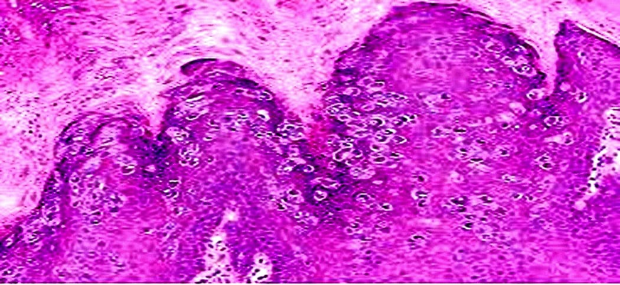
Fig 5 Myrmecia Highlighting Large eosinophilic intracytoplasmic inclusion bodies H&E 40X
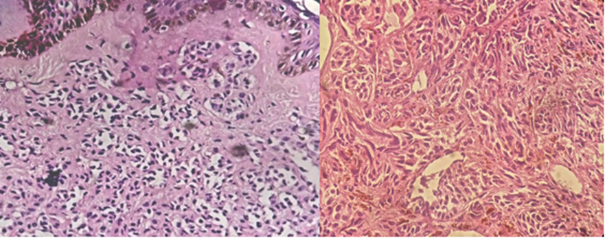
Fig 6 Intradermal Nevus H&E 10X. Fig 7 MELANOMA H&E 40X
DISCUSSION:
The present retrospective study highlights the histopathological spectrum of skin lesions encountered in a tertiary care hospital over a period of two years. A total of 112 cases were analyzed, demonstrating a predominance of non-neoplastic lesions (83.92%) over neoplastic lesions (8.92%), with a small proportion of inconclusive cases. This finding aligns with several previous studies, confirming that non-neoplastic dermatoses constitute the majority of skin biopsy diagnoses.
In this study, a male predominance (57.14%) was observed, which is consistent with studies by Padvi et al. and Dawande et al. This gender bias may be attributed to increased occupational exposure, environmental factors, and healthcare-seeking behavior among males.
The most commonly affected age group in the present study was 21–30 years, which is comparable to Dawande et al., who also reported a similar age distribution. However, some studies like Goswami et al. observed a higher prevalence in older age groups (>60 years), indicating that age distribution may vary depending on geographic, environmental, and demographic factors.
Among non-neoplastic lesions, leprosy (39.36%) was the most common finding in the present study. This is in concordance with studies conducted in endemic regions where infectious dermatoses remain highly prevalent. The high incidence of leprosy reflects ongoing public health challenges and emphasizes the need for early detection and treatment.
Other non-neoplastic lesions such as pityriasis rosea, lichen planus, lupus erythematosus, and granulomatous inflammation were observed in smaller proportions. The diversity of lesions highlights the importance of histopathological examination in differentiating clinically similar conditions.
Among neoplastic lesions, squamous cell carcinoma (50%) was the most common, followed by basal cell carcinoma (40%) and melanoma (10%). These findings are comparable with studies by Padvi et al. and Dawande et al., where squamous cell carcinoma was also the predominant malignancy. However, some studies have reported basal cell carcinoma as the most common, indicating regional variation in risk factors such as sun exposure and skin type.
The strong correlation between histopathological findings and clinical diagnosis reinforces the importance of biopsy as a diagnostic gold standard. Many lesions that appear similar clinically can only be accurately categorized through microscopic examination.
Overall, the present study reaffirms that histopathology plays a crucial role in the diagnosis, classification, and management of skin lesions and helps in guiding appropriate therapeutic interventions.
CONCLUSION:
This study demonstrates that non-neoplastic skin lesions constitute the majority of cases encountered in a tertiary care setting, with leprosy being the most prevalent condition. Among neoplastic lesions, squamous cell carcinoma was the most common malignancy.
The findings emphasize that clinical diagnosis alone is often insufficient for accurate characterization of skin lesions. Histopathological examination remains the gold standard for definitive diagnosis, enabling precise classification and guiding appropriate treatment.
Early biopsy and histopathological evaluation should be encouraged in all doubtful or persistent skin lesions to improve diagnostic accuracy and patient outcomes. Furthermore, the high prevalence of infectious dermatoses such as leprosy highlights the need for continued public health efforts, awareness, and early intervention strategies.
REFERENCES:
1. Kumar V, Abbas AK, Aster JC, Debnath J, Das A. Robbins, Cotran and Kumar Pathologic Basis of Disease. 11th ed. Philadelphia: Elsevier; 2025.
2. Singh BP, Bano S, Porte J, Ganguly S, Hirani N. Histomorphological spectrum of skin lesions in a tertiary care hospital: a 10-year retrospective study. Indian J Pathol Oncol. 2025;12(4):351–357.
3. Mithila KB, Priya GY, Anuradha V, Sreedhar V. Histopathological study of skin lesions in a tertiary care hospital. Int J Community Med Public Health. 2025;12(5):2113–2116.
4. Padvi AV, Patel DR, Shah YB. Histopathological study of spectrum of skin lesions in a tertiary care center. Indian J Pathol Oncol. 2024;11(1):52–57.
5. Dawande P, Kumbhare S, Wanjari K, Choudhary S. Histopathological spectrum of skin lesions: a retrospective study from a tertiary care center. Cureus. 2023;15(5):e38721.
6. Goswami BK, Barua M, Deka L, Das P. Histopathological spectrum of skin lesions in a tertiary care center: a cross-sectional study. J Family Med Prim Care. 2022;11(6):3025–3030.
7. Adhikari RC, Shah M, Jha AK. Histopathological spectrum of skin diseases in a tertiary care center. Nepal Med Coll J. 2019;21(1):10–14.
8. Grover S, Ranyal RK, Bedi MK. A cross section of skin diseases in rural Allahabad. Indian J Dermatol. 2008;53(4):179–181.
9. Kumar R, Kaur I, Kaur S. Histopathological study of skin lesions in a tertiary care hospital. J Clin Diagn Res. 2016;10(6):EC01–EC04.
10. Reddy BSN, Reddy BP, Reddy R. Clinicopathological study of skin lesions in a tertiary care hospital. Int J Res Med Sci. 2017;5(4):1503–1507.
11. Sharma R, Goswami KC, Barua M. Histopathological pattern of non-neoplastic skin lesions: a hospital-based study. Int J Res Dermatol. 2018;4(3):358–362.
12. Vora RV, Pilani AP, Kota RK, Singhal RR. Clinico-epidemiological study of skin diseases in a tertiary care center. Indian Dermatol Online J. 2015;6(6):423–426.