Intravascular Lithotripsy for Coronary Artery Calcification: Effectiveness and Safety in Complex Calcified Lesions Undergoing Percutaneous Coronary Intervention.
- Rahul, V , Assistant Professor and Consultant KLE JGMM Medical College, Hubballi Vihaan Heart Centre Hubli.
- Mohammed Naveed Nadaf , Consultant, Anesthesiology and Critical care VIHAAN HEART CARE HOSPITAL, Hubli, Karnataka.
- Sneha Karekar , Specialist in the Department of Cardiology Hospital-Manipal Hospital Old airport road Bangalore.
- Kishan Raj K , Associate Consultant Aster MIMS Kasargod.
Article Information:
Abstract:
Background: Coronary artery calcification remains a major challenge during percutaneous coronary intervention (PCI), often resulting in inadequate lesion preparation, suboptimal stent expansion, and increased procedural complications. Intravascular lithotripsy (IVL) has emerged as a novel calcium-modification technique designed to facilitate PCI in heavily calcified coronary lesions by generating acoustic pressure waves that selectively fracture calcified plaque while minimizing vessel injury. Objective: To evaluate the effectiveness and safety of intravascular lithotripsy in patients with complex calcified coronary artery lesions undergoing PCI. Materials and Methods: This prospective observational study was conducted at ___________ and included 70 consecutive patients with moderate-to-severe calcified coronary artery lesions who underwent IVL-assisted PCI. Baseline demographic, clinical, angiographic, procedural, and outcome data were collected and analyzed. The primary effectiveness endpoint was procedural success, defined as successful stent deployment with residual stenosis <30%, final TIMI grade 3 flow, and absence of in-hospital major adverse cardiovascular events (MACE). Results: The mean age of the study population was 68.4 ± 9.7 years, and 74.3% were male. Severe coronary calcification was present in 72.9% of lesions, while 70.0% were classified as ACC/AHA Type C lesions. Device success and angiographic success were achieved in 97.1% of patients, and procedural success was observed in 95.7%. Final TIMI grade 3 flow was achieved in 98.6% of cases. The mean residual stenosis following PCI was 8.4 ± 5.2%, and mean stent expansion was 92.6 ± 8.1% among patients undergoing intravascular imaging assessment. Conclusion: Intravascular lithotripsy is an effective and safe calcium-modification strategy for complex calcified coronary artery lesions undergoing PCI. High procedural success, excellent angiographic outcomes, favorable stent expansion, and low complication rates support the use of IVL as a valuable adjunctive technology in contemporary interventional cardiology.
Keywords:
Article :
INTRODUCTION:
Coronary artery calcification (CAC) represents one of the most challenging anatomical substrates encountered during percutaneous coronary intervention (PCI). Calcified coronary lesions are increasingly prevalent due to the aging population and the rising incidence of diabetes mellitus, chronic kidney disease, hypertension, and other cardiovascular risk factors. The presence of severe coronary calcification is associated with increased procedural complexity, reduced procedural success, and adverse clinical outcomes following PCI. Calcified plaques impair balloon expansion, hinder stent delivery, contribute to stent under-expansion and malapposition, and increase the risk of restenosis, stent thrombosis, myocardial infarction, and mortality [1,2].
The prevalence of coronary calcification among patients undergoing PCI has been reported to range from 18% to 31%, with the burden expected to increase substantially in contemporary interventional practice [3]. Severe calcification compromises vessel compliance and limits the effectiveness of conventional balloon angioplasty. Inadequate lesion preparation frequently results in suboptimal stent expansion, which remains one of the strongest predictors of long-term adverse cardiovascular events after PCI [4]. Consequently, effective calcium modification has become a critical component of successful revascularization in patients with heavily calcified coronary artery disease.
Several plaque-modification techniques have been developed to address calcified lesions, including cutting balloons, scoring balloons, rotational atherectomy, orbital atherectomy, and excimer laser coronary atherectomy. Although these technologies have improved outcomes in selected patients, they are associated with important limitations, including technical complexity, prolonged procedural times, distal embolization, vessel perforation, slow-flow or no-reflow phenomena, and a steep learning curve for operators [5]. Therefore, there has been considerable interest in developing safer and more effective strategies for calcium modification during PCI.
Intravascular lithotripsy (IVL) has emerged as a novel and promising technology for the treatment of heavily calcified coronary lesions. Adapted from extracorporeal lithotripsy used in the management of renal calculi, IVL utilizes a balloon-based catheter system that delivers localized pulsatile sonic pressure waves to fracture superficial and deep calcium deposits within the vessel wall while minimizing injury to surrounding soft tissues [6]. The device consists of multiple lithotripsy emitters mounted within a semi-compliant balloon inflated at low pressure. The generated acoustic shockwaves selectively disrupt calcium by creating microfractures, thereby increasing vessel compliance and facilitating optimal stent expansion [6,7].
The mechanism of action of IVL offers several theoretical and practical advantages over traditional atherectomy techniques. Unlike rotational and orbital atherectomy, IVL modifies both superficial and deep calcium without requiring high-speed rotational devices. Furthermore, IVL is associated with a lower risk of distal embolization, coronary dissection, perforation, and no-reflow events. The technology can be performed using standard PCI techniques and is considered relatively easy to adopt, making it attractive for widespread clinical use [8,9].
Clinical evidence supporting the use of IVL has expanded rapidly over the past several years. The DISRUPT CAD clinical program demonstrated high procedural success rates and favorable safety outcomes in patients with severely calcified coronary lesions undergoing PCI [10]. Intravascular imaging studies using optical coherence tomography (OCT) and intravascular ultrasound (IVUS) have confirmed that IVL effectively creates calcium fractures, increases luminal area, and improves stent expansion, which are key determinants of long-term procedural success [11]. Subsequent real-world registries and observational studies have further validated the effectiveness of IVL in a variety of complex clinical scenarios, including left main disease, bifurcation lesions, in-stent restenosis, chronic total occlusions, and acute coronary syndromes [12,13].
Recent evidence suggests that IVL achieves procedural success rates exceeding 90%, with low rates of major adverse cardiovascular events and procedural complications [12,14]. Furthermore, pooled analyses and registry data have demonstrated favorable outcomes even in lesions characterized by extensive calcium burden and calcified nodules, conditions traditionally associated with poor PCI outcomes [15]. These findings have positioned IVL as an important addition to the contemporary armamentarium for the management of calcified coronary artery disease.
Despite these encouraging results, questions remain regarding the effectiveness and safety of IVL in complex calcified lesions encountered in routine clinical practice. Real-world populations often present with multiple comorbidities, advanced lesion complexity, and challenging anatomical characteristics that may influence procedural outcomes. Therefore, further evaluation of IVL in diverse patient populations is essential to define its role in contemporary interventional cardiology.
The present study aims to evaluate the effectiveness and safety of intravascular lithotripsy in patients with complex calcified coronary lesions undergoing PCI. By assessing procedural success, angiographic outcomes, and periprocedural complications, this study seeks to contribute to the growing body of evidence supporting the use of IVL as a calcium-modification strategy in complex coronary interventions.
MATERIALS AND METHODS:
Study Design and Setting
This prospective observational study was conducted at ___________ between ___________ and ___________. The study evaluated the effectiveness and safety of intravascular lithotripsy (IVL) in patients with complex calcified coronary artery lesions undergoing percutaneous coronary intervention (PCI). The study was performed in accordance with the principles outlined in the Declaration of Helsinki and adhered to Good Clinical Practice guidelines.
Study Population
A total of 70 patients with angiographically significant calcified coronary artery disease who underwent PCI with IVL-assisted lesion preparation were included in the study.
Inclusion Criteria
Patients were eligible for inclusion if they met the following criteria:
1. Age ≥18 years.
2. Presence of symptomatic coronary artery disease, including stable angina, unstable angina, or acute coronary syndrome requiring PCI.
3. Angiographic evidence of moderate-to-severe coronary artery calcification as determined by fluoroscopy, intravascular ultrasound (IVUS), or optical coherence tomography (OCT).
4. Lesions requiring calcium modification before stent implantation.
5. Treatment using a coronary intravascular lithotripsy system as part of the PCI procedure.
6. Provision of informed consent.
Exclusion Criteria
Patients were excluded if they had:
1. Cardiogenic shock at presentation.
2. Known contraindications to dual antiplatelet therapy.
3. Severe contrast allergy not amenable to premedication.
4. Life expectancy less than one year due to non-cardiac comorbidities.
5. Incomplete procedural or clinical data.
6. Previous enrollment in the study.
Definition of Coronary Calcification
Coronary calcification was classified according to contemporary interventional cardiology criteria. Severe calcification was defined as radiopacities noted without cardiac motion before contrast injection involving both sides of the arterial wall, while moderate calcification was defined as radiopacities visible only during cardiac motion before contrast injection. Intravascular imaging findings such as calcium arc >180°, calcium thickness >0.5 mm, and calcium length >5 mm were recorded when available.
Data Collection
Baseline demographic, clinical, laboratory, angiographic, procedural, and outcome data were prospectively collected using a standardized case report form.
Clinical Variables
The following variables were recorded:
• Age and sex
• Body mass index (BMI)
• Hypertension
• Diabetes mellitus
• Dyslipidemia
• Smoking status
• Chronic kidney disease
• Previous myocardial infarction
• Previous PCI
• Previous coronary artery bypass grafting
• Clinical presentation (stable angina, unstable angina, NSTEMI, STEMI)
Angiographic Variables
The following lesion characteristics were assessed:
• Target vessel location
• Lesion length
• Reference vessel diameter
• Degree of stenosis
• Calcification severity
• Presence of bifurcation lesions
• Left main involvement
• Chronic total occlusion
• American College of Cardiology/American Heart Association (ACC/AHA) lesion classification
PCI Procedure and Intravascular Lithotripsy
All procedures were performed by experienced interventional cardiologists according to standard PCI techniques.
After coronary angiography and lesion assessment, appropriate guide catheters and guidewires were used to cross the target lesion. Intravascular imaging with IVUS or OCT was performed at the operator’s discretion before and after lesion preparation.
Coronary intravascular lithotripsy was performed using the Shockwave C2 Coronary IVL System (Shockwave Medical, Santa Clara, California, USA). The IVL balloon was selected in a 1:1 ratio to the reference vessel diameter and advanced across the calcified lesion.
The balloon was inflated to 4 atmospheres to ensure vessel-wall apposition, followed by delivery of lithotripsy pulses. Each treatment cycle consisted of ten pulses delivered at a rate of one pulse per second. Additional cycles were administered based on lesion characteristics and operator judgment, up to the maximum recommended pulse limit. Following calcium modification, the balloon was inflated to nominal pressure to optimize lesion preparation.
Drug-eluting stents were subsequently implanted according to standard practice. Post-dilatation using non-compliant balloons was performed whenever necessary to achieve optimal stent expansion.
Outcome Measures
Primary Effectiveness Endpoint
The primary effectiveness endpoint was procedural success, defined as:
• Successful stent delivery and deployment,
• Residual stenosis <30% by visual estimation,
• Final TIMI grade 3 coronary flow,
• Absence of in-hospital major adverse cardiovascular events (MACE).
Primary Safety Endpoint
The primary safety endpoint was the occurrence of procedural complications, including:
• Coronary perforation,
• Flow-limiting dissection,
• Slow-flow/no-reflow phenomenon,
• Abrupt vessel closure,
• Emergency coronary artery bypass surgery,
• Procedure-related death.
Secondary Endpoints
Secondary endpoints included:
1. Angiographic success.
2. Device success.
3. Stent expansion assessed by IVUS/OCT when available.
4. In-hospital MACE.
5. Thirty-day MACE.
MACE was defined as a composite of:
• Cardiac death,
• Myocardial infarction,
• Target lesion revascularization,
• Target vessel revascularization,
• Definite or probable stent thrombosis.
Follow-up
All patients were monitored throughout hospitalization and underwent clinical follow-up at 30 days after PCI through outpatient visits or structured telephone interviews. Clinical events were independently verified using hospital records whenever available.
Statistical Analysis
Data were analyzed using IBM SPSS Statistics version 26.(IBM Corp., Armonk, NY, USA).
Continuous variables were tested for normality using the Shapiro–Wilk test. Normally distributed variables were expressed as mean ± standard deviation, while non-normally distributed variables were reported as median with interquartile range.
Categorical variables were presented as frequencies and percentages.
Comparisons between groups were performed using:
• Independent-samples t-test for normally distributed continuous variables,
• Mann–Whitney U test for non-normally distributed variables,
• Chi-square test or Fisher’s exact test for categorical variables.
Multivariable logistic regression analysis was performed to identify independent predictors of procedural success and adverse clinical outcomes. Variables with p < 0.10 in univariate analysis were entered into the multivariable model.
A two-sided p-value <0.05 was considered statistically significant.
RESULTS:
A total of 70 patients with complex calcified coronary artery lesions undergoing PCI with intravascular lithotripsy (IVL) were included in the study. The mean age was 68.4 ± 9.7 years, and the majority were male (74.3%).
Hypertension (81.4%), diabetes mellitus (55.7%), and dyslipidemia (70.0%) were the most prevalent cardiovascular risk factors. Stable angina was the most common clinical presentation (42.9%), followed by non-ST-elevation myocardial infarction (NSTEMI) (31.4%).
Table 1. Baseline Demographic and Clinical Characteristics (n = 70)
|
Variable |
Value |
|
Age (years), mean ± SD |
68.4 ± 9.7 |
|
Male sex, n (%) |
52 (74.3) |
|
BMI (kg/m²), mean ± SD |
27.1 ± 3.8 |
|
Hypertension, n (%) |
57 (81.4) |
|
Diabetes mellitus, n (%) |
39 (55.7) |
|
Dyslipidemia, n (%) |
49 (70.0) |
|
Current smokers, n (%) |
18 (25.7) |
|
Chronic kidney disease, n (%) |
15 (21.4) |
|
Previous PCI, n (%) |
20 (28.6) |
|
Previous MI, n (%) |
16 (22.9) |
|
Stable angina, n (%) |
30 (42.9) |
|
Unstable angina, n (%) |
12 (17.1) |
|
NSTEMI, n (%) |
22 (31.4) |
|
STEMI, n (%) |
6 (8.6) |
Table 1 summarizes the baseline demographic and clinical characteristics of the study population. A total of 70 patients with complex calcified coronary artery lesions underwent IVL-assisted PCI. The mean age of the cohort was 68.4 ± 9.7 years, indicating that the study predominantly involved elderly patients. Males constituted 74.3% of the study population.
Cardiovascular risk factors were highly prevalent, with hypertension observed in 81.4%, dyslipidemia in 70.0%, and diabetes mellitus in 55.7% of patients. Chronic kidney disease was present in 21.4% of cases, reflecting a population at increased risk for severe coronary calcification. Stable angina was the most common clinical presentation (42.9%), followed by NSTEMI (31.4%), indicating that IVL was utilized across a broad spectrum of coronary artery disease presentations.
Table 2. Angiographic and Lesion Characteristics
|
Variable |
Value |
|
LAD lesions, n (%) |
30 (42.9) |
|
RCA lesions, n (%) |
22 (31.4) |
|
LCX lesions, n (%) |
12 (17.1) |
|
Left main lesions, n (%) |
6 (8.6) |
|
Lesion length (mm), mean ± SD |
28.6 ± 10.4 |
|
Reference vessel diameter (mm), mean ± SD |
3.18 ± 0.52 |
|
Diameter stenosis (%), mean ± SD |
82.4 ± 8.1 |
|
Moderate calcification, n (%) |
19 (27.1) |
|
Severe calcification, n (%) |
51 (72.9) |
|
Bifurcation lesions, n (%) |
17 (24.3) |
|
Left main disease, n (%) |
9 (12.9) |
|
Chronic total occlusion, n (%) |
7 (10.0) |
|
ACC/AHA Type C lesions, n (%) |
49 (70.0) |
Table 2 presents the angiographic and lesion-related characteristics of the target vessels. The left anterior descending artery was the most frequently treated vessel (42.9%), followed by the right coronary artery (31.4%). The mean lesion length was 28.6 ± 10.4 mm, indicating the presence of relatively long and complex lesions. Severe coronary calcification was observed in 72.9% of lesions, confirming the challenging nature of the study cohort. Furthermore, 70.0% of lesions were classified as ACC/AHA Type C lesions.
Additional high-risk anatomical features included bifurcation lesions (24.3%), left main coronary artery involvement (12.9%), and chronic total occlusions (10.0%). These findings demonstrate that IVL was predominantly used in anatomically complex and heavily calcified lesions.
Table 3. Procedural Characteristics
|
Variable |
Value |
|
Radial access, n (%) |
52 (74.3) |
|
Femoral access, n (%) |
18 (25.7) |
|
IVUS-guided PCI, n (%) |
41 (58.6) |
|
OCT-guided PCI, n (%) |
11 (15.7) |
|
IVL balloon diameter (mm), mean ± SD |
3.20 ± 0.48 |
|
Number of IVL pulses, mean ± SD |
58.3 ± 18.5 |
|
Drug-eluting stent implantation, n (%) |
70 (100) |
|
Post-dilatation performed, n (%) |
61 (87.1) |
|
Procedural time (minutes), mean ± SD |
71.5 ± 21.3 |
|
Contrast volume (mL), mean ± SD |
182.4 ± 54.8 |
Table 3 outlines the procedural details of IVL-assisted PCI. Radial access was used in the majority of patients (74.3%), reflecting contemporary interventional practice. Intravascular imaging guidance was frequently employed, with IVUS utilized in 58.6% and OCT in 15.7% of procedures.
The average number of lithotripsy pulses delivered was 58.3 ± 18.5, indicating adequate calcium modification before stent implantation. Drug-eluting stents were implanted in all patients, while post-dilatation was performed in 87.1% of cases to optimize stent expansion. The mean procedural duration was 71.5 ± 21.3 minutes, with an average contrast volume of 182.4 ± 54.8 mL. These findings suggest that IVL can be successfully integrated into routine PCI workflows for the treatment of severely calcified lesions.
Table 4. Effectiveness Outcomes
|
Outcome |
Value |
|
Device success, n (%) |
68 (97.1) |
|
Procedural success, n (%) |
67 (95.7) |
|
Angiographic success, n (%) |
68 (97.1) |
|
Final residual stenosis (%) |
8.4 ± 5.2 |
|
TIMI Grade 3 flow, n (%) |
69 (98.6) |
|
Stent expansion (%)* |
92.6 ± 8.1 |
|
In-hospital MACE, n (%) |
2 (2.9) |
Table 4 demonstrates the effectiveness of IVL in facilitating PCI in complex calcified coronary lesions. Device success was achieved in 97.1% of cases, while angiographic success was also observed in 97.1% of patients. The overall procedural success rate was 95.7%, indicating that IVL effectively enabled successful stent delivery and deployment with acceptable residual stenosis and restoration of coronary blood flow.
The mean residual stenosis following PCI was only 8.4 ± 5.2%, reflecting excellent lesion preparation and stent expansion. TIMI grade 3 flow was achieved in 98.6% of patients, demonstrating effective reperfusion. Among patients who underwent intravascular imaging assessment, mean stent expansion reached 92.6 ± 8.1%, supporting the ability of IVL to improve vessel compliance and optimize stent deployment. In-hospital MACE occurred in only 2.9% of patients, further supporting the procedural effectiveness of IVL.
Table 5. Safety Outcomes and 30-Day Clinical Events
|
Variable |
Value |
|
Coronary dissection, n (%) |
3 (4.3) |
|
Flow-limiting dissection, n (%) |
1 (1.4) |
|
Slow-flow/no-reflow, n (%) |
2 (2.9) |
|
Coronary perforation, n (%) |
0 (0.0) |
|
Abrupt vessel closure, n (%) |
0 (0.0) |
|
Emergency CABG, n (%) |
0 (0.0) |
|
In-hospital death, n (%) |
0 (0.0) |
|
30-day cardiac death, n (%) |
1 (1.4) |
|
30-day myocardial infarction, n (%) |
1 (1.4) |
|
Target lesion revascularization, n (%) |
1 (1.4) |
|
Definite/probable stent thrombosis, n (%) |
0 (0.0) |
|
Overall 30-day MACE, n (%) |
3 (4.3) |
Table 5 summarizes procedural safety and short-term clinical outcomes following IVL-assisted PCI. Procedural complications were infrequent. Coronary dissection occurred in 4.3% of patients, while flow-limiting dissection was observed in only 1.4%. Slow-flow or no-reflow phenomena occurred in 2.9% of cases. Notably, no cases of coronary perforation, abrupt vessel closure, emergency coronary artery bypass graft surgery, or in-hospital mortality were reported.
During 30-day follow-up, cardiac death, myocardial infarction, and target lesion revascularization each occurred in only one patient (1.4%). No definite or probable stent thrombosis was observed. The overall 30-day major adverse cardiovascular event rate was 4.3%, indicating a favorable short-term safety profile of IVL in the management of heavily calcified coronary lesions.
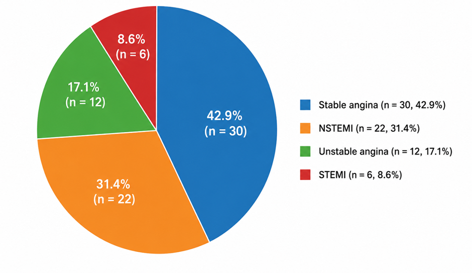
Figure 1. Distribution of clinical presentation among patients undergoing IVL-assisted PCI
Figure 1 illustrates the distribution of clinical presentations among the 70 patients who underwent intravascular lithotripsy (IVL)-assisted percutaneous coronary intervention (PCI). Stable angina was the most common clinical presentation, accounting for 42.9% (n = 30) of patients.
This was followed by non-ST-elevation myocardial infarction (NSTEMI), which was observed in 31.4% (n = 22) of cases. Unstable angina constituted 17.1% (n = 12) of the study population, while ST-elevation myocardial infarction (STEMI) was the least frequent presentation, occurring in 8.6% (n = 6) of patients.
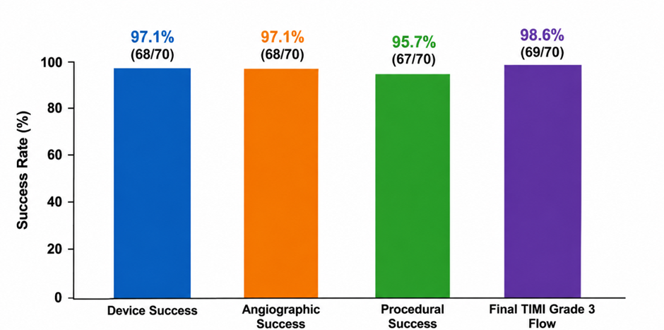
Figure 2. Procedural success, angiographic success, and device success rates following intravascular lithotripsy
Figure 2 illustrates the procedural outcomes following intravascular lithotripsy (IVL)-assisted percutaneous coronary intervention in patients with complex calcified coronary artery lesions. Device success was achieved in 97.1% (68/70) of patients, indicating successful delivery and activation of the IVL system across the target lesions. Angiographic success was also observed in 97.1% (68/70) of cases, reflecting effective lesion preparation and stent deployment with residual stenosis less than 30% and restoration of adequate coronary blood flow.
The overall procedural success rate was 95.7% (67/70), demonstrating a high rate of successful PCI without in-hospital major adverse cardiovascular events (MACE). Final TIMI grade 3 flow was achieved in 98.6% (69/70) of patients, indicating excellent post-procedural coronary perfusion. Collectively, these findings highlight the high effectiveness of IVL in facilitating optimal lesion modification, stent expansion, and successful revascularization in heavily calcified coronary lesions.
DISCUSSION:
The present study evaluated the effectiveness and safety of intravascular lithotripsy (IVL) in 70 patients with complex calcified coronary artery lesions undergoing percutaneous coronary intervention (PCI). The principal findings were: (1) a high procedural success rate of 95.7%, (2) angiographic and device success rates of 97.1%, (3) achievement of TIMI grade 3 flow in 98.6% of patients, and (4) a low incidence of procedural complications and 30-day major adverse cardiovascular events (MACE). These findings support the growing evidence that IVL is an effective and safe calcium-modification strategy for heavily calcified coronary lesions.
Coronary artery calcification remains one of the most important predictors of adverse procedural and long-term outcomes following PCI. Severe calcification limits lesion compliance, impairs balloon expansion, and contributes to stent under-expansion, which is strongly associated with restenosis and stent thrombosis [16,17]. In the present study, severe calcification was observed in 72.9% of lesions, while 70.0% were classified as ACC/AHA Type C lesions, highlighting the complexity of the study population. Despite these challenging anatomical characteristics, excellent procedural outcomes were achieved, emphasizing the ability of IVL to effectively modify calcified plaques and facilitate optimal stent deployment.
The procedural success rate of 95.7% observed in our study is comparable to that reported in the DISRUPT CAD clinical program. In the DISRUPT CAD II study, Ali et al. reported a procedural success rate exceeding 92% among patients with severely calcified coronary lesions treated with IVL-assisted PCI [18]. Our findings further validate these results in a real-world cohort that included complex lesion subsets such as bifurcation lesions, left main disease, and chronic total occlusions. The slightly higher success rate observed in our study may be attributable to the frequent use of intravascular imaging guidance and careful lesion preparation before stent implantation.
Device success and angiographic success were each achieved in 97.1% of patients. These results are consistent with observations from contemporary registries evaluating IVL in routine clinical practice. Recent studies have demonstrated high device deliverability, effective calcium fracture formation, and favorable angiographic outcomes in heavily calcified coronary lesions treated with IVL [12,13]. The high angiographic success rate observed in the current study likely reflects the ability of IVL-generated acoustic pressure waves to create fractures within both superficial and deep calcified plaques, thereby improving vessel compliance and facilitating optimal stent expansion.
Optimal coronary blood flow restoration remains a critical determinant of PCI success. In the present study, TIMI grade 3 flow was achieved in 98.6% of patients. This finding is comparable to previous IVL investigations, which consistently reported TIMI grade 3 flow rates above 95% following lesion preparation and stent implantation [11,22]. Unlike atherectomy-based calcium modification techniques, IVL does not rely on plaque ablation and therefore minimizes distal embolization and microvascular obstruction. This mechanism may explain the exceptionally high rates of final TIMI grade 3 flow and the low incidence of slow-flow/no-reflow phenomena observed in our cohort.
The safety profile of IVL in our study was favorable. Coronary dissection occurred in only 4.3% of patients, while flow-limiting dissection was observed in 1.4%. Importantly, no cases of coronary perforation, abrupt vessel closure, emergency coronary artery bypass graft surgery, or in-hospital mortality were recorded. These findings are consistent with previous investigations demonstrating the safety advantages of IVL compared with conventional atherectomy techniques [23]. By selectively fracturing calcium while preserving surrounding soft tissue integrity, IVL reduces the risk of vessel injury and provides a predictable and controlled method of plaque modification.
The incidence of 30-day MACE in our study was 4.3%, including one cardiac death, one myocardial infarction, and one target lesion revascularization. These outcomes are similar to those reported in pooled analyses of the DISRUPT CAD studies and contemporary meta-analyses, which have demonstrated low rates of mortality, myocardial infarction, and repeat revascularization following IVL-assisted PCI [11,14]. The favorable short-term outcomes observed in our study may be attributable to effective lesion preparation, excellent stent expansion, and minimal procedural complications.
The widespread use of intravascular imaging in the present study may have also contributed to the observed procedural success. IVUS or OCT guidance was utilized in approximately three-quarters of patients. Contemporary evidence supports the use of intracoronary imaging for accurate assessment of calcium morphology, optimization of lesion preparation, and evaluation of stent expansion [25]. Studies have demonstrated that calcium fracture formation identified by OCT following IVL is associated with greater luminal gain and improved stent expansion. In our cohort, mean stent expansion exceeded 90%, further supporting the effectiveness of IVL in achieving optimal procedural results.
The present study adds to the growing body of evidence supporting the role of IVL in the management of complex calcified coronary artery disease. Unlike earlier studies that primarily evaluated selected lesion subsets, our investigation included a broad spectrum of challenging lesions commonly encountered in routine clinical practice. The high success rates and low complication rates observed suggest that IVL can be safely incorporated into contemporary PCI strategies, particularly in patients with severe coronary calcification who may otherwise be at increased risk of procedural failure.
Several limitations should be acknowledged. First, the study was conducted at a single center with a relatively small sample size of 70 patients, which may limit the generalizability of the findings. Second, the observational design precludes direct comparison with alternative calcium-modification techniques such as rotational or orbital atherectomy. Third, longer-term follow-up data were not available, preventing assessment of late clinical outcomes. Future multicenter studies with larger populations and extended follow-up periods are warranted to further establish the long-term efficacy and safety of IVL in complex coronary interventions.
Overall, the findings of this study reinforce the growing clinical evidence that intravascular lithotripsy provides effective calcium modification, facilitates optimal stent deployment, and achieves favorable short-term clinical outcomes in patients with heavily calcified coronary artery disease undergoing PCI.
CONCLUSION:
Intravascular lithotripsy (IVL) demonstrated high effectiveness and an excellent safety profile in the treatment of complex calcified coronary artery lesions undergoing percutaneous coronary intervention. Despite the high prevalence of severe calcification and complex lesion morphology in the study population, IVL achieved high device success, angiographic success, and procedural success rates, while maintaining low rates of procedural complications and short-term major adverse cardiovascular events.
The technology effectively facilitated lesion preparation, optimized stent deployment, and resulted in excellent restoration of coronary blood flow, as evidenced by the high incidence of final TIMI grade 3 flow. Furthermore, the absence of major procedural complications such as coronary perforation, abrupt vessel closure, or emergency coronary artery bypass surgery underscores the procedural safety of IVL in heavily calcified lesions.
REFERENCES:
1. Genereux P, Madhavan MV, Mintz GS, Maehara A, Palmerini T, Lasalle L, et al. Ischemic outcomes after coronary intervention of calcified vessels in acute coronary syndromes: pooled analysis from the HORIZONS-AMI and ACUITY trials. J Am CollCardiol. 2014;63(18):1845-54.
2. Bourantas CV, Zhang YJ, Garg S, Iqbal J, Valgimigli M, Windecker S, et al. Prognostic implications of coronary calcification in patients with obstructive coronary artery disease treated by percutaneous coronary intervention. Eur Heart J. 2014;35(32):2185-94.
3. Oliveira C, Faria D, Morais P, Ferreira A, Teixeira R, Gonçalves L. Coronary intravascular lithotripsy effectiveness and safety in real-world calcified coronary lesions. J Clin Med. 2024;13(8):2296.
4. Fujino A, Mintz GS, Matsumura M, Lee T, Kim SY, Hoshino M, et al. A new optical coherence tomography-based calcium scoring system to predict stent underexpansion. EuroIntervention. 2018;13(18):e2182-e2189.
5. Barbato E, Shlofmitz E, Milkas A, et al. State of the art: evolving concepts in the treatment of heavily calcified and undilatable coronary stenoses. EuroIntervention. 2017;13(6):696-705.
6. Dini CS, Tomberli B, Mattesini A, Ristalli F, Valente S, Stolcova M, et al. Intravascular lithotripsy for calcific coronary and peripheral artery stenoses. EuroIntervention. 2019;15(8):714-721.
7. Butt N, Patel V, Chhabra L. Intravascular Lithotripsy. StatPearls [Internet]. Treasure Island (FL): StatPearls Publishing; 2023.
8. Kochiashvili G, Devadze N, Kalandadze I, et al. Intravascular lithotripsy: approach to advanced calcified coronary artery disease. Cureus. 2024;16(8):e67656.
9. Honton B, Banning AP, Chieffo A, et al. Coronary best practice in intravascular lithotripsy. IntervCardiol Rev. 2022;17:e18.
10. Ali ZA, Nef H, Escaned J, Werner N, Banning AP, Hill JM, et al. Safety and effectiveness of coronary intravascular lithotripsy for treatment of severely calcified coronary stenoses: the Disrupt CAD II study. CircCardiovascInterv. 2019;12(10):e008434.
11. Ali ZA, Hill JM, Ben-Yehuda O, et al. Intravascular lithotripsy for treatment of severely calcified coronary lesions: patient-level pooled analysis of the Disrupt CAD studies. J Am CollCardiolIntv. 2021;14(12):1337-1348.
12. Oliveri F, Verheye S, et al. Intravascular lithotripsy for the treatment of calcified coronary lesions: contemporary evidence and outcomes. J IntervCardiol. 2025;2025:Article ID 9982145.
13. Oliveri F, Verheye S, et al. Intravascular lithotripsy for calcific coronary bifurcations: procedural success and clinical outcomes from the BENELUX-IVL registry. Catheter CardiovascInterv. 2025.
14. Sagris M, et al. Efficacy of intravascular lithotripsy in coronary stenosis: a systematic review and meta-analysis. Eur Heart J. 2023;44(Suppl 2):ehad655.2179.
15. Ali ZA, Maehara A, Généreux P, et al. Outcomes of coronary intravascular lithotripsy for the treatment of calcified nodules: a pooled analysis of the Disrupt CAD studies. EuroIntervention. 2023;19(2):e123-e131.
16. Mintz GS, Popma JJ, Pichard AD, Kent KM, Satler LF, Chuang YC, et al. Patterns of calcification in coronary artery disease. A statistical analysis of intravascular ultrasound and coronary angiography in 1155 lesions. Circulation. 1995;91(7):1959-65.
17. Moussa I, Ellis SG, Jones M, Kereiakes DJ, McMartin D, Rutherford B, et al. Impact of coronary culprit lesion calcium in patients undergoing paclitaxel-eluting stent implantation. Am J Cardiol. 2005;96(9):1242-7.
18. Ali ZA, Nef H, Escaned J, Werner N, Banning AP, Hill JM, et al. Safety and effectiveness of coronary intravascular lithotripsy for treatment of severely calcified coronary stenoses: DISRUPT CAD II. CircCardiovascInterv. 2019;12(10).
19. Oliveri F, Verheye S, et al. Intravascular lithotripsy for the treatment of calcified coronary lesions: contemporary evidence and outcomes. J IntervCardiol. 2025;2025:Article ID 9982145.
20. Oliveri F, Verheye S, et al. BENELUX-IVL Registry Investigators. Clinical outcomes of intravascular lithotripsy in complex calcified coronary lesions. Catheter CardiovascInterv. 2024.
21. Hill JM, Kereiakes DJ, Shlofmitz RA, Klein AJ, Riley RF, Price MJ, et al. Intravascular lithotripsy for treatment of severely calcified coronary artery disease. J Am CollCardiol. 2020;76(22):2635-2646.
22. Chambers JW, Behrens AN, Martinsen BJ. Atherectomy devices for coronary artery disease. IntervCardiolClin. 2016;5(2):143-151.
23. Sagris M, Karamasis GV, et al. Outcomes of coronary intravascular lithotripsy: systematic review and meta-analysis. Eur Heart J. 2023;44(Suppl 2):ehad655.2179.
24. Räber L, Mintz GS, Koskinas KC, Johnson TW, Holm NR, Onuma Y, et al. Clinical use of intracoronary imaging. Eur Heart J. 2018;39(35):3281-3300.
25. Maehara A, Matsumura M, Ali ZA, Mintz GS, Stone GW. IVUS- and OCT-guided calcium modification and stent optimization in calcified coronary lesions. JACC Cardiovasc Imaging. 2020;13(10):2207-2223.