Anatomical and Morphometric Study of Pedicles in Adult Human Lumbar Vertebrae.
- Charu Taneja , Associate Professor, Department of Anatomy, Government Medical College, Nagaur, Rajasthan, India.
- Parth Roat , Department of Anatomy, Geetanjali Medical College & Hospital, Udaipur, Rajasthan, India.
- Devesh Kumar Sharma , Assistant Professor, Department of Anatomy, JIET Medical College and Hospital, Jodhpur, Rajasthan, India.
- Shushil Kudi , Senior Resident, Department of Community Medicine, Government Medical College, Nagaur, Rajasthan, India.
- Basant Kumar , Assistant Professor, Department of Biochemistry, JIET Medical College and Hospital, Jodhpur, Rajasthan, India.
- Isha Choudhary , Senior Resident, Department of Psychiatry, Government Medical College, Nagaur, Rajasthan, India.
Article Information:
Abstract:
Background: Lumbar pedicle dimensions are directly relevant to transpedicular fixation, yet these dimensions vary across vertebral levels and populations. Region-specific osteometric data can therefore assist anatomical teaching and operative planning. Objective: To compare pedicle width, pedicle height and interpedicular distance in typical and atypical adult human lumbar vertebrae. Materials and methods: A descriptive cross-sectional osteological study was carried out in the Department of Anatomy, Government Medical College, Nagaur, Rajasthan. One hundred dry adult human lumbar vertebrae of unknown age and sex were examined, including 50 typical and 50 atypical vertebrae. Fully ossified and intact specimens were included, whereas damaged, deformed or pathologically altered vertebrae were excluded. Right and left pedicle width and height and the interpedicular distance were measured with a digital Vernier caliper. Data were expressed as mean ± standard deviation, and p<0.05 was considered statistically significant. Results: No significant right-left difference was observed in pedicle width or height within either vertebral group. Atypical vertebrae had wider pedicles than typical vertebrae on the right (10.52 ± 2.82 mm vs 8.29 ± 1.76 mm) and left (10.75 ± 2.77 mm vs 8.31 ± 1.92 mm), with p<0.001 for both comparisons. Pedicle height was similar between groups on the right (14.06 ± 2.23 mm vs 13.89 ± 1.49 mm; p=0.65) and left (14.16 ± 2.24 mm vs 14.00 ± 1.35 mm; p=0.66). Interpedicular distance was greater in atypical vertebrae (18.71 ± 3.99 mm vs 17.34 ± 2.07 mm; p=0.003). Conclusion: Atypical lumbar vertebrae were characterised mainly by greater transverse pedicle dimensions, while pedicle height remained comparable with that of typical vertebrae. These measurements provide useful regional anatomical information for implant selection and pedicle screw placement, although patient-specific imaging remains essential in clinical practice.
Keywords:
Article :
INTRODUCTION:
The lumbar vertebrae bear much of the axial load of the trunk while permitting movement and protecting the neural structures within the vertebral canal. The pedicles form short, strong bridges between the vertebral body and posterior elements, and their dimensions influence the shape of the vertebral canal as well as the size of the osseous corridor available for spinal instrumentation.1,2
Pedicle screws are widely used for posterior stabilisation because fixation through the pedicle provides access to all three spinal columns. Their safety, however, depends on an accurate understanding of pedicle anatomy. A screw that is too large or incorrectly directed may breach the cortex and injure adjacent neural or vascular structures, whereas a screw that is too small may provide inadequate purchase. The cortical and subcortical structure of the pedicle also contributes substantially to screw stability.3,4
Lumbar pedicle morphology is not uniform. It varies with vertebral level, sex, body size, ancestry and the method used for measurement. Studies from Asian and Indian populations have generally shown a progressive increase in transverse pedicle diameter toward the lower lumbar region, but the absolute values differ across populations and study techniques.5-10The type of motion available at a joint formed by it also varies according to age, sex and the structure of joint .12
The fifth lumbar vertebra is structurally adapted to the lumbosacral junction and is conventionally described as atypical. Although several reports have documented level-wise lumbar measurements, data from western Rajasthan remain limited. In our study, pedicle width, pedicle height and interpedicular distance were measured in typical and atypical adult human lumbar vertebrae to assess bilateral symmetry and identify morphometric differences of potential surgical relevance.11
MATERIALS AND METHODS:
Study design and specimens
This descriptive cross-sectional osteological study was conducted over one year in the Department of Anatomy, Government Medical College, Nagaur, Rajasthan, India. The study material comprised 100 dry adult human lumbar vertebrae of unknown age and sex from the departmental osteology collection. Fifty specimens were classified as typical lumbar vertebrae and 50 as atypical lumbar vertebrae on the basis of standard anatomical features. Fully ossified vertebrae with intact pedicles and clearly defined landmarks were included. Specimens that were broken, incompletely ossified, deformed, eroded or affected by an evident pathological process were excluded.
Morphometric measurements
All measurements were recorded in millimetres with a digital Vernier caliper. Pedicle width was measured at the narrowest transverse part of each pedicle between its medial and lateral cortices. Pedicle height was measured at the narrowest superior-inferior part between the superior and inferior cortices. Both parameters were recorded separately on the right and left sides. Interpedicular distance was measured as the transverse distance between the medial surfaces of the two pedicles. The same anatomical landmarks and measurement technique were used for all specimens.
Statistical analysis
Continuous variables were summarised as mean ± standard deviation (SD). Right-left measurements were compared within each vertebral group, and the corresponding dimensions of typical and atypical vertebrae were compared between groups. All comparisons were two-sided, and a p value below 0.05 was considered statistically significant.
RESULTS:
A total of 100 adult dried lumbar vertebrae were evaluated, with equal numbers of typical and atypical specimens. The measurements of pedicle width, pedicle height and interpedicular distance are summarised below.
Bilateral measurements
Typical lumbar vertebrae showed nearly identical measurements on the two sides. Mean pedicle width was 8.29 ± 1.76 mm on the right and 8.31 ± 1.92 mm on the left (p=0.96), while mean pedicle height was 13.89 ± 1.49 mm and 14.00 ± 1.35 mm, respectively (p=0.70). Atypical vertebrae showed the same pattern: right and left pedicle widths were 10.52 ± 2.82 mm and 10.75 ± 2.77 mm (p=0.68), and the corresponding heights were 14.06 ± 2.23 mm and 14.16 ± 2.24 mm (p=0.82). None of the bilateral comparisons was statistically significant.
Table 1. Side-to-side comparison of pedicle dimensions in typical and atypical lumbar vertebrae
|
Parameter |
Typical right |
Typical left |
p value |
Atypical right |
Atypical left |
p value |
|
Width (mm) |
8.29 ± 1.76 |
8.31 ± 1.92 |
0.96 |
10.52 ± 2.82 |
10.75 ± 2.77 |
0.68 |
|
Height (mm) |
13.89 ± 1.49 |
14.00 ± 1.35 |
0.70 |
14.06 ± 2.23 |
14.16 ± 2.24 |
0.82 |
Values are expressed as mean ± SD.
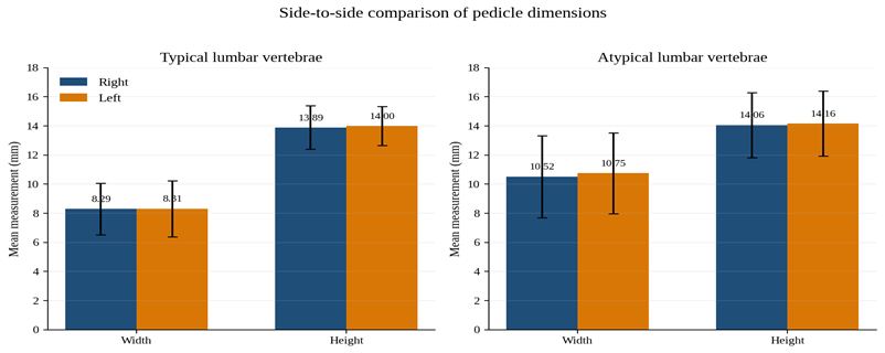
Figure 1. Side-to-side comparison of pedicle width and height in typical and atypical lumbar vertebrae. Error bars represent SD.
Comparison of typical and atypical vertebrae
The main between-group difference was seen in pedicle width. On the right side, atypical vertebrae had a mean width of 10.52 ± 2.82 mm compared with 8.29 ± 1.76 mm in typical vertebrae, a difference of 2.23 mm (p<0.001). On the left side, the corresponding values were 10.75 ± 2.77 mm and 8.31 ± 1.92 mm, a difference of 2.44 mm (p<0.001). Pedicle height did not differ significantly between the groups. The right-sided means were 14.06 ± 2.23 mm and 13.89 ± 1.49 mm (p=0.65), while the left-sided means were 14.16 ± 2.24 mm and 14.00 ± 1.35 mm (p=0.66) in atypical and typical vertebrae, respectively.
Table 2. Comparison of pedicle dimensions between typical and atypical lumbar vertebrae.
|
Parameter |
Side |
Typical |
Atypical |
Mean difference |
p value |
|
Pedicle width (mm) |
Right |
8.29 ± 1.76 |
10.52 ± 2.82 |
2.23 |
<0.001 |
|
Pedicle width (mm) |
Left |
8.31 ± 1.92 |
10.75 ± 2.77 |
2.44 |
<0.001 |
|
Pedicle height (mm) |
Right |
13.89 ± 1.49 |
14.06 ± 2.23 |
0.17 |
0.65 |
|
Pedicle height (mm) |
Left |
14.00 ± 1.35 |
14.16 ± 2.24 |
0.16 |
0.66 |
Values are expressed as mean ± SD.
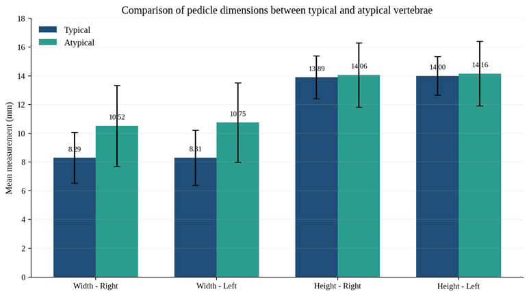
Figure 2. Comparison of pedicle width and height between typical and atypical lumbar vertebrae. Error bars represent SD.
Interpedicular distance
Mean interpedicular distance measured 17.34 ± 2.07 mm in typical vertebrae and 18.71 ± 3.99 mm in atypical vertebrae. The mean difference was 1.37 mm and was statistically significant (p=0.003). Taken together, the findings show that atypical vertebrae differed mainly in their transverse dimensions, whereas pedicle height remained similar in the two groups.
Table 3. Comparison of interpedicular distance between typical and atypical lumbar vertebrae
|
Measurement |
Typical |
Atypical |
p value |
|
Interpedicular distance (mm) |
17.34 ± 2.07 |
18.71 ± 3.99 |
0.003 |
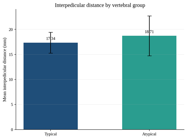
Figure 3. Mean interpedicular distance in typical and atypical lumbar vertebrae. Error bars represent SD.
DISCUSSION:
In our study, pedicle width and height were bilaterally symmetrical in both typical and atypical lumbar vertebrae. The clearest difference between the groups was a greater pedicle width in atypical vertebrae on both sides. Interpedicular distance was also greater in the atypical group, whereas pedicle height remained essentially unchanged. This pattern suggests that the transition to atypical lower lumbar morphology is expressed more through transverse widening than through an increase in vertical pedicle dimension.
The lack of a significant right-left difference is in keeping with earlier quantitative studies of the lumbar spine. Panjabi et al. described broadly reproducible bilateral vertebral geometry, and more recent pooled analyses have also found little clinically meaningful side-to-side variation in pedicle dimensions. Bilateral symmetry is useful during anatomical assessment and surgical planning because the opposite pedicle may serve as a reference when one side is difficult to delineate. It should not, however, be taken to mean that every individual vertebra is perfectly symmetrical.1,11
The significantly larger pedicle width in atypical vertebrae agrees with the well-recognised caudal increase in transverse pedicle diameter. Mitra et al. reported level-related changes in Indian specimens relevant to pedicle screw fixation, while Acharya et al. documented progressive widening in a large series of lower dorsal and lumbar vertebrae. Similar trends have been reported in more recent Indian osteological studies and in systematic reviews comparing different geographic populations.4,6-10
Pedicle height behaved differently from pedicle width. Mean height remained close to 14 mm in every side-group comparison, and the differences between typical and atypical vertebrae were small and statistically non-significant. This is consistent with published evidence that vertical pedicle dimensions show a less uniform level-wise pattern than transverse dimensions. The greater interpedicular distance in atypical vertebrae is anatomically compatible with the broader posterior configuration of the lumbosacral junction and with widening of the lower lumbar canal.10,11
These measurements have practical relevance to transpedicular instrumentation. The wider pedicles of atypical vertebrae may provide a larger osseous corridor, whereas the narrower pedicles of typical vertebrae may allow less margin for error. Screw diameter and trajectory should therefore be selected with attention to vertebral level and local anatomy rather than from a single generic reference value. These studies seek to provide information regarding the size, shape, and orientation of the pedicles, which are crucial for surgical procedures involving the lumbar spine (Mitra et al., 2002; Abiodun et al., 2020)4,13. At the same time, measurements from dried bones are best regarded as anatomical guidance; preoperative computed tomography and intraoperative imaging remain necessary because clinically important variation can occur between individuals.3-5,8,9.
CONCLUSION:
Atypical adult human lumbar vertebrae in our study had significantly wider pedicles on both sides and a greater interpedicular distance than typical vertebrae, while pedicle height was comparable between the groups. Right-left measurements were symmetrical in both groups. The findings identify transverse widening as the principal pedicle-related feature of atypical lumbar vertebrae and provide regional anatomical data that may assist teaching, implant selection and planning of pedicle screw fixation. Individual preoperative imaging remains essential before applying these measurements in clinical practice.
REFERENCES:
1. Panjabi MM, Goel V, Oxland T, Takata K, Duranceau J, Krag M, et al. Human lumbar vertebrae: quantitative three-dimensional anatomy. Spine (Phila Pa 1976). 1992;17(3):299-306. doi:10.1097/00007632-199203000-00010.
2. Berry JL, Moran JM, Berg WS, Steffee AD. A morphometric study of human lumbar and selected thoracic vertebrae. Spine (Phila Pa 1976). 1987;12(4):362-367. doi:10.1097/00007632-198705000-00010.
3. Hirano T, Hasegawa K, Takahashi HE, Uchiyama S, Hara T, Washio T, et al. Structural characteristics of the pedicle and its role in screw stability. Spine (Phila Pa 1976). 1997;22(21):2504-2510. doi:10.1097/00007632-199711010-00007.
4. Mitra SR, Datir SP, Jadhav SO. Morphometric study of the lumbar pedicle in the Indian population as related to pedicular screw fixation. Spine (Phila Pa 1976). 2002;27(5):453-459. doi:10.1097/00007632-200203010-00004.
5. Tan SH, Teo EC, Chua HC. Quantitative three-dimensional anatomy of lumbar vertebrae in Singaporean Asians. Eur Spine J. 2002;11(2):152-158. doi:10.1007/s00586-001-0365-7.
6. Acharya S, Dorje T, Srivastava A. Lower dorsal and lumbar pedicle morphometry in Indian population: a study of four hundred fifty vertebrae. Spine (Phila Pa 1976). 2010;35(10):E378-E384. doi:10.1097/BRS.0b013e3181cb7f2b.
7. Ashish S, Kalluraya P, Pai M, Murlimanju BV, Rao Y, Prabhu LV, et al. Morphometric study of the lumbar vertebrae in dried anatomical collections. F1000Res. 2023;11:1408. doi:10.12688/f1000research.126879.4.
8. Verma V, Agrawal U. Lumbar pedicle morphometry of dry vertebral columns in relation to transpedicular fixation: a cross-sectional study from Central India. Cureus. 2023;15(4):e38108. doi:10.7759/cureus.38108.
9. Priya A, Narayan RK, Ghosh SK, Sarangi PK. Analysing lumbar pedicle morphometry observed via traditional and recent modalities. J Orthop. 2023;43:17-24. doi:10.1016/j.jor.2023.07.022.
10. Wang Y, Kahaer A, Shi W, Guo H, Rexiti P, et al. Morphometric measurement of lumbar pedicle in different regions: a systematic review. J Orthop Surg Res. 2023;18(1):30. doi:10.1186/s13018-023-03499-w.
11. Bonczar M, Koszewski J, Czarnota W, Dziedzic M, Ostrowski P, Mozdzen K, et al. The morphology of the lumbar vertebrae: a systematic review with meta-analysis of 1481 individuals with implications for spine surgery. Surg Radiol Anat. 2025;47(1):22. doi:10.1007/s00276-024-03509-4.
12. Taneja C. A study of Range of motion at TMJ in adult population of western Rajasthan.Ind J of Anat.2017;6(1):17-20.
13. Abiodon,A.A.,Awoniran,P.O., Komolafe,O.A and Tanimowo,A.O. Cadaveric Evaluation of Lumbar Pedicle Morphometry among Nigerians. Journal of Biosciences and Medicines, 2020; 8: 1-8.