STUDY OF THE CLINICAL PROFILE OF HEPATIC ENCEPHALOPATHY IN PATIENTS WITH LIVER CIRRHOSIS PRESENTING TO A TERTIARY CARE CENTRE.
- Shahbaz Khan , Junior resident, Department of General Medicine, MGMCH Jaipur.
- Kishore Moolrajani , Professor, Department of General Medicine, MGMCH Jaipur.
- Sumeet Garg , Associate Professor, Department of General Medicine, Prince Medical College And Hospital Sikar, Rajasthan.
Article Information:
Abstract:
INTRODUCTION: Hepatic encephalopathy (HE) represents a spectrum of potentially reversible neuropsychiatric and neuromuscular disturbances that arise as a consequence of advanced hepatic dysfunction and/or portosystemic shunting. AIM: To study the clinical profile of hepatic encephalopathy in patients with liver cirrhosis presenting to a tertiary care centre. METHODOLOGY: This was a hospital-based prospective observational study carried out over a period of 18 months from April 2024 to July 2025 after obtaining approval from the Institutional Ethics Committee. RESULT: Among 200 patients with liver cirrhosis, 88 (44%) developed hepatic encephalopathy, with Grade II being the most common severity (38.6%) and altered sensorium present in all cases. Hepatic encephalopathy was significantly associated with higher MELD score, elevated serum ammonia, hyponatraemia, infection, gastrointestinal bleeding, renal dysfunction, and other precipitating factors (p < 0.001).CONCLUSION: Hepatic encephalopathy is a common and serious complication of liver cirrhosis, closely associated with advanced liver disease and potentially reversible precipitating factors. Early identification and prompt correction of risk factors such as hyperammonaemia, hyponatraemia, infection, and gastrointestinal bleeding improve patient outcomes and reduce disease severity.
Keywords:
Article :
INTRODUCTION:
Hepatic encephalopathy (HE) represents a spectrum of potentially reversible neuropsychiatric and neuromuscular disturbances that arise as a consequence of advanced hepatic dysfunction and/or portosystemic shunting1. Clinically, HE is broadly categorized into overt hepatic encephalopathy (OHE) and minimal hepatic encephalopathy (MHE). HE is highly prevalent among patients with cirrhosis.Although the exact pathogenesis of HE is complex and multifactorial, hyperammonemia remains central to disease development2. Impaired hepatic clearance of ammonia, combined with portosystemic shunting, leads to elevated systemic ammonia levels.
Ammonia crosses the blood-brain barrier and is metabolized within astrocytes, resulting in glutamine accumulation, osmotic imbalance, astrocyte swelling, cerebral edema, and neurotransmission abnormalities.3,4 The global burden of cirrhosis and its complications remains substantial. In the United States, chronic liver disease and cirrhosis were reported among the leading causes of death, ranking 10th among men and 12th among women, causing approximately 27,000 deaths annually.5 In India, where chronic alcohol use, viral hepatitis, and metabolic liver disease are major contributors to cirrhosis, the onset of HE is widely recognized as a marker of poor survival.
Studies have shown that following the first episode of HE, the 1-year and 3-year survival rates may fall to 42% and 23%, respectively, in the absence of liver transplantation.6 In addition to mortality, HE imposes considerable socioeconomic burden through repeated hospital admissions, prolonged treatment, loss of productivity, caregiver stress, and increased healthcare expenditure7. Several precipitating factors are known to trigger HE in patients with underlying cirrhosis. Common reversible causes include gastrointestinal bleeding, constipation, infections such as spontaneous bacterial peritonitis or urinary tract infection, electrolyte imbalance, dehydration, excessive diuretic use, sedative medications, renal dysfunction, and high protein load.8,9 The present study was therefore undertaken to evaluate the clinical profile and risk factors associated with adverse outcomes in patients presenting with hepatic encephalopathy secondary to cirrhosis of the liver. The specific objectives were to assess the severity and common precipitating factors of HE, determine the association of prognostic indicators such as Child-Turcotte-Pugh (CTP) and Model for End-Stage Liver Disease (MELD) scores with outcomes, study the underlying etiology of cirrhosis, identify predictors of poor clinical outcome, and review treatment protocols employed in such patients.
AIM
To study the clinical profile of hepatic encephalopathy in patients with liver cirrhosis presenting to a tertiary care centre.
METHODOLOGY:
This was a hospital-based prospective observational study carried out over a period of 18 months from April 2024 to July 2025 after obtaining approval from the Institutional Ethics Committee. The study was conducted in the Departments of General Medicine and Gastroenterology at Mahatma Gandhi Medical College and Hospital, Jaipur. Patients with chronic liver disease attending the outpatient department (OPD) or admitted to the inpatient department (IPD) under the Departments of General Medicine and Gastroenterology were screened for eligibility. Patients aged 18–65 years with a confirmed diagnosis of chronic liver disease or liver cirrhosis who were willing to participate and provided written informed consent were included in the study. Patients who were unwilling to participate, had known psychotic disorders, or were receiving psychoactive drugs, psychotropic medications, or illegal substances were excluded from the study.
RESULTS:
Table.1: Distribution of Study Participants According to Age Group (Years)
|
Age Group (Years) |
No. of Patients |
(%) |
|
18-27 |
41 |
20.50% |
|
28-37 |
45 |
22.50% |
|
38-47 |
45 |
22.50% |
|
48-57 |
39 |
19.50% |
|
58-67 |
30 |
15.00% |
|
Total |
200 |
|
|
Mean ± SD |
41.01 ± 13.55 |
|
The present study included a total of 200 patients with a mean age of 41.01 ± 13.55 years, indicating that the study population was predominantly middle-aged.
Table.2: Distribution of Study Participants According to History of Alcohol Consumption
|
History of alcohol |
No. of Patients |
(%) |
|
Yes |
142 |
71.00% |
|
No |
58 |
29.00% |
|
Total |
200 |
|
Out of the total 200 patients, 142 (71.0%) had a history of alcohol consumption, while 58 (29.0%) had no such history.
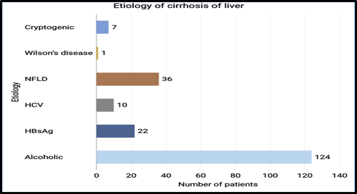
Figure 1: Distribution of Patients According to Etiology of liver Cirrhosis.
Viral causes included hepatitis B surface antigen (HBsAg) positivity in 22 patients (11%) and hepatitis C virus (HCV) infection in 10 patients (5%). Nonalcoholic Fatty Liver Disease (NAFLD) was observed in 36 patients (18%), representing a significant non-alcoholic metabolic cause.Less common etiologies included Wilson’s disease in 1 patient (0.5%), while 7 patients (3.5%) were classified as cryptogenic, where no definite cause could be identified.
Table 3: Grade of Hepatic Encephalopathy (n=88)
|
Grade |
Number of Patients |
% |
|
Grade I |
24 |
27.3 |
|
Grade II |
34 |
38.6 |
|
Grade III |
20 |
22.7 |
|
Grade IV |
10 |
11.4 |
Among patients with hepatic encephalopathy, the distribution of severity grades showed that Grade II was the most common, accounting for 34 patients (38.6%), followed by Grade I in 24 patients (27.3%). More advanced stages were less frequent, with Grade III observed in 20 patients (22.7%) and Grade IV in 10 patients (11.4%).
Table 4. MELD Score Distribution
|
MELD Score |
Number of Patients |
% |
|
<10 |
26 |
13 |
|
10–19 |
72 |
36 |
|
20–29 |
64 |
32 |
|
≥30 |
38 |
19 |
A total of 72 patients (36%) had MELD scores between 10–19, representing the largest subgroup, followed by 64 patients (32%) with scores 20–29, indicating moderately severe hepatic dysfunction. Patients with MELD ≥30, constituted 38 patients (19%). In contrast, only 26 patients (13%) had MELD scores <10.
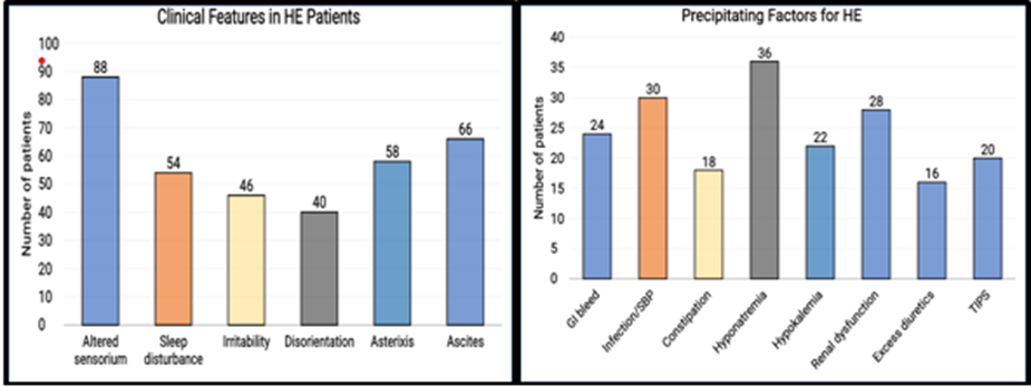
Figure 2,3:. Common Clinical Features and Precipitating Factors for HE (n=88)
Among patients with hepatic encephalopathy, altered sensorium was the most consistent clinical feature, observed in all patients (100%). Other common manifestations included ascites in 66 patients (75.0%) and asterixis in 58 patients (65.9%), indicating significant underlying liver dysfunction.
Sleep disturbance was reported in 54 patients (61.4%), while irritability and disorientation were present in 46 (52.3%) and 40 (45.5%) patients, respectively. Hyponatremia observed in 36 patients (40.9%), followed by infection/spontaneous bacterial peritonitis (SBP) in 30 patients (34.1%) and renal dysfunction in 28 patients (31.8%).
Gastrointestinal bleeding was identified in 24 patients (27.3%), while hypokalemia was present in 22 patients (25.0%). Other contributing factors included constipation in 18 patients (20.5%) and excess diuretic use in 16 patients (18.2%). TIPS was also noted as a risk factor in 20 patients (percentage to be calculated based on total sample).
Table 5. Comparison Between HE and Non-HE Patients
|
Variable |
Number of Patients (%) |
p value |
|
|
HE Present (n=88) |
HE Absent (n=112) |
||
|
MELD ≥20 |
68 (77.3%) |
34 (30.4%) |
<0.001 |
|
GI bleed |
24 (27.3%) |
12 (10.7%) |
0.002 |
|
Infection |
30 (34.1%) |
16 (14.3%) |
0.001 |
|
Hyponatremia |
36 (40.9%) |
18 (16.1%) |
<0.001 |
|
Creatinine >1.5 mg/dL |
28 (31.8%) |
14 (12.5%) |
0.003 |
|
Serum Ammonia >80 µmol/L |
70 (79.5%) |
20 (17.9%) |
<0.001 |
|
TIPS present |
14 (15.9%) |
6 (5.4%) |
0.01 |
A markedly higher proportion of patients with HE had a MELD score ≥20 (77.3% vs 30.4%, p < 0.001) and elevated serum ammonia levels >80 µmol/L (79.5% vs 17.9%, p < 0.001), indicating strong associations with disease severity and metabolic derangement. Hyponatremia (40.9% vs 16.1%, p < 0.001), infection (34.1% vs 14.3%, p = 0.001), and renal dysfunction as indicated by creatinine >1.5 mg/dL (31.8% vs 12.5%, p = 0.003) were also significantly more common in the HE groups. Additionally, gastrointestinal bleeding (27.3% vs 10.7%, p = 0.002) and the presence of TIPS (15.9% vs 5.4%, p = 0.01) were observed more frequently among patients with HE.
Table.6: Number of Patients with Raised Laboratory Parameters Above Cut-off Values
|
Parameter (Cut-off) |
Number of Patients (%) |
p value |
||
|
HE Present (n=88) |
HE Absent (n=112) |
Total (n=200) |
||
|
Total Bilirubin >3 mg/dL |
64 (66.67) |
32 (33.33) |
96 |
<0.001 |
|
Albumin <3 g/dL |
70 (64.81) |
38 (35.19) |
108 |
<0.001 |
|
INR >1.5 |
58 (69.05) |
26 (30.95) |
84 |
<0.001 |
|
Sodium <130 mEq/L |
36 (66.67) |
18 (33.33) |
54 |
<0.001 |
|
Serum Ammonia >80 µmol/L |
70 (92.11) |
6 (7.89) |
76 |
<0.001 |
|
SGOT/AST >80 IU/L |
49 (51.58) |
46 (48.42) |
95 |
>0.05 |
|
SGPT/ALT >60 IU/L |
42 (51.85) |
39 (48.15) |
81 |
>0.05 |
A significantly higher proportion of patients with HE had elevated total bilirubin >3 mg/dL (66.67% vs 33.33%), hypoalbuminemia <3 g/dL (64.81% vs 35.19%), elevated INR >1.5 (69.05% vs 30.95%), hyponatremia <130 mEq/L (66.67% vs 33.33%), and raised serum ammonia >80 µmol/L (92.11% vs 7.89%) compared to those without HE (p < 0.001 for all).In contrast, although a higher proportion of patients with HE had elevated SGOT (AST) >80 IU/L (51.58% vs 48.42%) and SGPT (ALT) >60 IU/L (51.85% vs 48.15%), these differences were not statistically significant (p > 0.05).
Table 8. Multivariate Analysis for Predictors of HE
|
Variable |
Odds Ratio |
95% CI |
p value |
|
MELD ≥20 |
4.2 |
2.4–7.1 |
<0.001 |
|
Hyponatremia |
3.1 |
1.8–5.2 |
<0.001 |
|
Serum Ammonia >80 µmol/L |
3.8 |
2.1–6.4 |
<0.001 |
|
Infection |
2.6 |
1.4–4.6 |
0.002 |
|
GI bleed |
2.2 |
1.2–4.0 |
0.01 |
|
TIPS present |
2.4 |
1.1–4.8 |
0.02 |
MELD score ≥20 was the strongest predictor (OR = 4.2, 95% CI: 2.4–7.1, p < 0.001), followed by serum ammonia >80 µmol/L (OR = 3.8, 95% CI: 2.1–6.4, p < 0.001) and hyponatremia (OR = 3.1, 95% CI: 1.8–5.2, p < 0.001). Infection (OR = 2.6, p = 0.002), TIPS presence (OR = 2.4, p = 0.02), and gastrointestinal bleeding (OR = 2.2, p = 0.01) were also significantly associated with increased odds of hepatic encephalopathy.
DISCUSSION:
The mean age of patients in the present study was 41.01 ± 13.55 years, with the highest proportions in the 28–37 years and 38–47 years age groups (22.5% each).This contrasts with studies from Western and high-income settings, where cirrhotic patients tend to be older. Bohra et al.10 (2020) reported a median age of 57 years (IQR 50–65) in their multicentric HE cohort.
Viral hepatitis remained a significant contributor in the present study, with 16% of patients having either HBV or HCV infection. This is broadly consistent with Pervin et al.11(2024) finding serological evidence of viral hepatitis (B, C, or both) in 63.6% of their patients, reflecting the higher viral hepatitis burden in South Asia.
The largest subgroup comprised patients with MELD scores of 10–19 (36%), followed by 20–29 (32%), ≥30 (19%), and <10 (13%). Bohra et al.410 (2020) reported a median MELD score of 25 (IQR 18–31) in their multicentric HE cohort, indicating a comparably advanced disease burden.
Among the 88 patients with hepatic encephalopathy in the present study, Grade II was the most common severity (38.6%), followed by Grade I (27.3%), Grade III (22.7%), and Grade IV (11.4%). The predominance of Grade II HE indicates that a majority of patients presented with moderate neuropsychiatric dysfunction Bawankule S et al.12 (2019) reported that 45% of patients presented with Grade III HE and 44% with Grade II, suggesting that their cohort had a somewhat higher burden of severe disease.
Altered sensorium was the universal presenting feature, observed in all 88 patients (100%), confirming its status as the defining neuropsychiatric manifestation of overt HE. Ascites was present in 75.0% of HE patients, asterixis in 65.9%, sleep disturbance in 61.4%, irritability in 52.3%, and disorientation in 45.5%. In the present study, hyponatraemia was the most common precipitating factor (40.9%), followed by infection/SBP (34.1%), renal dysfunction (31.8%), gastrointestinal bleeding (27.3%), hypokalaemia (25.0%), constipation (20.5%), excess diuretic use (18.2%), and TIPS-related shunting (15.9% of HE patients).
The markedly elevated serum ammonia in the HE group (96 ± 28 vs 58 ± 18 µmol/L; p < 0.001) in the present study, with 79.5% of HE patients having ammonia >80 µmol/L compared to only 17.9% without HE (p < 0.001), confirms the central role of hyperammonaemia in the pathogenesis of HE. Importantly, elevated ammonia was the second strongest independent predictor of HE on multivariate analysis in the present study (OR = 3.8, 95% CI: 2.1–6.4; p < 0.001), substantiating its pathophysiological centrality. The significantly lower serum sodium in HE patients (128 vs 134 mEq/L; p < 0.001) is consistent with the well-established contribution of hyponatraemia to HE pathogenesis.
In the present study, hyponatraemia (<130 mEq/L) was significantly more prevalent in HE patients (66.67% vs 33.33%; p < 0.001) and was an independent predictor of HE on multivariate analysis (OR = 3.1, 95% CI: 1.8–5.2; p < 0.001). This finding is corroborated by Pervin et al.11 (2024), who identified electrolyte imbalance as the most common precipitating factor (54.4%), and by multiple other studies that have established hyponatraemia as a potent modulator of HE severity and a target for therapeutic intervention.
The non-significant difference in transaminase levels (AST and ALT) between HE and non-HE groups in the present study is an important and clinically relevant finding.
On univariate comparison,The identification of MELD score ≥20 as the strongest independent predictor of HE (OR = 4.2) in the present study is strongly supported by the published literature. Sonia et al.13 (2020) confirmed a significant positive correlation between MELD score and in-hospital mortality in HE patients. The strong association between serum ammonia >80 µmol/L and HE (OR = 3.8) is mechanistically consistent with the neurotoxic role of ammonia in HE pathogenesis, as discussed above. Hyponatraemia as an independent predictor (OR = 3.1) is consistent with multiple published studies. Pervin et al.11 (2024) found electrolyte imbalance to be the most frequent precipitating factor in their cohort.
The independent association of infection with HE (OR = 2.6) underscores the synergistic role of systemic inflammation and bacterial translocation in precipitating neuropsychiatric decompensation in cirrhosis. Bohra et al. (2020) identified infection as the most common trigger for HE in their multicentric study (43%)10.Gastrointestinal bleeding as an independent predictor (OR = 2.2) reflects the classic role of blood in the gut as a nitrogen load that drives ammonia production by colonic bacteria, overwhelming the liver's detoxification capacity in the setting of cirrhosis. GI bleeding was identified as the leading HE precipitant by Mondal et al.14 (2006).
CONCLUSION:
The present study demonstrated that hepatic encephalopathy (HE) is a common and serious complication among patients with liver cirrhosis, affecting 44% of the study population. The majority of affected patients were middle-aged males, with alcohol-related liver disease being the most common underlying etiology. Grade II hepatic encephalopathy was the most frequent clinical presentation, while altered sensorium was observed universally among HE patients, followed by ascites, asterixis, sleep disturbance, irritability, and disorientation.hepatic encephalopathy remains a frequent complication of cirrhosis and is closely linked to disease severity and reversible precipitating factors. Early recognition of high-risk patients, prompt correction of triggering factors, and aggressive management of advanced liver dysfunction may significantly reduce morbidity and improve outcomes in cirrhotic patients presenting with hepatic encephalopathy.
REFERENCES:
1. Liere V, Sandhu G, DeMorrow S. Recent advances in hepatic encephalopathy. F1000Res. 2017;6:1637.
2. Wijdicks EFM. Hepatic encephalopathy. N Engl J Med. 2016;375(17):1660-70.
3. Anderson RN, Smith BL. Deaths: leading causes for 2001. Natl Vital Stat Rep. 2003;52(9):1-85.
4. Saunders JB, Walters JRF, Davies AP, Paton A. A 20-year prospective study of cirrhosis. BMJ. 1981;282(6260):263-6.
5. Neff G, Zachry W 3rd. Systematic review of the economic burden of overt hepatic encephalopathy and pharmacoeconomic impact of rifaximin. Pharmacoeconomics. 2018;36(7):809-22.
6. Weissenborn K. Diagnosis of minimal hepatic encephalopathy. J Clin Exp Hepatol. 2015;5(Suppl 1):S54-9.
7. Agrawal S, Umapathy S, Dhiman RK, et al. Minimal hepatic encephalopathy impairs quality of life. J Clin Exp Hepatol. 2015;5(Suppl 1):S42-8.
8. 8.Bajaj JS. Minimal hepatic encephalopathy matters in daily life. World J Gastroenterol. 2008;14(23):3609-15.
9. Sidhu SS, Sharma BC, Goyal O, et al. L-ornithine L-aspartate in bouts of overt hepatic encephalopathy. Hepatology. 2018;67(2):700-10.
10. Bohra A, Worland T, Hui S, Terbah R, Farrell A, Robertson M. Prognostic significance of hepatic encephalopathy in patients with cirrhosis treated with current standards of care. World J Gastroenterol 2020; 26(18): 2221-2231
11. Pervin MW, Choudhury MRK, Sadi MR, Sarker A. Immediate prognosis of hepatic encephalopathy in a tertiary hospital. Saudi J Med. 2024;9(2):61-67.
12. Bawankule S, Kumar S, Gaidhane A, Quazi M, Singh AP. Clinical profile of patients with hepatic encephalopathy in cirrhosis of liver. J Datta Meghe Inst Med Sci Univ. 2019;14(3):130-136.
13. Sonia, Nagpal S, Mittal A. Spectrum of hepatic encephalopathy admitted in a tertiary care center. Indian J Med Sci. doi:10.25259/IJMS_55_2020.
14. Mondal SK, Chakrabarti S, Bhattacharya R, Bandyopadhyay D, Chakraborty PP, Nath U, Bandyopadhyay R, Mandal L. Observations of hepatic encephalopathy profile in a tertiary care centre. J Indian Med Assoc. 2006 Sep;104(9):516-8, 524. PMID: 17388010.