Decoding Coronary Artery Disease: A Diagnostic Contrast Between Cardiac CT and MRI.
- Sathish Babu , Professor and HOD, Department of Radiodiagnosis, Sree Mookambika Institute of Medical Sciences.
- C. Sylviya , Junior Resident, Department of Radiodiagnosis, Sree Mookambika Institute of Medical Sciences.
- Bharath Chandran , Assistant Professor, Department of Radiodiagnosis, Sree Mookambika Institute of Medical Sciences.
Article Information:
Abstract:
Background: Coronary artery disease (CAD) is a leading cause of morbidity and mortality worldwide. Non-invasive imaging modalities such as Cardiac Computed Tomography (CCT) and Cardiac Magnetic Resonance Imaging (CMR) have gained prominence in evaluating CAD. While CCT provides high-resolution anatomical information on coronary artery stenosis, CMR offers functional and tissue-level insights including myocardial perfusion, ischemia, and viability. Comparative studies evaluating the diagnostic performance of these modalities remain essential to optimize clinical decision-making. Aim: To compare the diagnostic accuracy and clinical utility of Cardiac CT and Cardiac MRI in the evaluation of patients with suspected coronary artery disease. Materials and Methods: This prospective comparative study was conducted over 10 months in the Department of Radiology. A total of 20 patients with suspected CAD were enrolled. All patients underwent both Cardiac CT (128-slice CT scanner, ECG-gated protocol) and Cardiac MRI (1.5T scanner, cine imaging, stress perfusion, and late gadolinium enhancement) within one week. A final composite diagnosis based on clinical, biochemical, and invasive coronary angiography (when available) served as the reference standard. Diagnostic performance of CCT and CMR was compared in terms of sensitivity, specificity, and ability to provide anatomical versus functional assessment. Results: Among 20 patients, significant CAD was detected in 12 (60%). CCT correctly identified 11/12 cases (sensitivity 91.6%, specificity 87.5%), whereas CMR detected 10/12 cases (sensitivity 83.3%, specificity 90%). CCT was superior in detecting coronary artery stenosis, while CMR was superior in characterizing myocardial perfusion and viability. Both modalities demonstrated complementary roles in the comprehensive evaluation of CAD. Conclusion: Cardiac CT is highly effective in detecting coronary artery stenosis, whereas Cardiac MRI adds significant value in functional and tissue characterization. Their complementary use enhances diagnostic accuracy, guiding precise patient management in suspected CAD.
Keywords:
Article :
INTRODUCTION:
Coronary artery disease (CAD) remains the most common cause of mortality globally, accounting for nearly 17.9 million deaths each year, representing 32% of all global deaths[1]. The disease burden is projected to rise, particularly in low- and middle-income countries, owing to urbanization, sedentary lifestyle, and increasing prevalence of risk factors such as diabetes, hypertension, and dyslipidemia[2]. In India, CAD contributes to almost one-quarter of all deaths, with the prevalence steadily increasing among younger populations, thereby posing a significant public health challenge[3].
The diagnosis of CAD traditionally relies on invasive coronary angiography, considered the gold standard for assessing coronary stenosis. However, invasive techniques carry procedural risks and are resource-intensive, highlighting the need for accurate non-invasive modalities[4]. Cardiac Computed Tomography (CCT) has emerged as a reliable imaging tool, providing high-resolution anatomical visualization of coronary arteries and enabling detection of luminal stenosis with excellent negative predictive value[5]. On the other hand, Cardiac Magnetic Resonance Imaging (CMR) offers unique advantages, including assessment of myocardial perfusion, tissue viability, and fibrosis, thereby addressing the functional consequences of ischemia rather than merely anatomical narrowing[6].
Several comparative studies have highlighted the complementary strengths of these two modalities. While CCT excels in anatomical delineation, CMR contributes to understanding myocardial pathophysiology[7]. Recent meta-analyses suggest that the combined use of CCT and CMR may improve diagnostic accuracy and prognostic stratification in CAD, particularly in patients with intermediate pre-test probability[8].
Given the growing burden of CAD in India and worldwide, there is a strong need for locally relevant comparative studies that evaluate both anatomical and functional imaging strategies. Such evidence will help clinicians adopt optimal, patient-tailored diagnostic algorithms that improve outcomes while minimizing unnecessary invasive procedures.
AIM AND OBJECTIVES
Aim
To compare the diagnostic utility of Cardiac Computed Tomography (CCT) and Cardiac Magnetic Resonance Imaging (CMR) in the evaluation of patients with suspected coronary artery disease.
Objectives
1) To assess and compare the sensitivity, specificity, positive predictive value, and negative predictive value of CCT and CMR in detecting significant coronary artery disease using a composite reference standard.
2) To evaluate the relative strengths of CCT (anatomical assessment) and CMR (functional and viability assessment) in comprehensive characterization of coronary artery disease.
MATERIALS AND METHODS:
Study Design and Duration:
This was a prospective, comparative study conducted in the Department of Radiology over a period of 10 months.
Study Population:
A total of 20 patients with clinical suspicion of coronary artery disease (CAD), referred for non-invasive imaging, were included in the study. Patients with contraindications to MRI (e.g., pacemakers, metallic implants), renal dysfunction (eGFR < 30 ml/min/1.73m²), history of allergic reaction to contrast agents, or hemodynamic instability were excluded.
Imaging Protocols:
1. Cardiac CT (CCT):
· Performed using a 128-slice CT scanner with ECG-gated acquisition.
· Intravenous iodinated contrast (70–90 ml) was administered at 5 ml/sec, followed by a saline flush.
· Images were reconstructed with 0.6 mm slice thickness.
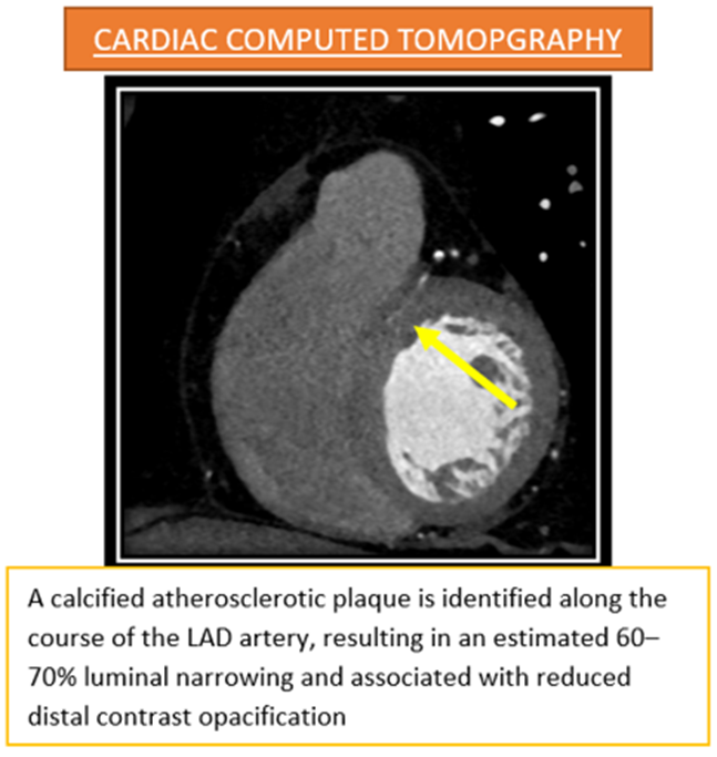
· Coronary artery stenosis was graded, and luminal narrowing ≥50% was considered significant.
2. Cardiac MRI (CMR):
· Conducted using a 1.5 Tesla MRI scanner.
· Standard protocol included:
· Cine sequences for cardiac morphology and function.
· Stress perfusion imaging using adenosine (140 µg/kg/min).
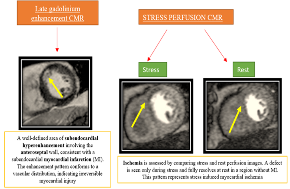
· Late gadolinium enhancement (0.2 mmol/kg) for myocardial viability assessment.
· Significant ischemia was defined as reversible perfusion defect, and infarction was defined by late gadolinium enhancement.
Reference Standard:
A composite diagnosis derived from clinical profile, biochemical markers (including troponins), and invasive coronary angiography (where available) was used as the reference standard for confirmation of CAD.
Data Analysis:
· Sensitivity, specificity, positive predictive value (PPV), and negative predictive value (NPV) of Cardiac CT and Cardiac MRI were calculated against the reference standard.
· Agreement between both modalities was assessed using kappa statistics.
· Data were analyzed using SPSS version 25.0.
Ethical Considerations:
The study protocol was approved by the Institutional Ethics Committee. Informed written consent was obtained from all participants prior to inclusion.
RESULTS:
Table 1: Baseline Characteristics of Study Participants (n = 20)
|
Characteristic |
Value / Frequency |
Percentage (%) |
|
Mean age (years) |
56.8 ± 9.2 |
– |
|
Male |
14 |
70.0 |
|
Female |
6 |
30.0 |
|
Hypertension |
12 |
60.0 |
|
Diabetes mellitus |
10 |
50.0 |
|
Dyslipidemia |
8 |
40.0 |
|
Smoking history |
6 |
30.0 |
|
Family history of CAD |
4 |
20.0 |
Table 2: Distribution of Patients Based on Final Composite Diagnosis
|
Diagnosis |
Frequency (n) |
Percentage (%) |
|
Significant CAD (≥50% stenosis) |
12 |
60.0 |
|
Non-significant CAD (<50%) |
5 |
25.0 |
|
Normal coronary arteries |
3 |
15.0 |
|
Total |
20 |
100.0 |
Table 3: Diagnostic Performance of Cardiac CT and Cardiac MRI
|
Modality |
Sensitivity (%) |
Specificity (%) |
Positive Predictive Value (PPV) (%) |
Negative Predictive Value (NPV) (%) |
Diagnostic Accuracy (%) |
|
Cardiac CT |
91.6 |
87.5 |
91.6 |
87.5 |
90.0 |
|
Cardiac MRI |
83.3 |
90.0 |
90.9 |
81.8 |
85.0 |
Table 4: Comparative Detection of Coronary Artery Disease
|
Condition Detected |
Composite Diagnosis (n) |
Detected by CT (n) |
Detected by MRI (n) |
|
Left Anterior Descending (LAD) stenosis |
7 |
7 |
6 |
|
Right Coronary Artery (RCA) stenosis |
3 |
3 |
2 |
|
Left Circumflex (LCx) stenosis |
2 |
1 |
2 |
|
Multivessel disease |
4 |
4 |
3 |
|
Myocardial ischemia/viability defect |
5 |
3 |
5 |
Table 5: Impact of Imaging Findings on Clinical Decision-Making
|
Clinical Impact |
Frequency (n) |
Percentage (%) |
|
Diagnosis confirmed |
8 |
40.0 |
|
Earlier diagnosis aided |
4 |
20.0 |
|
Management altered (medical/surgical) |
3 |
15.0 |
|
Surgical/intervention planning aided |
3 |
15.0 |
|
No additional impact |
2 |
10.0 |
|
Total |
20 |
100.0 |
DISCUSSION:
In this study, the diagnostic performance of Cardiac CT (CCT) and Cardiac MRI (CMR) in patients with suspected coronary artery disease (CAD) was evaluated. The findings demonstrated that CCT had higher sensitivity for detecting significant coronary stenosis, while CMR showed comparable specificity and additional strength in functional assessment such as perfusion and viability. These results align with the current body of evidence suggesting that both modalities play complementary roles in the evaluation of CAD.
In our study, CCT detected CAD in 91.6% of patients with significant stenosis, consistent with the ACCURACY trial, which reported a sensitivity of 95% and specificity of 83% for 64-slice CCT angiography in patients without prior CAD[5]. Similarly, Meijboom et al. demonstrated that multislice CT achieved a sensitivity of 99% for detecting stenoses compared to invasive angiography, highlighting its role as a reliable non-invasive gatekeeper[9]. With respect to CMR, our study found a sensitivity of 83.3% and specificity of 90% in identifying CAD. These findings are comparable to the MR-IMPACT II trial, which reported sensitivity and specificity values of 75% and 59% respectively, confirming the utility of CMR perfusion imaging in detecting ischemia[10]. Furthermore, Nagel et al. demonstrated the superiority of CMR perfusion over SPECT in terms of diagnostic accuracy, strengthening the case for its routine use in functional assessment[6].
Importantly, our results showed that CCT was superior for anatomical delineation, while CMR contributed uniquely to tissue characterization and viability assessment. This agrees with the CE-MARC study, which concluded that CMR provided superior diagnostic accuracy compared to SPECT and also offered prognostic value[11]. The ability of CMR to detect myocardial fibrosis through late gadolinium enhancement provides an added layer of information not achievable with CCT, making it indispensable for comprehensive CAD evaluation. Meta-analyses also support the complementary nature of both modalities. Danad et al. found that CCT had the highest sensitivity (94%) for anatomical stenosis, while CMR offered better functional assessment when compared to fractional flow reserve, the physiological gold standard[8,12]. This suggests that integrating both techniques in selected patients may optimize diagnostic accuracy and reduce unnecessary invasive angiography.
The strength of our study lies in its prospective comparative design, enabling a direct evaluation of CCT and CMR within the same patient cohort. However, the limitations include a relatively small sample size and single-center setting, which may limit the generalizability of results. Future multicentric studies with larger populations are warranted to establish the most cost-effective diagnostic algorithms in resource-limited settings such as India.
CONCLUSION:
Both Cardiac CT and Cardiac MRI demonstrate high diagnostic value in the evaluation of suspected coronary artery disease, but each modality has distinct strengths. Cardiac CT provides excellent anatomical delineation of coronary artery stenosis with high sensitivity, while Cardiac MRI offers superior insights into myocardial perfusion, ischemia, and tissue characterization. Rather than being competitive, these modalities are complementary, and their combined use may enhance overall diagnostic accuracy and patient management in clinical practice.
REFERENCES:
1. Cardiovascular diseases (CVDs) [Internet]. [cited 2025 Nov 16];Available from: https://www.who.int/news-room/fact-sheets/detail/cardiovascular-diseases-(cvds)
2. Roth GA, Mensah GA, Johnson CO, Addolorato G, Ammirati E, Baddour LM, et al. Global Burden of Cardiovascular Diseases and Risk Factors, 1990–2019. Journal of the American College of Cardiology 2020;76(25):2982–3021.
3. Prabhakaran D, Jeemon P, Sharma M, Roth GA, Johnson C, Harikrishnan S, et al. The changing patterns of cardiovascular diseases and their risk factors in the states of India: the Global Burden of Disease Study 1990–2016. The Lancet Global Health 2018;6(12):e1339–51.
4. Patel MR, Peterson ED, Dai D, Brennan JM, Redberg RF, Anderson HV, et al. Low Diagnostic Yield of Elective Coronary Angiography. N Engl J Med 2010;362(10):886–95.
5. Budoff MJ, Dowe D, Jollis JG, Gitter M, Sutherland J, Halamert E, et al. Diagnostic Performance of 64-Multidetector Row Coronary Computed Tomographic Angiography for Evaluation of Coronary Artery Stenosis in Individuals Without Known Coronary Artery Disease. Journal of the American College of Cardiology 2008;52(21):1724–32.
6. Nagel E, Greenwood JP, McCann GP, Bettencourt N, Shah AM, Hussain ST, et al. Magnetic Resonance Perfusion or Fractional Flow Reserve in Coronary Disease. N Engl J Med 2019;380(25):2418–28.
7. Knuuti J, Wijns W, Saraste A, Capodanno D, Barbato E, Funck-Brentano C, et al. 2019 ESC Guidelines for the diagnosis and management of chronic coronary syndromes. European Heart Journal 2020;41(3):407–77.
8. Danad I, Szymonifka J, Twisk JWR, Norgaard BL, Zarins CK, Knaapen P, et al. Diagnostic performance of cardiac imaging methods to diagnose ischaemia-causing coronary artery disease when directly compared with fractional flow reserve as a reference standard: a meta-analysis. Eur Heart J 2016;ehw095.
9. Meijboom WB, Meijs MFL, Schuijf JD, Cramer MJ, Mollet NR, Van Mieghem CAG, et al. Diagnostic Accuracy of 64-Slice Computed Tomography Coronary Angiography. Journal of the American College of Cardiology 2008;52(25):2135–44.
10. Schwitter J, Wacker CM, Wilke N, Al-Saadi N, Sauer E, Huettle K, et al. MR-IMPACT II: Magnetic Resonance Imaging for Myocardial Perfusion Assessment in Coronary artery disease Trial: perfusion-cardiac magnetic resonance vs. single-photon emission computed tomography for the detection of coronary artery disease: a comparative multicentre, multivendor trial. European Heart Journal 2013;34(10):775–81.
11. Greenwood JP, Maredia N, Younger JF, Brown JM, Nixon J, Everett CC, et al. Cardiovascular magnetic resonance and single-photon emission computed tomography for diagnosis of coronary heart disease (CE-MARC): a prospective trial. The Lancet 2012;379(9814):453–60.
Takx RAP, Blomberg BA, Aidi HE, Habets J, De Jong PA, Nagel E, et al. Diagnostic Accuracy of Stress Myocardial Perfusion Imaging Compared to Invasive Coronary Angiography With Fractional Flow Reserve Meta-Analysis. Circ: Cardiovascular Imaging 2015;8(1):e002666.