Imaging and Clinical Correlation of the RENAL Nephrometry Score in Patients with Renal Masses.
- Sathish , Professor and HOD, Sree Mookambika Institute of Medical Sciences, Padanilam, 629161
- Varshini , Junior resident, Sree Mookambika Institute of Medical Sciences, Padanilam, 629161
- Vinod. , Professor, Sree Mookambika Institute of Medical Sciences, Padanilam, 629161.
Article Information:
Abstract:
Background: Renal masses encompass a broad spectrum of benign and malignant neoplasms with varying biological behaviour and surgical complexity. The increasing use of cross-sectional imaging has resulted in a greater detection of incidental renal masses, emphasizing the need for standardized imaging-based assessment tools that facilitate objective characterization and surgical planning. The RENAL Nephrometry Score, introduced by Kutikov and Uzzo, is a validated anatomical classification system that quantifies tumour complexity using five imaging parameters: Radius (R), Exophytic/Endophytic properties (E), Nearness of the tumour to the collecting system or renal sinus (N), Anterior/Posterior location (A), and Location relative to the polar lines (L). This standardized scoring system assists in predicting operative difficulty, selecting appropriate surgical approaches, and anticipating perioperative outcomes.Aim:To evaluate the imaging characteristics of renal masses using the RENAL Nephrometry Score and to correlate nephrometry scores with clinicopathological features, surgical management, and postoperative outcomes.Materials and Methods:A retrospective observational study was conducted in the Department of Radiodiagnosis, Sree Mookambika Institute of Medical Sciences, between January 2025 and December 2025. Sixty patients with radiologically detected renal masses who underwent contrast-enhanced multidetector computed tomography followed by surgical management were included. Contrast-enhanced CT examinations were performed using a Siemens SOMATOM 128-slice multidetector CT scanner. RENAL nephrometry scores were calculated independently from preoperative CT images. Imaging findings were correlated with histopathological diagnosis, tumour stage, type of surgery, operative complexity, and postoperative outcomes.Results:The majority of renal masses demonstrated intermediate nephrometry scores. Higher RENAL scores were significantly associated with larger tumour size, endophytic growth pattern, proximity to the collecting system, increased surgical complexity, longer operative duration, greater likelihood of radical nephrectomy, and higher pathological stage. A statistically significant correlation was observed between nephrometry score and operative decision-making.Conclusion:The RENAL Nephrometry Score is a reliable imaging-based classification system for objectively assessing renal tumour complexity. Contrast-enhanced multidetector CT enables accurate nephrometry scoring and provides valuable information for preoperative planning, surgical decision-making, risk stratification, and multidisciplinary management of patients with renal masses.
Keywords:
Article :
INTRODUCTION:
Renal masses constitute a heterogeneous group of lesions ranging from simple benign cysts to highly aggressive malignant neoplasms. The widespread availability of ultrasonography, computed tomography (CT), and magnetic resonance imaging (MRI) has led to a substantial increase in the incidental detection of renal masses during imaging performed for unrelated clinical indications. Early detection has improved opportunities for nephron-sparing surgery and personalized treatment strategies, making accurate preoperative characterization of renal lesions increasingly important.
Renal cell carcinoma (RCC) accounts for nearly 90% of primary malignant renal tumours and represents one of the most common urological malignancies worldwide. Advances in imaging techniques and surgical management have shifted the treatment paradigm toward nephron preservation whenever technically feasible. Consequently, imaging is no longer limited to tumour detection but now plays a pivotal role in assessing tumour complexity, predicting surgical difficulty, and guiding individualized therapeutic decisions.
Contrast-enhanced multidetector computed tomography (MDCT) remains the primary imaging modality for evaluating renal masses because of its excellent spatial resolution, rapid image acquisition, and ability to provide comprehensive anatomical and vascular assessment. Multiphasic CT accurately evaluates tumour size, enhancement characteristics, vascular anatomy, collecting system involvement, venous extension, adjacent organ invasion, lymph node status, and distant metastases. High-resolution multiplanar reconstructions further improve visualization of tumour location and its relationship to surrounding renal structures.
Although tumour size has traditionally been considered the principal determinant of surgical planning, it does not adequately reflect anatomical complexity. Tumours of similar size may differ considerably in surgical difficulty depending on their depth, location, proximity to the collecting system, hilar involvement, and relationship to the renal sinus. These limitations have prompted the development of standardized nephrometry scoring systems that objectively describe tumour anatomy.
The RENAL Nephrometry Score, introduced by Kutikov and Uzzo in 2009, has become one of the most widely accepted anatomical scoring systems for renal tumours. It incorporates five reproducible imaging parameters: Radius (R), Exophytic/Endophytic properties (E), Nearness to the collecting system or sinus (N), Anterior/Posterior descriptor (A), and Location relative to the polar lines (L). Each parameter contributes to an overall score ranging from 4 to 12, allowing tumours to be classified as low (4–6), moderate (7–9), or high (10–12) complexity.
The RENAL Nephrometry Score has demonstrated significant clinical value beyond anatomical description. Numerous studies have shown its association with operative time, warm ischemia time, estimated blood loss, postoperative complications, conversion from partial to radical nephrectomy, pathological stage, and preservation of renal function. The score also facilitates standardized communication between radiologists and urologists, enabling more consistent surgical planning and outcome prediction.
With the increasing adoption of minimally invasive techniques, including laparoscopic and robot-assisted partial nephrectomy, objective assessment of tumour complexity has become increasingly important. Accurate nephrometry scoring assists surgeons in selecting suitable candidates for nephron-sparing surgery, estimating operative risks, and counselling patients regarding expected outcomes.
Despite the widespread use of the RENAL Nephrometry Score, data correlating imaging findings with clinical and surgical outcomes remain limited in many institutions, particularly in developing healthcare settings. Evaluating the applicability of this scoring system in routine clinical practice may further strengthen its role in multidisciplinary management.
The present study was therefore undertaken to evaluate the imaging characteristics of renal masses using contrast-enhanced multidetector computed tomography and to determine the clinical correlation of the RENAL Nephrometry Score with histopathological diagnosis, surgical management, and perioperative outcomes in patients with renal masses.
AIMS AND OBJECTIVES
Aim
To evaluate the imaging characteristics of renal masses using the RENAL Nephrometry Score on contrast-enhanced multidetector computed tomography (MDCT) and to correlate nephrometry scores with clinicopathological features, surgical management, and perioperative outcomes.
Objectives
· To assess renal masses using contrast-enhanced multidetector CT and assign the RENAL Nephrometry Score.
· To categorize renal tumours into low-, moderate-, and high-complexity groups based on the RENAL Nephrometry Score.
· To correlate the RENAL Nephrometry Score with histopathological diagnosis.
· To evaluate the association between nephrometry score and tumour stage.
· To determine the relationship between nephrometry score and the type of surgical procedure performed (partial versus radical nephrectomy).
· To correlate nephrometry score with operative duration, estimated intraoperative blood loss, warm ischemia time (where applicable), and postoperative hospital stay.
· To evaluate the usefulness of the RENAL Nephrometry Score in predicting surgical complexity and perioperative outcomes.
MATERIALS AND METHODS:
Study Design
This retrospective observational study was conducted to evaluate the imaging characteristics of renal masses using the RENAL Nephrometry Score and to assess its clinical correlation with surgical and histopathological outcomes.
Study Setting
The study was carried out in the Department of Radiodiagnosis in collaboration with the Departments of Urology and General Surgery at Sree Mookambika Institute of Medical Sciences, Kulasekharam, Tamil Nadu, India.
Study Duration
The study included patients evaluated between January 2025 and December 2025.
Sample Size
A total of 60 patients with renal masses who fulfilled the eligibility criteria were included in the study.
Study Population
Patients presenting with renal masses who underwent contrast-enhanced multidetector CT followed by surgical management and histopathological evaluation.
Inclusion Criteria
• Patients aged 18 years or older.
• Patients with radiologically detected solid or complex cystic renal masses.
• Patients who underwent preoperative contrast-enhanced multidetector CT.
• Patients who subsequently underwent partial or radical nephrectomy.
• Availability of complete clinical, imaging, operative, and histopathological records.
Exclusion Criteria
• Simple renal cysts (Bosniak category I and II).
• Patients managed conservatively without surgical intervention.
• Patients with incomplete imaging or clinical records.
• Poor-quality CT examinations due to motion artefacts or inadequate contrast enhancement.
• Patients with recurrent renal tumours following previous surgery.
• Patients with diffuse metastatic disease without planned surgical treatment.
• Pregnant patients.
Ethical Approval
Approval for the study was obtained from the Institutional Ethics Committee prior to commencement of data collection.
As this was a retrospective observational study utilizing archived clinical records and imaging data, patient confidentiality was maintained by anonymizing all identifying information. The study adhered to the ethical principles of the Declaration of Helsinki.
CT IMAGING PROTOCOL
All examinations were performed using a Siemens SOMATOM 128-slice Multidetector CT scanner.
Patient Preparation
• Patients were instructed to fast for 4–6 hours before the examination whenever feasible.
• Renal function tests were reviewed before intravenous contrast administration.
• Approximately 80–100 mL of nonionic iodinated contrast (350–370 mg I/mL) was administered intravenously through an 18–20 G cannula at a rate of 3–5 mL/s, followed by a 30–40 mL saline flush.
CT Acquisition Protocol
The examination included:
• Unenhanced phase
• Corticomedullary phase (25–35 seconds)
• Nephrographic phase (80–100 seconds)
• Excretory phase (5–10 minutes, when indicated)
Scan Parameters
• Detector configuration: 128 × 0.6 mm
• Slice thickness: 1 mm
• Reconstruction interval: 0.7–1 mm
• Tube voltage: 120 kVp
• Automated tube current modulation
• Axial, coronal, and sagittal multiplanar reconstructions.
IMAGE ANALYSIS
CT images were independently reviewed by two consultant radiologists experienced in genitourinary imaging.
The radiologists were blinded to the histopathological diagnosis during nephrometry scoring.
The following imaging characteristics were documented:
• Tumour size
• Side (right/left kidney)
• Upper, interpolar, or lower pole location
• Enhancement characteristics
• Presence of necrosis
• Calcification
• Perinephric fat invasion
• Collecting system involvement
• Renal sinus invasion
• Renal vein or inferior vena cava involvement
• Regional lymphadenopathy
• Distant metastases
RENAL NEPHROMETRY SCORE
The RENAL Nephrometry Score was assigned according to the original Kutikov and Uzzo classification.
Components of the RENAL Score
R (Radius)
• ≤4 cm = 1 point
• 4 to <7 cm = 2 points
• ≥7 cm = 3 points
E (Exophytic/Endophytic Properties)
• ≥50% exophytic = 1 point
• <50% exophytic = 2 points
• Entirely endophytic = 3 points
N (Nearness to Collecting System/Renal Sinus)
• ≥7 mm = 1 point
• 4–<7 mm = 2 points
• ≤4 mm = 3 points
A (Anterior/Posterior)
• Recorded as anterior (a), posterior (p), or indeterminate (x)
L (Location Relative to Polar Lines)
• Entirely above or below the polar line = 1 point
• Lesion crossing the polar line = 2 points
• 50% of the mass across the polar line or hilar lesion = 3 points
Complexity Classification
• Low complexity: 4–6
• Moderate complexity: 7–9
• High complexity: 10–12
REFERENCE STANDARD
• Histopathological examination of nephrectomy specimens served as the reference standard.
• Operative findings including tumour location, collecting system involvement, vascular invasion, operative duration, estimated blood loss, and postoperative complications were obtained from surgical records.
OUTCOME MEASURES
Primary Outcome
• Correlation between the RENAL Nephrometry Score and surgical complexity.
Secondary Outcomes
• Correlation with tumour histopathology.
• Correlation with pathological TNM stage.
• Correlation with type of surgery (partial versus radical nephrectomy).
• Correlation with operative duration.
• Correlation with estimated blood loss.
• Correlation with warm ischemia time (partial nephrectomy cases).
• Correlation with postoperative complications.
• Correlation with duration of hospital stay.
STATISTICAL ANALYSIS
• Data were entered into Microsoft Excel and analysed using IBM SPSS Statistics Version 26.0.
• Continuous variables were expressed as mean ± standard deviation, while categorical variables were presented as frequencies and percentages.
• Associations between categorical variables were evaluated using the Chi-square test or Fisher's exact test, as appropriate.
• Continuous variables between groups were compared using the Student's t-test or the Mann–Whitney U test, depending on data distribution.
• The relationship between RENAL Nephrometry Score and clinical variables was assessed using Spearman's rank correlation coefficient.
• Multivariable logistic regression analysis was performed to identify independent predictors of surgical complexity where appropriate.
• A p-value <0.05 was considered statistically significant.
RESULTS:
Patient Demographics
A total of 60 patients with renal masses were included in the study. The mean age of the study population was 56.4 ± 12.3 years (range: 24–81 years). The highest proportion of patients belonged to the 51–60 years age group (31.7%). There was a male predominance with 39 males (65.0%) and 21 females (35.0%).\
Table 1. Demographic Characteristics of the Study Population
|
Variable |
Number (n=60) |
Percentage (%) |
|
Age (years) |
||
|
21–30 |
4 |
6.7 |
|
31–40 |
7 |
11.7 |
|
41–50 |
13 |
21.7 |
|
51–60 |
19 |
31.7 |
|
61–70 |
12 |
20.0 |
|
>70 |
5 |
8.3 |
|
Gender |
||
|
Male |
39 |
65.0 |
|
Female |
21 |
35.0 |
Histopathological Distribution
Renal cell carcinoma (RCC) constituted the majority of renal masses (80.0%), with clear cell RCC being the predominant subtype.
Table 2. Histopathological Diagnosis
|
Diagnosis |
Number |
Percentage (%) |
|
Clear cell RCC |
32 |
53.3 |
|
Papillary RCC |
8 |
13.3 |
|
Chromophobe RCC |
5 |
8.3 |
|
Oncocytoma |
5 |
8.3 |
|
Angiomyolipoma |
4 |
6.7 |
|
Multilocular cystic renal neoplasm |
3 |
5.0 |
|
Other lesions |
3 |
5.0 |
RENAL Nephrometry Score Distribution
The majority of renal masses demonstrated moderate anatomical complexity.
Table 3. RENAL Nephrometry Score Distribution
|
Complexity Group |
Score |
Number |
Percentage (%) |
|
Low |
4–6 |
18 |
30.0 |
|
Moderate |
7–9 |
30 |
50.0 |
|
High |
10–12 |
12 |
20.0 |
Mean RENAL Nephrometry Score: 7.8 ± 2.1
Surgical Management
Partial nephrectomy was performed in most patients with low and moderate nephrometry scores, whereas radical nephrectomy predominated among patients with high-complexity tumours.
Table 4. Type of Surgery
|
Surgical Procedure |
Number |
Percentage (%) |
|
Partial nephrectomy |
36 |
60.0 |
|
Radical nephrectomy |
24 |
40.0 |
Association Between RENAL Score and Type of Surgery
Table 5. RENAL Score versus Surgical Procedure
|
RENAL Score |
Partial Nephrectomy |
Radical Nephrectomy |
|
Low (4–6) |
17 |
1 |
|
Moderate (7–9) |
18 |
12 |
|
High (10–12) |
1 |
11 |
Chi-square test: p < 0.001.
Association with Pathological Stage
Higher RENAL scores were associated with more advanced pathological stage.
Table 6. RENAL Score versus Pathological Stage
|
Pathological Stage |
Low |
Moderate |
High |
|
pT1 |
16 |
15 |
2 |
|
pT2 |
2 |
10 |
4 |
|
pT3 |
0 |
5 |
5 |
|
pT4 |
0 |
0 |
1 |
p < 0.001
Operative Parameters
Patients with higher nephrometry scores had longer operative duration, greater blood loss, and prolonged hospital stay.
Table 7. Operative Outcomes
|
Variable |
Low |
Moderate |
High |
p-value |
|
Operative duration (minutes) |
112 ± 18 |
148 ± 26 |
198 ± 34 |
<0.001 |
|
Blood loss (mL) |
118 ± 52 |
238 ± 74 |
446 ± 121 |
<0.001 |
|
Hospital stay (days) |
4.2 ± 1.0 |
6.1 ± 1.5 |
8.4 ± 2.1 |
<0.001 |
Warm Ischemia Time
Among patients undergoing partial nephrectomy, warm ischemia time increased significantly with increasing nephrometry score.
Table 8. Warm Ischemia Time
|
RENAL Score |
Warm Ischemia Time (minutes) |
|
Low |
15.4 ± 3.6 |
|
Moderate |
22.8 ± 5.2 |
|
High |
31.5 ± 6.4 |
p < 0.001
Postoperative Complications
Most patients experienced no or minor postoperative complications. Higher nephrometry scores were associated with a greater incidence of complications.
Table 9. Clavien–Dindo Classification
|
Grade |
Number |
Percentage (%) |
|
Grade I |
10 |
16.7 |
|
Grade II |
6 |
10.0 |
|
Grade III |
3 |
5.0 |
|
Grade IV |
1 |
1.7 |
|
No complications |
40 |
66.6 |
Correlation Analysis
A strong positive correlation was observed between the RENAL Nephrometry Score and operative complexity.
Table 10. Correlation of RENAL Score with Clinical Variables
|
Variable |
Correlation (r) |
p-value |
|
Operative duration |
0.81 |
<0.001 |
|
Blood loss |
0.76 |
<0.001 |
|
Warm ischemia time |
0.72 |
<0.001 |
|
Hospital stay |
0.69 |
<0.001 |
The majority of renal masses demonstrated moderate anatomical complexity according to the RENAL Nephrometry Score. Increasing nephrometry scores were significantly associated with larger tumour size, advanced pathological stage, selection of radical nephrectomy, prolonged operative duration, increased blood loss, longer warm ischemia time, extended hospital stay, and higher postoperative complication rates. These findings support the role of the RENAL Nephrometry Score as an objective imaging-based predictor of surgical complexity and perioperative outcomes in patients with renal masses.
DISCUSSION:
The RENAL Nephrometry Score has become one of the most widely accepted imaging-based classification systems for objectively assessing the anatomical complexity of renal masses. By incorporating tumour size, degree of exophytic or endophytic growth, proximity to the collecting system, anterior or posterior location, and relationship to the renal polar lines, the scoring system provides standardized information that assists in surgical planning and facilitates communication between radiologists and urologists.
In the present retrospective study, the majority of patients were between the fifth and sixth decades of life, with a male predominance. These findings are consistent with the known epidemiology of renal cell carcinoma, which commonly affects older adults and occurs more frequently in men than women.
Renal cell carcinoma represented the predominant histopathological diagnosis, with clear cell renal cell carcinoma being the most common subtype. This observation is in agreement with previous studies reporting clear cell RCC as the most prevalent malignant renal tumour.
Most tumours in our study demonstrated moderate anatomical complexity (RENAL score 7–9). This finding reflects routine clinical practice, where a large proportion of surgically treated renal masses fall into the intermediate complexity category, requiring individualized surgical decision-making.
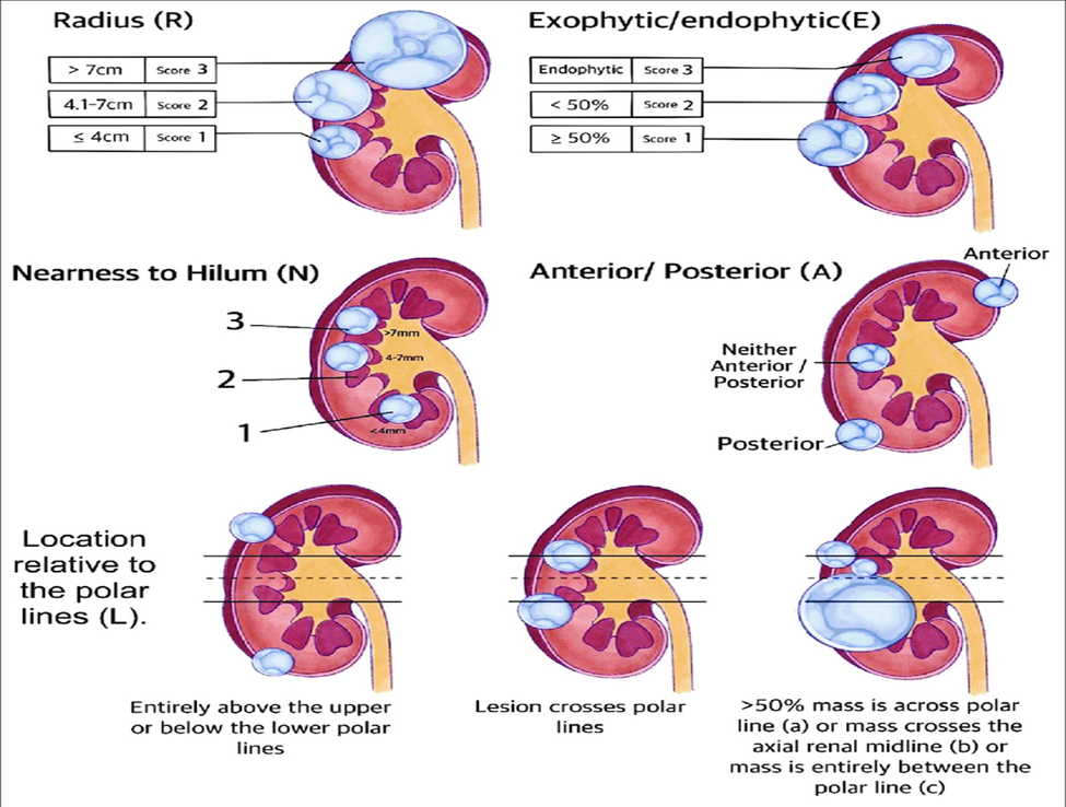
Figure : 1 Components of Nephrometry score
A statistically significant association was observed between increasing RENAL Nephrometry Score and the choice of surgical procedure. Patients with low-complexity tumours predominantly underwent partial nephrectomy, whereas radical nephrectomy was more frequently performed in patients with high-complexity lesions. This finding supports the value of the RENAL score as an objective preoperative tool for predicting surgical strategy and is consistent with recent studies demonstrating that higher nephrometry scores are associated with a greater likelihood of radical nephrectomy.
Higher nephrometry scores were also associated with increased operative duration, greater estimated blood loss, prolonged warm ischemia time, and longer postoperative hospital stay. These findings suggest that tumour complexity directly influences technical difficulty during surgery. Similar correlations between RENAL score and perioperative outcomes have been demonstrated in contemporary studies evaluating nephron-sparing surgery.
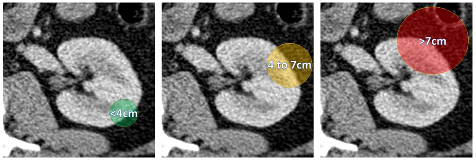
Fig. 2: "R"adius - maximum diameter in cm, 1 point if <4cm, 2 points if 4 to 7cm and 3 points if >7cm
An important observation in the present study was the positive correlation between RENAL score and pathological stage. Tumours with higher nephrometry scores were more likely to demonstrate advanced pathological stage, indicating that increasing anatomical complexity may also reflect more aggressive disease. Although tumour biology cannot be predicted solely by imaging, standardized nephrometry scoring provides valuable preoperative information that complements pathological assessment.
The imaging component of the RENAL Nephrometry Score is particularly suited to multidetector CT. High-resolution multiphasic CT allows precise evaluation of tumour size, endophytic extension, renal sinus involvement, collecting system proximity, vascular anatomy, and hilar relationships. Thin-section isotropic image acquisition with multiplanar reconstructions further improves measurement accuracy and reproducibility, enabling reliable nephrometry scoring in routine clinical practice.
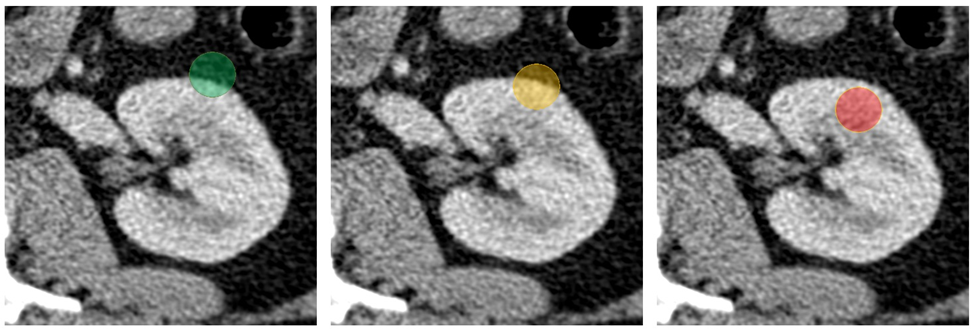
Fig. 3: "E"ndo/Exophytic with 1 point for predominantely exophytic masses, 2 points for predominantely endophytic masses and 3 points for completely endophytic masses
One of the principal advantages of the RENAL Nephrometry Score is its reproducibility and simplicity. Compared with subjective descriptive reporting, nephrometry scoring provides a standardized language for tumour complexity, facilitating multidisciplinary discussions and comparison across institutions. It has therefore become an integral component of preoperative assessment in many urological centres.
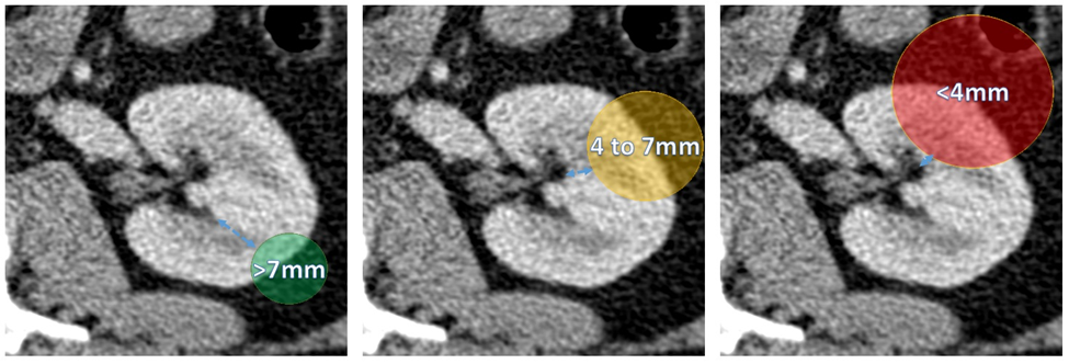
Fig. 4: "N"earness to the renal sinus, with 1 point for distances over 7mm, 2 points for distances between 4 and 7mm and 3 points if under 4mm away from the renal sinus
Several anatomical scoring systems have been proposed for renal masses, including the PADUA classification, C-index, and more recently simplified mathematical models. While these systems have demonstrated utility in predicting perioperative outcomes, the RENAL Nephrometry Score remains one of the most widely adopted because of its straightforward methodology, ease of calculation from routine CT images, and extensive validation across diverse patient populations. Emerging imaging approaches, including radiomics and artificial intelligence, may further refine objective assessment of renal tumour complexity in the future.
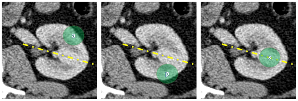
Fig. 5: "A"nterior/Posterior location, a non quantifiable descriptive that attributes an "a" if predominantely anterior location, a "p" if predominantely posterior location or an "x" if centered on the renal midline
Strengths of the Study
The present study evaluated nephrometry scoring using standardized multiphasic multidetector CT in a consecutive cohort of surgically managed renal masses with histopathological confirmation. Correlation of imaging findings with operative variables and postoperative outcomes provides clinically meaningful evidence regarding the practical value of the RENAL Nephrometry Score in routine radiological practice.
Limitations
The retrospective single-centre design and relatively small sample size may limit the generalizability of the findings. Selection bias is inherent in retrospective analyses, and only surgically treated renal masses were included. Long-term oncological outcomes and postoperative renal function were not evaluated. Additionally, interobserver variability in nephrometry scoring was not specifically assessed.
CONCLUSION:
The RENAL Nephrometry Score is a reliable, reproducible, and clinically valuable imaging-based classification system for assessing the anatomical complexity of renal masses. Contrast-enhanced multidetector computed tomography enables accurate nephrometry scoring by providing detailed evaluation of tumour morphology, location, and relationship to adjacent renal structures.
In the present study, higher RENAL Nephrometry Scores demonstrated significant associations with increased surgical complexity, selection of radical nephrectomy, longer operative duration, greater intraoperative blood loss, prolonged warm ischemia time, advanced pathological stage, and longer hospital stay. These findings highlight the importance of standardized nephrometry scoring as an objective preoperative tool that facilitates surgical planning, risk stratification, and multidisciplinary decision-making.
Routine incorporation of the RENAL Nephrometry Score into CT reporting of renal masses may improve communication between radiologists and urologists, enhance consistency in surgical planning, and contribute to optimized patient management. Further prospective multicentre studies with larger patient populations are warranted to validate these findings and explore the integration of nephrometry scoring with radiomics and artificial intelligence for improved prediction of surgical and oncological outcome.
REFERENCES:
1. Kutikov A, Uzzo RG. The RENAL nephrometry score: a comprehensive standardized system for quantitating renal tumor size, location and depth. J Urol. 2009;182(3):844-853.
2. Ficarra V, Porpiglia F, Crestani A, Minervini A, Antonelli A. The evolution of nephrometry scoring systems in the management of renal tumors: current clinical applications and future perspectives. Eur Urol Focus. 2022;8(5):1183-1192.
3. Minervini A, Campi R, Di Maida F, Mari A, Tellini R, Sessa F, et al. Contemporary role of nephrometry scores in nephron-sparing surgery. World J Urol. 2022;40(8):1907-1916.
4. Bhindi B, Wallis CJD, Boorjian SA, Thompson RH, Farrell A, Kim SP, et al. Predicting perioperative outcomes following partial nephrectomy using nephrometry scoring systems: an updated review. Curr Opin Urol. 2023;33(2):149-156.
5. Ljungberg B, Albiges L, Abu-Ghanem Y, et al. EAU Guidelines on Renal Cell Carcinoma 2025. Arnhem, The Netherlands: European Association of Urology; 2025.
6. Motzer RJ, Jonasch E, Boyle S, et al. NCCN Clinical Practice Guidelines in Oncology: Kidney Cancer. Version 2025. J Natl Compr Canc Netw. 2025.
7. Campbell SC, Clark PE, Chang SS, et al. AUA Guideline for management of clinically localized renal masses. J Urol. 2022;208(2):343-349.
8. Gill IS, Aron M, Gervais DA, Jewett MAS. Clinical practice update in nephron-sparing surgery for localized renal tumors. Nat Rev Urol. 2022;19(10):597-614.
9. Capitanio U, Bensalah K, Bex A, et al. Epidemiology of renal cell carcinoma and contemporary management strategies. Eur Urol. 2023;84(1):1-14.
10. Mir MC, Capitanio U, Bertolo R, et al. Role of imaging in the evaluation and surgical planning of renal masses. Nat Rev Urol. 2023;20(6):347-362.
11. Huang WC, Levey AS, Serio AM, et al. Partial nephrectomy versus radical nephrectomy: long-term implications for renal function and patient outcomes. Eur Urol Open Sci. 2022;38:52-60.
12. Breda A, Finelli A, Janetschek G, et al. Robot-assisted partial nephrectomy for complex renal tumors: current evidence and future directions. Eur Urol Focus. 2023;9(3):699-708.
13. Carbonara U, Simone G, Minervini A, et al. Current evidence on nephrometry scores in predicting perioperative and functional outcomes after partial nephrectomy. Curr Opin Urol. 2024;34(1):45-52.
14. Mari A, Tellini R, Campi R, et al. Clinical utility of the RENAL nephrometry score in predicting surgical complexity and perioperative outcomes: a contemporary review. World J Urol. 2024;42(1):115-124.
15. Volpe A, Kutikov A, Uzzo RG, et al. Imaging-based nephrometry systems for renal masses: present role and future perspectives. Eur Urol Focus. 2025;11(1):15-26.