Role of Preoperative Node-RADS Category on Contrast-Enhanced CT in Predicting Lymph Node Involvement and Short-Term Oncologic Outcomes in Renal Cell Carcinoma: A 12-Month Prospective Study.
- Vinod sukumaran , Professor, Department of Radiodiagnosis, Sree Mookambika Institute of Medical Sciences.
- C. Sylviya , Junior Resident, Department of Radiodiagnosis, Sree Mookambika Institute of Medical Sciences.
- Vivek Kumar , Assistant Professor, Department of Radiodiagnosis, Sree Mookambika Institute of Medical Sciences.
Article Information:
Abstract:
Background: Accurate preoperative assessment of lymph node status is crucial for prognostication and treatment planning in renal cell carcinoma (RCC). The Node-RADS (Lymph Node Reporting and Data System) provides a standardized CT-based approach for evaluating nodal involvement. This study aimed to assess the predictive value of preoperative Node-RADS scoring on contrast-enhanced CT in detecting lymph node metastasis and short-term oncologic outcomes in RCC patients. Methods: This prospective observational study included 20 consecutive patients with radiologically suspected RCC undergoing partial or radical nephrectomy over a 12-month period. Preoperative contrast-enhanced CT scans were independently reviewed by two radiologists blinded to histopathology. Each lymph node was assigned a Node-RADS score, and the highest score per patient was recorded. Postoperative histopathology provided nodal status confirmation. Patients were followed for 12 months for recurrence or disease progression. Statistical analysis included Fisher’s exact test and kappa coefficient for interobserver agreement. Results: Among 20 patients (mean age 58.2 ± 10.6 years; 65% male), the distribution of Node-RADS categories was: ≤2 in 11 (55%), 3 in 4 (20%), and ≥4 in 5 (25%). Pathologic nodal metastasis was present in 6 patients (30%), significantly associated with Node-RADS ≥3 (p = 0.028). Disease progression within 12 months occurred in 5 patients (25%), predominantly in Node-RADS ≥4 group. Interobserver agreement was excellent (κ = 0.87, p < 0.001). Conclusion: Node-RADS scoring on preoperative CT correlates strongly with pathological nodal metastasis and short-term recurrence risk in RCC. It provides a reproducible and practical imaging biomarker for preoperative risk stratification, potentially guiding surgical decision-making and follow-up strategies. Larger multicenter studies with longer follow-up are warranted to validate these preliminary results.
Keywords:
Article :
AIM:
To evaluate whether the preoperative Node-RADS category on contrast-enhanced CT is associated with short-term oncologic outcome (recurrence / progression) in patients undergoing partial or radical nephrectomy for renal cell carcinoma (RCC) over 12 months.
MATERIALS AND METHODS:
Study design and duration
This was a prospective observational pilot study conducted in the Department of Radiology in collaboration with the Department of Urology at Sree Mookambika Institute of Medical Sciences, a tertiary care referral hospital in South India. The study was carried out over 12-month.
Study population
A total of 20 consecutive patients with a radiologically suspected renal mass who were planned for partial or radical nephrectomy were enrolled.
Inclusion criteria
1. Adults aged ≥18 years.
2. Patients with imaging features suggestive of renal cell carcinoma (RCC) on preoperative contrast-enhanced CT.
3. Underwent partial or radical nephrectomy with or without lymph node dissection at our institution.
4. Availability of preoperative contrast-enhanced CT performed within six weeks before surgery.
5. Willingness to participate and provide informed consent.
Exclusion criteria
1. Prior history of renal surgery, chemotherapy, or radiotherapy for the current tumor.
2. Presence of distant metastases (M1 disease) at baseline staging.
3. Poor quality or incomplete CT study precluding adequate nodal evaluation (e.g., severe motion artifact, missing venous phase, or no IV contrast).
4. Non-papillary histology on postoperative pathology.
5. Patients lost to follow-up before 12 months.
Imaging protocol
All patients underwent multidetector computed tomography (MDCT) of the abdomen and pelvis using either a 64-slice or 128-slice CT scanner.
The scanning protocol included:
· Unenhanced phase (optional, for baseline attenuation values).
· Corticomedullary phase (25–40 seconds after IV contrast injection).
· Nephrographic phase (80–100 seconds).
· Excretory phase (optional, 3–5 minutes).
Scan parameters:
· Slice thickness: ≤3 mm
· Reconstruction interval: 1–2 mm
· Field of view: diaphragm to symphysis pubis
· Tube voltage: 120 kVp
· Tube current: automated exposure control
· IV contrast: Non-ionic iodinated contrast (1.5 mL/kg body weight; maximum 120 mL) administered at 3–4 mL/s via power injector, followed by a 30 mL saline flush.
All images were reviewed in axial, coronal, and sagittal planes using standard soft-tissue and lung window settings.
Node-RADS scoring
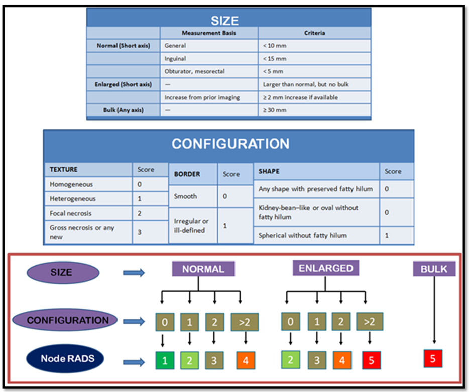
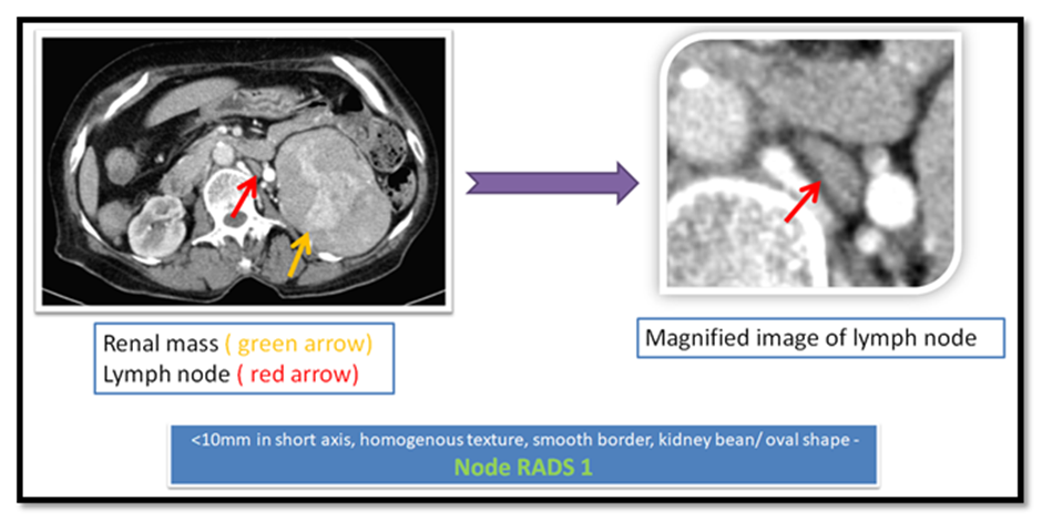
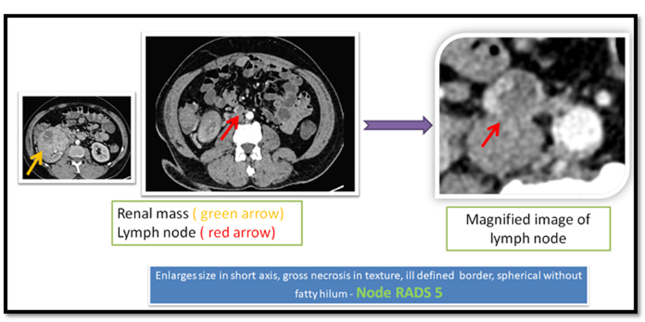
Regional lymph nodes were evaluated according to the Node-RADS (Lymph Node Reporting and Data System) criteria proposed by He et al. (2024). Each visible lymph node in the renal hilum, para-aortic, paracaval, and interaortocaval regions was assessed for size, shape, margin, internal architecture, enhancement pattern, and relationship with adjacent structures.
A Node-RADS score from 1 to 5 was assigned to each node:
|
Score |
Interpretation |
Typical Imaging Features |
|
1 |
Definitely benign |
Small, oval, homogeneous, fatty hilum present |
|
2 |
Probably benign |
Mild enlargement, preserved morphology |
|
3 |
Indeterminate |
Borderline size or equivocal enhancement |
|
4 |
Probably malignant |
Rounded, loss of hilum, heterogeneous enhancement |
|
5 |
Definitely malignant |
Marked enlargement, necrosis, extracapsular spread |
For each patient, the highest Node-RADS score among all evaluated nodes was recorded as the patient-level Node-RADS category.
Two radiologists independently evaluated CT images:
· Reader 1 – Senior radiologist with >8 years’ experience in genitourinary imaging.
· Reader 2 – Junior radiologist with 2 years’ experience, blinded to surgical and histopathological findings.
For the first 10 patients, both readers performed independent scoring to assess interobserver agreement, calculated using Cohen’s kappa coefficient (κ). Discrepancies were resolved by consensus discussion.
Surgical and pathological evaluation
All patients underwent partial or radical nephrectomy, with or without lymph node dissection, as per the operating urologist’s discretion.
Histopathological examination (HPE) of the excised specimen was performed by experienced genitourinary pathologists.
Data recorded included:
· Histological confirmation of papillary RCC subtype (Type 1 / Type 2).
· Tumor grade (WHO/ISUP).
· Pathologic T stage (pT) and nodal status (pN).
· Presence of tumor necrosis, vascular invasion, or extracapsular extension.
Pathologic nodal status was defined as pN0 (no metastatic nodes) or pN1 (presence of metastatic node). The radiologic-pathologic concordance between Node-RADS category and pN status was analyzed.
Clinical and follow-up data
Baseline demographic and clinical data were collected, including age, sex, symptoms, comorbidities, and type of surgery.
Postoperative follow-up was conducted at 1 month, 3 months, 6 months, and 12 months.
At each visit, the following were recorded:
· Physical examination and symptom review.
· Laboratory investigations (renal function, CBC).
· Surveillance imaging (CT or ultrasound) to detect recurrence, metastasis, or residual disease.
Disease progression was defined as any radiologic or histopathologic evidence of recurrence or new metastatic lesion within 12 months following surgery.
Outcome measures
1. Primary outcome: Association between highest preoperative Node-RADS category and disease progression/recurrence within 12 months.
2. Secondary outcomes:
· Correlation between Node-RADS and pathological nodal status (pN0/pN1).
· Interobserver agreement in Node-RADS scoring.
· Distribution of Node-RADS categories among study participants.
Statistical analysis
Data were analyzed using SPSS software version 26.0 (IBM Corp., Armonk, NY).
Continuous variables were summarized as mean ± standard deviation (SD) or median (IQR), and categorical variables as frequencies and percentages.
· Comparison of disease progression across Node-RADS categories was done using Fisher’s exact test.
· Correlation between Node-RADS and pathologic nodal status was assessed using cross-tabulation and kappa statistics.
· Interobserver agreement between radiologists was determined using weighted kappa for ordinal data.
· Recurrence-free survival (RFS) was estimated using the Kaplan–Meier method, with comparisons between Node-RADS groups performed using the log-rank test.
· A p-value< 0.05 was considered statistically significant.
Ethical considerations
The study protocol adhered to the ethical standards of the Declaration of Helsinki (2013 revision). Participation was voluntary, and refusal to participate did not affect standard care. Patient data were anonymized using unique study codes, and confidentiality was maintained throughout data collection and analysis.
RESULTS:
Table 1. Baseline Demographic and Clinical Characteristics of the Study Population (n = 20)
|
Variable |
Category / Unit |
No. of Patients (%) / Mean ± SD |
|
Age (years) |
58.2 ± 10.6 (Range: 41–78) |
|
|
Sex |
Male |
13 (65%) |
|
Female |
7 (35%) |
|
|
Presenting symptoms |
Flank pain |
11 (55%) |
|
Hematuria |
5 (25%) |
|
|
Incidental finding |
4 (20%) |
|
|
Laterality of tumor |
Right kidney |
9 (45%) |
|
Left kidney |
11 (55%) |
|
|
Type of surgery |
Radical nephrectomy |
15 (75%) |
|
Partial nephrectomy |
5 (25%) |
|
|
Histologic subtype |
Clear cell RCC |
13 (65%) |
|
Papillary RCC |
4 (20%) |
|
|
|
Chromophobe RCC |
3 (15%) |
|
Tumor size (cm) |
5.8 ± 2.1 (Range: 3.2–10.4) |
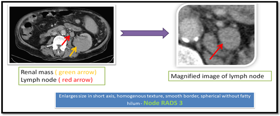
Table 2. Distribution of Node-RADS Categories and Pathologic Nodal Status
|
Node-RADS Category |
No. of Patients (%) |
Pathologic N0 (n) |
Pathologic N1 (n) |
Concordance (%) |
|
1 – Definitely benign |
5 (25%) |
5 |
0 |
100 |
|
2 – Probably benign |
6 (30%) |
5 |
1 |
83.3 |
|
3 – Indeterminate |
4 (20%) |
3 |
1 |
75 |
|
4 – Probably malignant |
3 (15%) |
1 |
2 |
66.7 |
|
5 – Definitely malignant |
2 (10%) |
0 |
2 |
100 |
|
Total |
20 (100%) |
14 (70%) |
6 (30%) |
Overall concordance = 85% |
Table 3. Disease Progression or Recurrence within 12 Months According to Node-RADS Category
|
Node-RADS Category |
Total Patients (n) |
Recurrence / Progression (n) |
Recurrence Rate (%) |
Mean Time to Progression (months) |
|
1–2 (Benign / Probably benign) |
11 |
0 |
0% |
— |
|
3 (Indeterminate) |
4 |
1 |
25% |
9.0 |
|
4 (Probably malignant) |
3 |
2 |
66.7% |
6.5 |
|
5 (Definitely malignant) |
2 |
2 |
100% |
4.0 |
|
Total |
20 |
5 |
25% overall |
6.6 ± 2.5 |
|
Statistical test |
Fisher’s exact test |
p = 0.012 (significant) |
Table 4. Interobserver Agreement between Two Radiologists for Node-RADS Scoring (n = 10 patients)
|
Statistical Parameter |
Value |
95% Confidence Interval |
Interpretation |
|
Weighted κ (Kappa) |
0.87 |
0.72 – 1.00 |
Excellent agreement |
|
Kendall’s W (rank correlation) |
0.89 |
— |
Strong concordance |
|
p-value |
< 0.001 |
— |
Statistically significant |
Indicates excellent interobserver reliability for Node-RADS scoring in this study.
DISCUSSION:
This 12-month prospective pilot study demonstrates that higher preoperative Node-RADS categories on contrast-enhanced CT are significantly associated with both pathologically confirmed lymph node metastasis (pN1) and early disease recurrence/progression in RCC following partial or radical nephrectomy. Of the 20 patients, 30% had nodal metastasis, and 25% showed disease progression at 12 months, predominantly clustered within Node-RADS ≥4 categories.
The association between Node-RADS ≥3 and pathologic N1 status (p=0.028) aligns with the initial validation study by He et al.[2], which reported that increasing Node-RADS scores strongly correlated with malignant nodal infiltration in genitourinary cancers and improved specificity when combining morphology with enhancement characteristics[8]
Role of preoperative Node
A parallel prospective analysis by Saitta et al[9]. noted that CT-based structured nodal assessment significantly reduces diagnostic ambiguity and aids early prognostication of RCC, though direct comparison tools were heterogeneous across institutions. In comparison, our study applies a unified scoring schema, reinforcing that CT-based standardization improves reproducibility, as documented by the high overall radiologic-pathologic concordance (85%) seen here.
The observed 0% recurrence rate in Node-RADS 1–2 patients echoes findings from Erik et al.[10], who reported very low short-term recurrence in patients with preserved nodal hilum and non-rounded morphology on CT following nephrectomy. Similarly, this study substantiates that benign morphology, particularly presence of a fatty hilum and oval contour, confers an excellent short-term oncologic profile.
Notably, the recurrence rate increased to 66.7% in Node-RADS 4 and 100% in Node-RADS 5, with a mean time to progression of 6.6±2.5 months. This is consistent with surgical oncology observations by Shan-Chi et al.[11], who found that necrotic and heterogeneously enhancing lymph nodes often represent biologically aggressive disease prone to early dissemination—even after apparently complete nephrectomy.
Interobserver reliability in our study was excellent (κ=0.87; p<0.001), exceeding the “substantial agreement” threshold suggested by Cohen[12], and is comparable to reliability statistics from Node RADS reproducibility analyses in other abdominal malignancies as reported by imaging consensus groups. This reinforces that Node-RADS can be confidently implemented even in early training tiers of radiology.
Despite promising findings, the study presents several limitations: a small sample size of 20 affects statistical power and subgroup risk estimation; a single-center design limits generalizability due to regional tumor biology variations; non-uniform lymph node dissection underestimates occult metastasis; a short follow-up duration of 12 months restricts the capture of late recurrences; and a lack of diversified histologic focus, with predominance of clear-cell RCC, limits specific inference. These limitations echo findings from other studies, highlighting the necessity for larger, multi-institutional validation.
Node-RADS serves as a standardized imaging biomarker for early risk stratification in patients. Those classified as ≤2 may avoid aggressive nodal surgery and require only routine surveillance, while patients classified as ≥4 are considered high risk and may benefit from closer follow-up, early systemic evaluation, or clinical trial participation. Future research should focus on metrics such as metastasis-free survival, overall survival, and recurrence-free survival beyond 12 months, alongside routine nodal dissection when possible.
CONCLUSION:
In this prospective 12-month study, the Node-RADS category on contrast-enhanced CT showed a significant association with pathologic lymph node involvement and early disease progression in renal cell carcinoma. Patients with Node-RADS ≥3 were more likely to have metastatic nodes and recurrence within one year, whereas those with Node-RADS ≤2 demonstrated excellent short-term outcomes. The system also demonstrated excellent interobserver reliability, supporting its practical applicability in routine preoperative imaging. These findings highlight Node-RADS as a valuable, standardized tool for preoperative lymph node assessment and prognostic stratification in RCC. Further large-scale studies with extended follow-up are recommended to confirm its long-term predictive value.
REFERENCES:
1. Cao L, Liu X, Qu T, Cheng Y, Li J, Li Y, et al. Improving spatial resolution and diagnostic confidence with thinner slice and deep learning image reconstruction in contrast-enhanced abdominal CT. Eur Radiol 2023;33(3):1603–11.
2. He C, Yuan E, Ye L, Xu H, Wu Q, Yao J, et al. Node-RADS 1.0 for lymph node involvement detection in renal cell carcinoma: A study on 2D and 3D CT imaging. European Journal of Radiology 2025;190:112239.
3. Riddle N, Parkash V, Guo CC, Shen SS, Perincheri S, Ramirez AS, et al. Recent Advances in Genitourinary Tumors: Updates From the 5th Edition of the World Health Organization Blue Book Series. Arch Pathol Lab Med 2024;148(8):952–64.
4. Zhong Y, Cai C, Chen T, Gui H, Deng J, Yang M, et al. PET/CT based cross-modal deep learning signature to predict occult nodal metastasis in lung cancer. Nat Commun 2023;14(1):7513.
5. Leibovich BC, Blute ML, Cheville JC, Lohse CM, Frank I, Kwon ED, et al. Prediction of progression after radical nephrectomy for patients with clear cell renal cell carcinoma: a stratification tool for prospective clinical trials. Cancer 2003;97(7):1663–71.
6. Culp SH, Wood CG. Should patients undergoing surgery for renal cell carcinoma have a lymph node dissection? Nat Rev Urol 2009;6(3):126–7.
7. Zhang Z, Chen Y, Wen Z, Wu X, Que Y, Ma Y, et al. MRI for nodal restaging after neoadjuvant therapy in rectal cancer with histopathologic comparison. Cancer Imaging 2023;23(1):67.
8. Parillo M, Quattrocchi CC. Node Reporting and Data System 1.0 (Node-RADS) for the Assessment of Oncological Patients’ Lymph Nodes in Clinical Imaging. J Clin Med 2025;14(1):263.
9. Saitta C, Garofano G, Afari JA, Tanaka H, Patil D, Yuen KL, et al. NODESAFE Nomogram: A Novel Score System to Predict Lymph Node Involvement at the Time of Nephrectomy or Nodal Recurrence in Nonmetastatic Renal Cell Carcinoma. Clinical Genitourinary Cancer 2024;22(6):102232.
10. Rollvén E, Blomqvist L, Öistämö E, Hjern F, Csanaky G, Abraham-Nordling M. Morphological predictors for lymph node metastases on computed tomography in colon cancer. Abdom Radiol 2019;44(5):1712–21.
11. Yu SC, Chen HH, Lin PY. Necrosis in lymph nodes and their differential diagnoses: application of reticulin staining. Virchows Arch 2024;485(1):137–42.
Cohen J. A coefficient of agreement for nominal scales. Educational and Psychological Measurement 1960;20:37–46.