HYSTEROSCOPIC EVALUATION IN PATIENTS WITH ABNORMAL UTERINE BLEEDING.
- Sneha Mainath , Junior resident, Department of Obstetrics and Gynecology, Government Medical college , Vizianagaram, Andhra Pradesh, India.
- N. Manasa , Assistant professor, Department of Obstetrics and Gynecology, Government Medical college, Vizianagaram, Andhra Pradesh, India.
- B.Rajani Kumari , Associate professor, Department of Obstetrics and Gynecology, Government Medical college, Vizianagaram, Andhra Pradesh, India.
- Aruna Subha Sree Rao , Professor and HOD , Department of Obstetrics and Gynecology, Government Medical college, Vizianagaram, Andhra Pradesh, India..
Article Information:
Abstract:
Background: Abnormal uterine bleeding (AUB) is one of the most common gynecological complaints affecting women of reproductive and perimenopausal age and significantly impacts their quality of life. The FIGO PALM–COEIN classification provides a standardized approach to identifying the structural and non-structural causes of AUB. Accurate diagnosis requires evaluation of the endometrial cavity, with hysteroscopy-guided endometrial biopsy offering superior diagnostic accuracy through direct visualization and targeted tissue sampling, while histopathological examination remains the gold standard for definitive diagnosis. Methods: This hospital-based prospective observational study was conducted over 18 months at a tertiary care center(GGH VZM) and included 60 women with abnormal uterine bleeding (AUB) or postmenopausal bleeding who met the inclusion criteria. All participants underwent hysteroscopic evaluation followed by directed endometrial biopsy, and the hysteroscopic findings were correlated with histopathological examination to determine the underlying etiology of AUB. Results: A total of 60 women with abnormal uterine bleeding (AUB) who fulfilled the inclusion criteria were included in the present study. The mean age of the participants was 42.75 ± 8.61 years, with the majority belonging to the 31–40 years (46.7%) age group. Heavy menstrual bleeding was the most common presenting complaint (75%), and a significant correlation was observed between hysteroscopic and histopathological findings (χ² = 60.0, p < 0.001). Conclusion: Hysteroscopy and histopathological examination are complementary diagnostic modalities in the evaluation of abnormal uterine bleeding. Their combined use provides accurate diagnosis and facilitates appropriate management by correlating direct visualization with definitive tissue diagnosis.
Keywords:
Article :
INTRODUCTION:
Abnormal uterine bleeding (AUB) is a common gynecological disorder characterized by abnormalities in the frequency, regularity, duration, or volume of menstrual bleeding unrelated to pregnancy and is a major cause of morbidity among women of reproductive and perimenopausal age. It significantly impairs quality of life, contributes to iron deficiency anemia, and imposes a substantial socioeconomic and healthcare burden. The prevalence of AUB is estimated to be 11–13% among women of reproductive age, increasing to 25–30% during the late reproductive and perimenopausal years.
According to the 2021 update by the International Federation of Gynecology and Obstetrics (FIGO), nearly 20% of women experience AUB during their reproductive lifespan, although prevalence varies with demographic characteristics and diagnostic criteria. In India, AUB accounts for approximately 17–30% of gynecological consultations, constituting nearly one-third of outpatient gynecology visits and more than 70% of consultations in the reproductive, perimenopausal, and postmenopausal age groups. Clinically, AUB is classified as acute, requiring immediate intervention to prevent excessive blood loss and hemodynamic instability, or chronic, characterized by abnormal bleeding patterns persisting for most of the preceding six months.
To facilitate standardized diagnosis and management, FIGO introduced the PALM–COEIN classification system, which categorizes the etiologies of AUB into structural causes—Polyp, Adenomyosis, Leiomyoma, and Malignancy/Hyperplasia—and non-structural causes including Coagulopathy, Ovulatory dysfunction, Endometrial disorders, Iatrogenic causes, and Not otherwise classified. Accurate identification of the underlying pathology is essential for appropriate management and is achieved through a combination of clinical evaluation, transvaginal ultrasonography, hysteroscopy, and endometrial sampling. While transvaginal ultrasonography serves as the initial imaging modality, hysteroscopy permits direct visualization of the uterine cavity and facilitates targeted biopsy of suspicious lesions, thereby offering superior diagnostic accuracy over blind dilatation and curettage for detecting focal intrauterine abnormalities.
Histopathological examination of endometrial tissue remains the gold standard for the diagnosis of endometrial hyperplasia and malignancy. Consequently, hysteroscopy-guided endometrial biopsy has emerged as an indispensable diagnostic tool, enabling precise evaluation of intrauterine pathology and guiding evidence-based management of women presenting with abnormal uterine bleeding.
MATERIALS AND METHODS:
A hospital-based prospective observational study was conducted over a period of 18 months from January 2023 to June 2024 in the Department of Obstetrics and Gynaecology at Government District General Hospital, Vizianagaram, Andhra Pradesh, a tertiary care referral center. The study included women presenting to the outpatient and inpatient services with abnormal uterine bleeding (AUB) or postmenopausal bleeding who fulfilled the predefined eligibility criteria and provided written informed consent. The primary objective was to evaluate the causes of AUB, identify endometrial lesions such as polyps and fibroids, and assess the endometrial surface for the presence of hyperplasia through hysteroscopic evaluation and histopathological correlation.
Inclusion criteria:
1) Women in the reproductive age group (18-40 yrs).
2) Women in peri menopausal age (>40 yrs).
3) Post menopausal bleeding.
4) Patients willing to given consent to participate in the study.
Exclusion criteria:
1) Pregnant women, abortions, pelvic infections, cervical cancer.
2) Surgical and medical conditions like uncontrolled diabetes. severe hypertension, Hypothyroidism, Hyperthyroidism, liver/ renal diseases.
A total of 60 patients were enrolled. The sample size was calculated using the formula n = [Z²p(1−p)]/d², where Z = 0.95, power (p) = 80%, and precision (d) = 5.5%, yielding a minimum required sample size of 57.7; therefore, 60 patients were considered adequate for the study. Pregnant women, patients with abortion, pelvic infections, cervical carcinoma, and those with significant medical or surgical comorbidities such as uncontrolled diabetes mellitus, severe hypertension, thyroid disorders, or hepatic and renal diseases were excluded. All enrolled patients underwent detailed clinical evaluation, hysteroscopic examination, and directed endometrial sampling, and the hysteroscopic findings were subsequently correlated with histopathological examination to establish the final diagnosis.
RESULTS:
A total of 60 women who fulfilled the study inclusion criteria were enrolled in the study.
Age distribution
TABLE 1: Distribution according to Age Group
|
S.No |
Age Group(Years) |
Number |
Percent |
|
01 |
21 to 30 |
01 |
01.7% |
|
02 |
31 to 40 |
28 |
46.7% |
|
03 |
41 to 50 |
23 |
38.4% |
|
04 |
51 to 60 |
04 |
06.6% |
|
05 |
>60 |
04 |
06.6% |
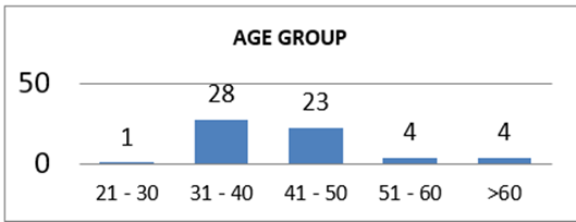
CHART I: Distribution according to age group
The majority of women in the study belonged to the 31–40 years age group, comprising 28 (46.7%) participants, followed by the 41–50 years age group with 23 (38.4%) participants. Women aged 51–60 years and those above 60 years each constituted 4 (6.6%) participants. Only 1 (1.7%) participant belonged to the 21–30 years age group. Overall, 85.1% of the study population was between 31 and 50 years of age, indicating that abnormal uterine bleeding was predominantly observed among women in the late reproductive and perimenopausal age groups
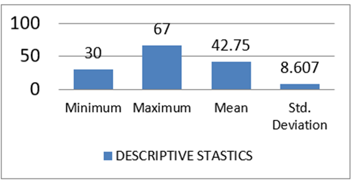
CHART II: Descriptive statistics (mean age)
TABLE II: Descriptive statistics (mean age)
|
S.No |
Minimum |
Maximum |
Mean |
Std.Deviation |
|
Age |
30 |
67 |
42.75 |
8.607 |
The age of the study participants ranged from 30 to 67 years, with a mean age of 42.75 ± 8.61 years. This indicates that the majority of women included in the study were in the late reproductive and perimenopausal age group, which is the period during which abnormal uterine bleeding is most commonly encountered. The standard deviation of 8.61 years reflects a moderate variation in the ages of the study population.
Table III - Distribution according to chief complaints
|
S.No |
Chief Complaint-1 |
Number |
Percent |
|
01 |
HMB |
45 |
75.00% |
|
02 |
Inter menstrual bleeding |
01 |
01.70% |
|
03 |
Inter menstrual spotting |
01 |
01.70% |
|
04 |
Polymenorrhoea |
08 |
13.30% |
|
05 |
Post menopausal bleeding |
05 |
08.30% |
|
S.No |
Chief Complaint-2 |
Number |
Percent |
|
01 |
Dysmenorrhea |
06 |
10.00% |
|
02 |
Dyspareunia |
04 |
06.70% |
|
03 |
HMB |
01 |
01.70% |
|
04 |
Passing Blood Clot |
11 |
18.30% |
|
05 |
Polymenorrhea |
01 |
01.70% |
|
06 |
No |
37 |
61.60% |
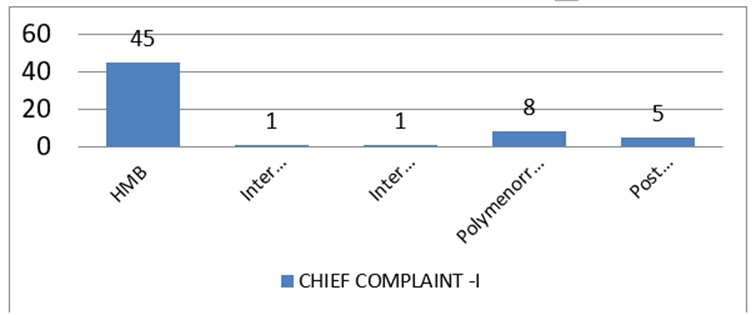
CHART IIA
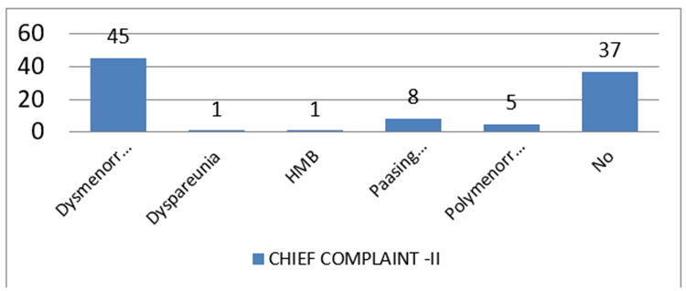
CHART IIIB:
The most common primary chief complaint (Chief Complaint-1) was heavy menstrual bleeding (HMB), reported by 45 (75.0%) women. This was followed by polymenorrhoea in 8 (13.3%) patients and postmenopausal bleeding in 5 (8.3%) patients. Intermenstrual bleeding and intermenstrual spotting were each observed in 1 (1.7%) patient.
Regarding the secondary chief complaint (Chief Complaint-2), 37 (61.6%) women did not report any additional complaint. Among those with associated symptoms, passing blood clots was the most frequent, occurring in 11 (18.3%) patients, followed by dysmenorrhea in 6 (10.0%) patients and dyspareunia in 4 (6.7%) patients. Heavy menstrual bleeding and polymenorrhoea were each reported as secondary complaints by 1 (1.7%) patient.
TABLE IV: Distribution according to P/A findings, L/E of EG, P/V findings,P/V (weeks)
|
S.No |
Findings |
Number |
Percent |
|
01 |
P/A NAD |
60 |
100.00% |
|
02 |
L/E of EG NAD |
60 |
100.00% |
|
03 |
P/V Anteverted |
50 |
83.30% |
|
04 |
P/V Retroverted |
10 |
16.70% |
|
P/V (weeks) |
|||
|
S.No |
P/V (Weeks) |
Number |
Percent |
|
01 |
06 |
03 |
05.00% |
|
02 |
08 |
12 |
20.00% |
|
03 |
10 |
06 |
10.00% |
|
04 |
12 |
01 |
01.70% |
|
05 |
Normal |
33 |
55.00% |
|
06 |
Small |
05 |
08.30% |
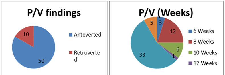
CHART IV
On general physical examination, all 60 (100%) women had no abnormality detected (NAD) on per abdominal (P/A) examination as well as on local examination of the external genitalia (L/E of EG).
On per vaginal (P/V) examination, the uterus was found to be anteverted in 50 (83.3%) women, while 10 (16.7%) women had a retroverted uterus.
With respect to uterine size on per vaginal examination, a normal-sized uterus was observed in 33 (55.0%) women, making it the most common finding. An 8-week-sized uterus was noted in 12 (20.0%) women, followed by a 10-week-sized uterus in 6 (10.0%) women. A small uterus was found in 5 (8.3%) women, while 3 (5.0%) and 1 (1.7%) women had uterine sizes corresponding to 6 weeks and 12 weeks gestation, respectively.
TABLE V: Distribution according to USG, hysteroscopy
|
S.No |
USG Findings |
Number |
Percent |
|
01 |
Normal |
26 |
43.30% |
|
02 |
Adenomyosis |
02 |
03.30% |
|
03 |
Bulky |
10 |
16.70% |
|
04 |
Intramural fibroid |
05 |
08.30% |
|
05 |
Polyp |
02 |
03.30% |
|
06 |
Submucous fibroid |
05 |
08.30% |
|
07 |
Thickened Endometrium |
10 |
16.70% |
|
Hysterscopy |
|||
|
S.No |
Hysterscopy finding |
Number |
Percent |
|
01 |
Endocervical canal - Normal |
60 |
100.00% |
|
02 |
Internal OS - Normal |
60 |
100.00% |
|
03 |
Tubal ostia - Patent |
60 |
100.00% |
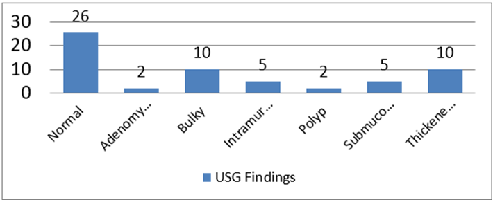
CHART V
On ultrasonographic (USG) evaluation, 26 (43.3%) women had normal findings, making it the most common observation. Among the abnormal findings, bulky uterus and thickened endometrium were each observed in 10 (16.7%) women. Intramural fibroids and submucous fibroids were each identified in 5 (8.3%) patients, while adenomyosis and endometrial polyps were each detected in 2 (3.3%) patients.
On diagnostic hysteroscopy, the endocervical canal was normal in all 60 (100%) women. Similarly, the internal os was normal in 100% of the study population, and bilateral tubal ostia were patent in all 60 (100%) patients.
TBALE VI: Distribution according to endometrial cavity
|
S.No |
Endometrial cavity |
Number |
Percent |
|
01 |
Atrophic endometrium |
04 |
06.70% |
|
02 |
Endocervical polyp |
01 |
01.70% |
|
03 |
Endometrial polyp |
01 |
01.70% |
|
04 |
Hyperplastic endometrium |
07 |
11.70% |
|
05 |
Intramural fibroid |
02 |
03.30% |
|
06 |
Proliferative endometrium |
23 |
38.30% |
|
07 |
Secretory Endometrium |
12 |
20.00% |
|
08 |
Submucous fibroid |
10 |
16.70% |
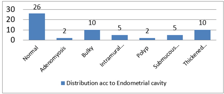
CHART VI
Hysteroscopic evaluation of the endometrial cavity revealed that proliferative endometrium was the most common finding, observed in 23 (38.3%) women. This was followed by secretory endometrium in 12 (20.0%) patients and submucous fibroids in 10 (16.7%) patients. Hyperplastic endometrium was identified in 7 (11.7%) women, while atrophic endometrium was observed in 4 (6.7%) patients. Intramural fibroids were detected in 2 (3.3%) women, whereas endometrial polyps and endocervical polyps were each identified in 1 (1.7%) patient.
TBALE VII: Distribution according to HPE
|
S.No |
HPE |
Endometrial cavity |
Number |
Percent |
|
01
|
Atrophic endometrium |
Yes |
04 |
06.70% |
|
No |
56 |
93.30% |
||
|
02 |
Leiomyoma |
Yes |
14 |
23.30% |
|
No |
46 |
76.70% |
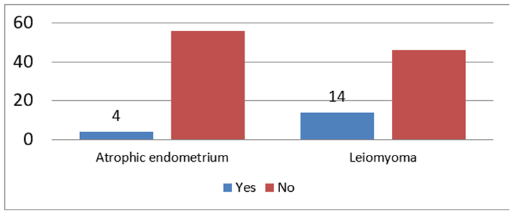
CHART VII
Histopathological examination (HPE) revealed atrophic endometrium in 4 (6.7%) women, while the remaining 56 (93.3%) women did not demonstrate atrophic endometrial changes.
With respect to leiomyoma, histopathology confirmed its presence in 14 (23.3%) women, whereas 46 (76.7%) women showed no evidence of leiomyoma.
TABLE VIII: Distribution according to HPE
|
S.No |
HPE |
Number |
Percent |
|
01 |
Atrophic endometrium |
01 |
01.70% |
|
02 |
Endocervical polyp |
03 |
05.00% |
|
03 |
Endometrial polyp |
01 |
01.70% |
|
04 |
Hyperplastic endometrium |
07 |
11.70% |
|
05 |
Intramural fibroid |
21 |
35.00% |
|
06 |
Proliferative endometrium |
23 |
38.30% |
|
07 |
Secretory Endometrium |
02 |
03.30% |
|
08 |
Submucous fibroid |
02 |
03.30% |
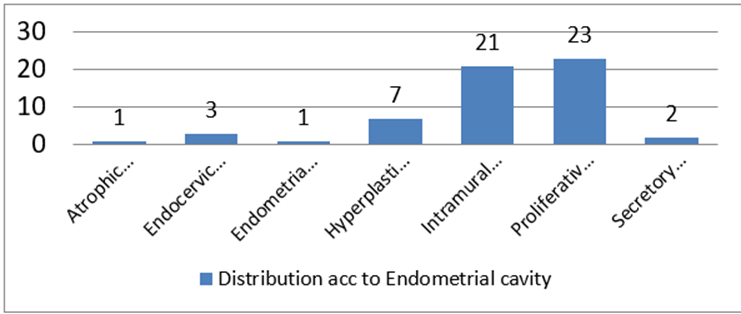
CHART VIII
Histopathological examination revealed that proliferative endometrium was the most common finding, observed in 23 (38.3%) women. This was followed by intramural fibroid (leiomyoma) in 21 (35.0%) patients. Hyperplastic endometrium was identified in 7 (11.7%) women, while endocervical polyps were present in 3 (5.0%) patients. Secretory endometrium and submucous fibroids were each observed in 2 (3.3%) women. Atrophic endometrium and endometrial polyps were the least common findings, each detected in 1 (1.7%) patient.
TABLE IX: Association of endometrial cavity with leiomyoma (HPE)
|
S.No |
Endometrial cavity |
Leiomyoma |
|
|
|
Yes |
No |
|
|
01 |
Atrophic endometrium |
0 |
4 |
|
02 |
Endocervical polyp |
1 |
0 |
|
03 |
Endometrial polyp |
1 |
0 |
|
04 |
Hyperplastic endometrium |
0 |
7 |
|
05 |
Intramural fibroid |
2 |
0 |
|
06 |
Proliferative endometrium |
0 |
23 |
|
07 |
Secretory Endometrium |
0 |
12 |
|
08 |
Submucous fibroid |
10 |
0 |
Chisquare value -60.00
P value - <0.0001
The association between hysteroscopic endometrial cavity findings and histopathological diagnosis of leiomyoma demonstrated that all women with submucous fibroids (10 cases) and intramural fibroids (2 cases) on hysteroscopy were confirmed to have leiomyoma on histopathological examination. In addition, one case each of endocervical polyp and endometrial polyp was also found to have leiomyoma on histopathology. In contrast, none of the women with atrophic endometrium (4 cases), hyperplastic endometrium (7 cases), proliferative endometrium (23 cases), or secretory endometrium (12 cases) showed histopathological evidence of leiomyoma.
The association between hysteroscopic findings and histopathological diagnosis was found to be highly statistically significant (Chi-square = 60.00, p < 0.001), indicating a strong relationship between hysteroscopic detection of fibroid lesions and histopathological confirmation of leiomyoma.
DISCUSSION:
In the present study, the correlation between hysteroscopic endometrial cavity findings and histopathological diagnosis of leiomyoma was evaluated to determine the diagnostic accuracy of hysteroscopy in identifying structural uterine lesions responsible for abnormal uterine bleeding (AUB). A highly statistically significant association was observed between the two modalities (χ² = 60.0, p < 0.001), indicating excellent agreement between hysteroscopic findings and histopathological examination.
Among the hysteroscopic findings, all 10 patients diagnosed with submucous fibroids and 2 patients diagnosed with intramural fibroids were subsequently confirmed as having leiomyoma on histopathological examination. This demonstrates the high sensitivity of hysteroscopy in detecting intracavitary and submucosal fibroid lesions. Furthermore, one patient each with hysteroscopic findings suggestive of an endocervical polyp and an endometrial polyp also demonstrated leiomyoma on histopathological examination. These observations may be explained by the presence of small or partially protruding leiomyomas that can mimic endometrial or endocervical polyps during hysteroscopic visualization, particularly when the lesion is covered by intact endometrium or projects minimally into the uterine cavity.
In contrast, none of the patients with hysteroscopic findings of proliferative endometrium (23 cases), secretory endometrium (12 cases), hyperplastic endometrium (7 cases), or atrophic endometrium (4 cases) showed histopathological evidence of leiomyoma. This finding suggests that these hysteroscopic endometrial patterns are generally not associated with underlying myometrial smooth muscle tumors and represent either physiological cyclical endometrial changes or non-structural endometrial pathology. The absence of leiomyoma in these categories further strengthens the specificity of hysteroscopy in differentiating structural uterine lesions from normal or benign endometrial conditions.
The highly significant association observed in the present study (χ² = 60.0, p < 0.001) confirms that hysteroscopy is a reliable and accurate diagnostic modality for the evaluation of women with abnormal uterine bleeding. Unlike transvaginal ultrasonography, which may occasionally fail to characterize the exact nature or intracavitary extent of a lesion, hysteroscopy allows direct visualization of the uterine cavity, enabling accurate identification of submucous fibroids, endometrial polyps, hyperplastic lesions, and other focal abnormalities. Moreover, hysteroscopy permits targeted biopsy from suspicious areas, thereby improving diagnostic yield and reducing the likelihood of missing focal lesions that may be overlooked by blind endometrial sampling.
The findings of the present study are consistent with those reported by Das S et al., who demonstrated excellent diagnostic accuracy of hysteroscopy in identifying intracavitary lesions, with histopathological examination confirming the majority of hysteroscopic diagnoses. Similarly, Khan TA et al. reported a strong concordance between hysteroscopic and histopathological findings, with hysteroscopy exhibiting high sensitivity, specificity, positive predictive value, and overall diagnostic accuracy in the evaluation of abnormal uterine bleeding. These studies, together with the present findings, reinforce the role of hysteroscopy as the gold standard investigation for the assessment of intracavitary pathology.
Overall, the present study demonstrates that hysteroscopy has excellent diagnostic performance in detecting leiomyomas, particularly submucous fibroids, and shows a high degree of agreement with histopathological examination. The statistically significant correlation observed between hysteroscopic findings and histopathological diagnosis highlights the clinical value of hysteroscopy as both a diagnostic and therapeutic tool. Its ability to provide direct visualization of the uterine cavity, facilitate targeted biopsy, and accurately identify focal lesions makes it an indispensable investigation in the comprehensive evaluation and management of women presenting with abnormal uterine bleeding.
CONCLUSION:
Hysteroscopy and histopathology should not be regarded as competing diagnostic modalities but rather as complementary investigations in the evaluation of abnormal uterine bleeding. Hysteroscopy provides accurate visualization and localization of intrauterine pathology, whereas histopathological examination confirms the diagnosis at the tissue level. The integration of both modalities offers the highest diagnostic accuracy and represents the optimal approach for the comprehensive evaluation and effective management of women with abnormal uterine bleeding.
REFERENCES:
1. Selvanathan S, Acharya N, Singhal S: Quality of life after hysterectomy and uterus-sparing hysteroscopic management of abnormal uterine bleeding or heavy menstrual bleeding. J Midlife Health. 2019;10:63-69.
2. Munro MG, Critchley HO, Fraser IS: The two FIGO systems for normal and abnormal uterine bleeding symptoms and classification of causes of abnormal uterine bleeding in the reproductive years: 2018 revisions.Int J Gynaecol Obstet. 2018;143:393-408.
3. Spencer CP, Whitehead MI. Endometrial assessment revisited. Br J Obstet Gynaecol. 1999;106:623-632.
4. ACOG committee opinion no. 557: Management of acute abnormal uterine bleeding in nonpregnant reproductive-aged women. Obstet Gynecol. 2013;121(4):891-896.
5. Whitaker L, Critchley HO. Abnormal uterine bleeding. Best Pract Res Clin Obstet Gynaecol. 2016;34:54-65.
6. Kairavi D, Patole KP, and Kathaley M. "Endometrial evaluation by histopathology in abnormal uterine bleeding in perimenopausal and postmenopausal patients." MVP Journal of Medical Science. 2014;1(2):75-79.
7. Razzes A, Shankar-ud-Din S, Soomro N. Role of diagnostic hysteroscopy in case of abnormal uterine bleeding. Pak J Surg. 2011;27(4):309–315.