When Medication Mimics Disease: Sevelamer-Associated Crystals in the Gastrointestinal Tract-A Case Report.
- Priyanka Dhingra , Consultant Pathologist, Department of Pathology, Mata Chanan Devi Hospital, Janakpuri, New Delhi, India.
- Rajneesh Gulati , Senior Consultant Gastroenterologist, Department of Gastroenterology, Mata Chanan Devi Hospital, Janakpuri, New Delhi, India.
- Lokesh Nath Jha , Associate Consultant Gastroenterologist, Department of Gastroenterology, Mata Chanan Devi Hospital, Janakpuri, New Delhi, India.
Article Information:
Abstract:
Background: Sevelamer is a non-absorbable phosphate-binding resin frequently prescribed for the treatment of hyperphosphatemia in patients with chronic kidney disease (CKD). Although gastrointestinal adverse effects are common, crystal-associated gastrointestinal injury is an uncommon and underrecognized complication. Histologically, sevelamer crystals possess a characteristic fish-scale appearance that is essential for diagnosis. Case Presentation: A 44-year-old female with end-stage renal disease on maintenance hemodialysis presented with lower gastrointestinal bleeding. She had been receiving sevelamer therapy for hyperphosphatemia for eight years before symptom onset. Colonoscopic biopsy revealed ulcerated colonic mucosa with inflammatory granulation tissue and dense mixed inflammatory infiltrates. Histopathological examination demonstrated yellow-brown crystalline deposits embedded within the ulcer bed, displaying the characteristic broad curvilinear fish-scale morphology of sevelamer. Foreign body giant cell reaction, acute inflammation, and focal hemorrhage were present. No granulomatous inflammation, dysplasia, or malignancy was identified. Correlation with the patient’s medication history established the diagnosis of sevelamer-associated crystal-induced gastrointestinal injury. Conclusion: Sevelamer-associated gastrointestinal injury should be considered in dialysis-dependent patients presenting with gastrointestinal bleeding or unexplained ulcerative lesions. Recognition of the characteristic crystal morphology is critical to avoid diagnostic pitfalls and facilitate timely therapeutic intervention.
Keywords:
Article :
INTRODUCTION:
Hyperphosphatemia is a common metabolic complication in patients with advanced chronic kidney disease and is associated with increased cardiovascular morbidity and mortality. Sevelamer hydrochloride and sevelamer carbonate are non-calcium phosphate-binding polymers commonly used to control serum phosphate levels in patients undergoing dialysis.
Although generally considered safe, sevelamer has increasingly been implicated in crystal-associated gastrointestinal mucosal injury. Since the first descriptions of sevelamer crystal deposition in gastrointestinal biopsies, reports have documented lesions involving the esophagus, stomach, small intestine, colon, and rectum. Clinical manifestations vary widely and include abdominal pain, diarrhea, gastrointestinal bleeding, bowel obstruction, perforation, and ischemic-like injury.
Recognition of sevelamer-induced injury is important because the resulting lesions can closely mimic inflammatory bowel disease, ischemic colitis, infectious colitis, and even gastrointestinal malignancy. Histopathological identification of the characteristic fish-scale crystalline material remains the cornerstone of diagnosis.
We report a case of sevelamer-associated colonic injury presenting with lower gastrointestinal bleeding in a patient receiving maintenance hemodialysis and highlight the diagnostic importance of clinicopathological correlation.
CASE PRESENTATION:
A 44-year-old female with end-stage renal disease secondary to chronic kidney disease was receiving maintenance hemodialysis. She presented with recurrent episodes of lower gastrointestinal bleeding.
The patient had been prescribed sevelamer for management of hyperphosphatemia approximately eight years before presentation. There was no documented history of inflammatory bowel disease, colorectal malignancy, or previous gastrointestinal surgery.
Colonoscopy demonstrated ulcerated mucosal lesions in the colon, and multiple biopsy samples were obtained for histopathological evaluation.
Pathological Findings
Gross Examination
Multiple grey-white soft tissue fragments measuring together 1.0 × 0.5 × 0.3 cm were received in formalin and processed routinely.
Microscopic Examination
Histological examination revealed fragments of ulcerated colonic mucosa with extensive replacement by inflammatory granulation tissue. Dense mixed inflammatory infiltrates composed predominantly of neutrophils, lymphocytes, plasma cells, and histiocytes were present within the ulcer bed.
Low-power examination demonstrated extensive mucosal ulceration associated with granulation tissue formation and inflammatory exudates. Residual colonic crypts were identified at the periphery of the lesion.
Within the ulcerated tissue were multiple irregular yellow-brown crystalline deposits exhibiting broad curvilinear contours and a characteristic fish-scale internal architecture. These crystals were embedded within inflammatory granulation tissue and were associated with prominent foreign body-type multinucleated giant cell reaction.
High-power examination highlighted the diagnostic fish-scale morphology of the crystalline material. The crystals were surrounded by acute inflammatory infiltrates and histiocytic response. Focal hemorrhage and vascular congestion were also identified.
No granulomatous inflammation, dysplasia, adenomatous change, or malignancy was present.
Following review of the patient’s medication history, the histological findings were considered diagnostic of sevelamer-associated crystal-induced colonic injury.
Diagnosis
Colon, biopsy: Ulcerated inflammatory granulation tissue with foreign body giant cell reaction and characteristic sevelamer crystal deposition, consistent with sevelamer-associated gastrointestinal mucosal injury.
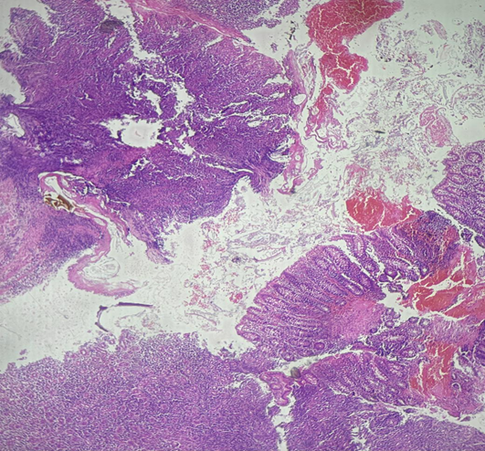
Figure 1. Low-power photomicrograph (H&E, ×40) showing extensive mucosal ulceration with inflammatory granulation tissue, hemorrhage, and dense mixed inflammatory infiltrates. Residual colonic crypts are present at the periphery. Sevelamer crystals are identified within the ulcer bed.
Figure 2. Intermediate-power photomicrograph (H&E, ×100) demonstrating irregular yellow-brown crystalline deposits embedded within inflammatory granulation tissue and associated foreign body-type inflammatory reaction.
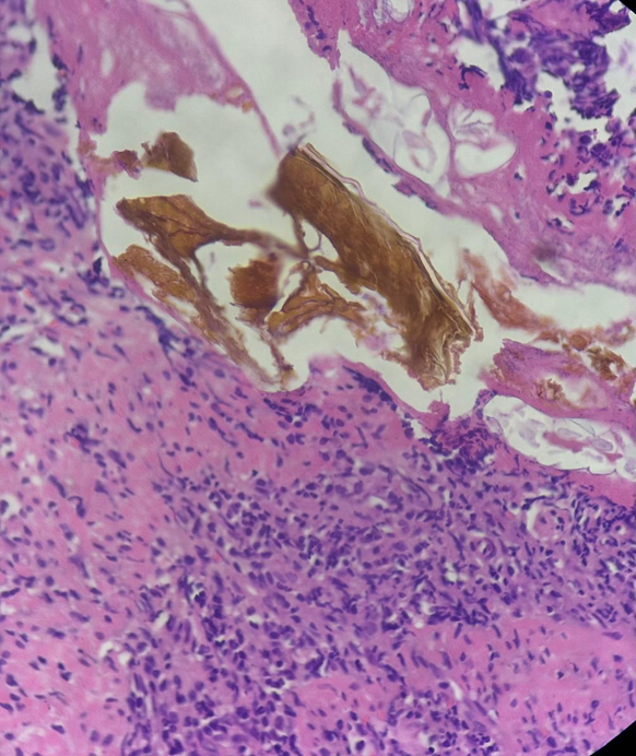
Figure 3. High-power photomicrograph (H&E, ×200–400) highlighting the characteristic broad curvilinear fish-scale internal architecture of sevelamer crystals surrounded by acute inflammatory infiltrates and histiocytic reaction.
RESULTS:
Table 1. Reported Histopathological Features of Sevelamer-Associated Gastrointestinal Injury
|
Histopathological Feature |
Frequency in Reported Cases |
|
Mucosal ulceration |
Very common |
|
Granulation tissue formation |
Common |
|
Acute inflammatory infiltrate |
Common |
|
Chronic inflammatory infiltrate |
Common |
|
Foreign body giant cell reaction |
Common |
|
Ischemic-type injury |
Occasional |
|
Transmural necrosis |
Rare |
|
Perforation |
Rare |
|
Pseudotumoral lesion formation |
Rare |
|
Characteristic fish-scale crystals |
Diagnostic feature |
Table 2. Differential Diagnosis of Medication-Associated Gastrointestinal Crystals
|
Feature |
Sevelamer |
Sodium Polystyrene Sulfonate (Kayexalate) |
Cholestyramine |
|
Color on H&E |
Yellow-brown to rusty pink |
Violet-purple |
Pale eosinophilic |
|
Shape |
Broad, curved |
Angulated |
Rhomboid |
|
Fish-scale pattern |
Prominent |
Present but narrower |
Absent or indistinct |
|
Associated injury |
Ulceration, bleeding, ischemia |
Necrosis, ulceration |
Usually minimal |
|
Typical patient |
CKD on dialysis |
Hyperkalemia treatment |
Hyperlipidemia/bile acid disorders |
DISCUSSION:
Sevelamer is a non-absorbable anion-exchange resin widely prescribed for the treatment of hyperphosphatemia in patients with advanced chronic kidney disease and end-stage renal disease undergoing dialysis. Although gastrointestinal adverse effects such as nausea, abdominal discomfort, constipation, and diarrhea are well recognized, crystal-associated gastrointestinal mucosal injury has emerged as an increasingly recognized but underdiagnosed complication.
Since the seminal report by Swanson et al., sevelamer crystals have been identified throughout the gastrointestinal tract, including the esophagus, stomach, small intestine, colon, and rectum. Histologically, these crystals display a characteristic broad curvilinear "fish-scale" internal architecture with yellow-brown to rusty pink staining on hematoxylin and eosin sections. Recognition of this distinctive morphology is essential because the associated inflammatory changes may obscure the underlying diagnosis.
The exact mechanism of sevelamer-associated injury remains incompletely understood. Proposed mechanisms include direct mucosal toxicity, mechanical epithelial injury, promotion of local ischemic changes, and perpetuation of inflammation through foreign body-type reactions. Crystal deposition is often observed within areas of pre-existing mucosal injury, suggesting that sevelamer may exacerbate ongoing tissue damage and impair mucosal healing.
The spectrum of gastrointestinal pathology associated with sevelamer is broad and ranges from incidental crystal deposition to severe ulceration, ischemic injury, inflammatory pseudotumor formation, bowel obstruction, perforation, and gastrointestinal hemorrhage. Several studies have documented clinically significant bleeding requiring endoscopic evaluation, emphasizing the potential morbidity associated with this entity.
The differential diagnosis includes other medication-associated crystalline deposits, particularly sodium polystyrene sulfonate (Kayexalate) and cholestyramine. While all three agents may exhibit a mosaic-like internal pattern, sevelamer crystals are typically yellow-brown and demonstrate broad irregular fish-scale markings. In contrast, Kayexalate crystals tend to be basophilic with narrower angulated scales, whereas cholestyramine crystals are generally pale eosinophilic and lack the characteristic morphology of sevelamer. Awareness of these distinctions is crucial for accurate pathological interpretation.
In the present case, a 44-year-old female with end-stage renal disease on maintenance hemodialysis presented with lower gastrointestinal bleeding approximately eight years after initiation of sevelamer therapy. Histopathological examination revealed extensive mucosal ulceration, inflammatory granulation tissue, mixed inflammatory infiltrates, and characteristic yellow-brown fish-scale crystals associated with foreign body giant cell reaction. No evidence of inflammatory bowel disease, granulomatous inflammation, dysplasia, or malignancy was identified. The temporal relationship between drug exposure and symptom onset, together with the characteristic histological findings, strongly supported a diagnosis of sevelamer-associated crystal-induced colonic injury. This case highlights the importance of meticulous medication review in patients with unexplained gastrointestinal ulceration and bleeding. Failure to recognize sevelamer-associated injury may result in misdiagnosis as inflammatory bowel disease, ischemic colitis, infectious colitis, or colorectal neoplasia, potentially leading to unnecessary investigations and inappropriate therapeutic interventions. Increased awareness among pathologists, gastroenterologists, and nephrologists is therefore essential for timely diagnosis and optimal patient management.
CONCLUSION:
Sevelamer-associated crystal-induced gastrointestinal injury is an uncommon but important cause of lower gastrointestinal bleeding in patients with chronic kidney disease receiving dialysis. Histopathological recognition of characteristic fish-scale crystals within ulcerated mucosa is essential for diagnosis. Awareness of this entity among pathologists and clinicians can prevent diagnostic errors and facilitate appropriate modification of phosphate-binding therapy.
Learning Points
1. Sevelamer crystals are an increasingly recognized cause of gastrointestinal mucosal injury in dialysis patients.
2. Lower gastrointestinal bleeding may be the presenting manifestation.
3. The characteristic yellow-brown fish-scale crystal morphology is highly diagnostic.
4. Foreign body giant cell reaction and ulceration are common associated histological findings.
5. Clinicopathological correlation and medication review are essential for accurate diagnosis.
REFERENCES:
1. Swanson BJ, Limketkai BN, Liu TC, et al. Sevelamer crystals in the gastrointestinal tract (GIT): a new entity associated with mucosal injury. Am J Surg Pathol. 2013;37(11):1686–1693.
2. Yuste C, Merida E, Hernandez E, et al. Gastrointestinal complications induced by sevelamer crystals. Clin Kidney J. 2017;10(4):539–544.
3. Chintamaneni M, Dasanu CA. Sevelamer-associated rectosigmoid ulcers. Clin Exp Gastroenterol. 2014;7:23–27.
4. Madan P, Bhayana S, Chandra P, Hughes JI. Lower gastrointestinal bleeding from sevelamer-associated mucosal injury. ACG Case Rep J. 2018;5:e35.
5. Nguyen T, Weixler B, Birk JW. Sevelamer-induced gastrointestinal injury presenting as colitis. Case Rep Gastrointest Med. 2019;2019:2489095.
6. Arnold MA, Cooper K. Medication-associated crystal injury in the gastrointestinal tract. Arch Pathol Lab Med. 2020;144(6):715–720.
7. González RS, Lagana SM, Szeto O, et al. Challenges in the diagnosis of medication-related gastrointestinal crystal deposition. Hum Pathol. 2019;84:188–197.
8. Abraham SC. Drug-related injuries of the gastrointestinal tract. Surg Pathol Clin. 2017;10(4):867–882.
9. Kramann R, Erpenbeck J, Schneider RK, et al. Clinical consequences of phosphate binders in chronic kidney disease. Kidney Int Rep. 2018;3(4):843–855.
10. Farris AB, Lauwers GY. Medication-induced gastrointestinal pathology. Adv Anat Pathol. 2021;28(5):315–326.
11. Rostoker G, Griuncelli M, Loridon C, et al. Crystal-associated gastrointestinal injury in dialysis patients. Nephrol Dial Transplant. 2019;34(7):1158–1165.
12. Harel Z, Harel S, Shah PS, et al. Gastrointestinal adverse events associated with phosphate binders in chronic kidney disease. Clin J Am Soc Nephrol. 2013;8(8):1402–1409.
13. Iesalnieks I, Kilger A, Glass H, et al. Sevelamer crystal-induced gastrointestinal lesions: clinicopathological features. Int J Colorectal Dis. 2016;31(6):1201–1207.
14. Gonzalez E, Gutierrez A, Fernandez C, et al. Colonic ulceration associated with sevelamer crystals in a hemodialysis patient. Rev Esp Enferm Dig. 2018;110(5):326–328.
15. Deepak P, Czapla N, Alayo QA, et al. Medication-associated crystal colitis: a diagnostic challenge. Dig Dis Sci. 2022;67(3):912–919.