3D Time-of-Flight MR Angiography Evaluation of Circle of Willis Variants: Prevalence, Anatomical Patterns, and Clinical Implications in an Adult Population.
- Sathish Babu , Head of the Department, Sree Mookambika Institute of Medical sciences, Kulasekharam.
- Dharshika R , Junior Resident, Sree Mookambika Institute of Medical sciences, Kulasekharam.
- Vinod , Associate Professor, Sree Mookambika Institute of Medical sciences, Kulasekharam.
Article Information:
Abstract:
Purpose: To evaluate the prevalence, distribution, and clinical implications of Circle of Willis (CoW) variants using three-dimensional time-of-flight magnetic resonance angiography (3D TOF-MRA) in an adult population, and to analyze age-related trends in CoW completeness. Materials and Methods: This retrospective cross-sectional study included 200 consecutive adult patients (aged 18–89 years) who underwent 3D TOF-MRA of the intracranial circulation on a 1.5T MRI scanner between August 2024 to march 2025. Standard TOF-MRA parameters were used with thin-slice acquisition (≤1 mm), high matrix resolution, and slab coverage through the CoW. Image analysis was performed using multiplanar and maximum-intensity projection reconstructions. Each arterial segment (A1, ACom, P1, PCom) was classified as normal, hypoplastic (diameter <1.0 mm), or absent. The presence of a fetal-type posterior cerebral artery (fPCA) was also recorded. Results: A complete (classical) CoW configuration was present in 15% (30/200) of subjects. The most frequent variations involved the posterior communicating arteries (PCoA): bilateral PCoA hypoplasia/aplasia occurred in 29% (58/200), and unilateral PCoA hypoplasia/aplasia in 20% (40/200). Additional variants included ACom abnormalities in 12% (24/200), A1 hypoplasia/aplasia in 9% (18/200), and fetal-type PCA in 10% (20/200). Incomplete CoW configurations were significantly more prevalent in individuals aged ≥60 years (68%) compared with those under 40 years (46%) (p < 0.01). The patterns were consistent that PCoA variations represent the most common CoW anomaly. Conclusion: 3D TOF-MRA provides high-resolution, contrast-free visualization of the Circle of Willis and its variants, enabling accurate anatomic assessment in routine clinical practice. Recognition of CoW variants is essential for understanding collateral circulation potential, assessing cerebrovascular risk, and planning surgical or endovascular interventions. The posterior communicating arteries are the most variable components, and CoW incompleteness increases with advancing age, underscoring the need for systematic evaluation in neuroimaging protocols.
Keywords:
Article :
INTRODUCTION:
The Circle of Willis (CoW) is a critical arterial anastomotic network located in the interpeduncular cistern at the base of the brain. It acts as a central vascular junction connecting the carotid and vertebrobasilar systems, ensuring adequate perfusion when one of the major feeding arteries becomes narrowed or occluded. Functionally, a complete and well-balanced CoW maintains stable cerebral hemodynamics, while developmental variations may reduce collateral potential, influencing vulnerability to ischemia, aneurysm formation, and outcomes of neurovascular interventions.
Anatomically, the CoW is formed by the union of the internal carotid and vertebrobasilar systems. The anterior circulation comprises the internal carotid arteries and their branches, the anterior cerebral arteries (ACAs), connected by the anterior communicating artery (ACom). The posterior circulation, derived from the vertebral arteries and the basilar artery, gives rise to the posterior cerebral arteries (PCAs), which are connected to the anterior system via the posterior communicating arteries (PCoAs). In its classical configuration, the CoW is a symmetrical polygonal ring formed by the A1 segments of the ACAs, the ACom, the P1 segments of the PCAs, and the bilateral PCoAs. However, numerous anatomic variants exist—such as hypoplasia, aplasia, fenestrations, or the presence of a fetal-type PCA—resulting in an incomplete or asymmetric circle. A complete CoW is seen in only about 10–30% of individuals, with posterior communicating artery variations being the most frequent.
From an embryologic perspective, the CoW develops during early gestation as a series of transient carotid–vertebrobasilar connections remodel to establish the adult configuration. Initially, the posterior circulation is supplied primarily through the internal carotid arteries via the primitive posterior communicating arteries. As the basilar and posterior cerebral arteries mature, the vertebrobasilar system assumes a dominant role, while the primitive communications regress. Incomplete regression or persistence of these embryonic channels results in the common variants observed in adults, such as a fetal-type PCA, hypoplastic P1 segments, or absent communicating arteries. Appreciating these embryologic transitions helps explain the diversity of CoW configurations encountered in adults.
Imaging of Circle of Willis
Several imaging modalities are used to evaluate the Circle of Willis, including digital subtraction angiography (DSA), computed tomography angiography (CTA), and magnetic resonance angiography (MRA). DSA remains the diagnostic gold standard owing to its excellent spatial and temporal resolution, but its invasive nature, radiation exposure, and contrast requirements limit its routine use. CTA provides high-quality vascular mapping but similarly involves ionizing radiation and iodinated contrast. In contrast, MRA—particularly three-dimensional time-of-flight (3D TOF) sequences—offers a non-invasive, contrast-free, and reproducible technique for visualizing intracranial arteries with excellent diagnostic accuracy.
In TOF-MRA, flow-related enhancement produces bright signal intensity in rapidly moving blood, while stationary tissue signal is suppressed. On 3D TOF images, a normal CoW appears as a continuous, symmetric arterial ring with homogeneous high signal in the A1, ACom, PCom, and P1 segments. Thin-section, high-resolution acquisition and multiplanar reconstruction allow for precise visualization of small communicating arteries and detection of hypoplastic or absent segments. These attributes make 3D TOF-MRA the preferred method for evaluating CoW anatomy in both clinical and research settings.
Given the high frequency of anatomical variants and their influence on cerebral hemodynamics, accurate assessment of the CoW is essential for risk evaluation in stroke, aneurysm planning, and other neurovascular pathologies. The present study aims to determine the prevalence and distribution of Circle of Willis variants using 3D TOF-MRA in a cohort of 200 adult patients, to evaluate age-related trends, and to emphasize the clinical importance of identifying these variants in routine neuroimaging practice.
MATERIALS AND METHODS:
Study Design
This retrospective, cross-sectional observational study was carried out in the Department of Radiology at Sree Mookambika Institute of medical sciences, kulasekharam to evaluate the prevalence and configuration patterns of Circle of Willis (CoW) variants using three-dimensional time-of-flight magnetic resonance angiography (3D TOF-MRA). The study adhered to the principles of the Declaration of Helsinki, and ethical clearance was obtained from the Institutional Ethics Committee prior to data collection. Patient records were anonymized to maintain confidentiality. The study period extended from August 2024 to March 2025, and all imaging data were reviewed retrospectively from the hospital’s Picture Archiving and Communication System (PACS).
Study Population and Sample Size
The study population comprised 200 consecutive adult patients who underwent non-contrast 3D TOF-MRA of the intracranial circulation for various clinical indications. The selected sample size provided adequate statistical power to determine the prevalence of CoW variants based on previously published radiologic cohort data. The study excluded pediatric cases to ensure anatomical uniformity, as cerebral arterial maturation varies significantly in childhood. All MRI scans were performed on a 1.5 Tesla superconducting scanner (e.g., GE, Siemens, or Philips) using a standard head coil and a uniform acquisition protocol to minimize inter-scan variability.
Each patient’s demographic data (age and sex) and clinical indication were recorded. None of the patients received intravenous contrast, and all studies were performed as part of their diagnostic evaluation for non-acute neurological symptoms.
Inclusion Criteria
Participants were included based on the following criteria:
1) Adults aged 18 years or older undergoing 3D TOF-MRA of the brain for evaluation of non-emergent neurological complaints such as headache, dizziness, transient ischemic attack, syncope, or suspected vascular anomaly.
2) High-quality imaging datasets with adequate flow-related enhancement and complete slab coverage through the Circle of Willis, permitting detailed visualization of its component vessels (A1, ACom, P1, and PCom segments).
3) Absence of any contraindication to MRI (e.g., metallic implants, pacemakers, or severe claustrophobia).
Exclusion Criteria
The following exclusion criteria were applied to eliminate confounding variables and ensure accurate anatomical interpretation:
1) Patients with a history of intracranial surgical or endovascular intervention, including aneurysm clipping, coiling, stenting, or bypass procedures, as these can alter the native vascular architecture.
2) Images demonstrating significant motion artifacts, partial coverage of the CoW, or poor signal-to-noise ratio, which impaired reliable evaluation of small communicating arteries.
3) Presence of intracranial pathologies such as large space-occupying lesions, hemorrhage, infarcts, or arteriovenous malformations that distorted or displaced the CoW anatomy.
Continuous variables, such as patient age, were expressed as mean ± standard deviation (SD). Categorical variables, including CoW segment presence, hypoplasia, or aplasia, were summarized as frequencies and percentages. The prevalence of each CoW variant was calculated, and age-related differences in CoW completeness were assessed using the Chi-square test, with p < 0.05 considered statistically significant. Graphical representations, including bar charts and pie diagrams, were used to illustrate the distribution of CoW variants across age groups and overall anatomical patterns.
RESULTS:
A total of 200 adult patients were included in the study, comprising 110 males (55%) and 90 females (45%), with a mean age of 52.4 ± 16.3 years (range, 18–89 years). A complete classical Circle of Willis (CoW) configuration was identified in 30 patients (15%), whereas 170 patients (85%) demonstrated one or more anatomical variations resulting in an incomplete CoW (Table 1). The most frequently observed variants involved the posterior communicating arteries (PCoA). Bilateral PCoA hypoplasia or aplasia was identified in 58 patients (29%), while unilateral PCoA hypoplasia or aplasia was present in 40 patients (20%). Variations of the anterior communicating artery (ACom) were observed in 24 patients (12%), A1 segment hypoplasia or aplasia in 18 patients (9%), and fetal-type posterior cerebral artery (fPCA) in 20 patients (10%). These findings indicate that the PCoA represents the most variable component of the Circle of Willis (Table 2).
Age-wise analysis demonstrated a progressive increase in the prevalence of incomplete Circle of Willis configurations with advancing age. Incomplete CoW was observed in 66.7% (40/60) of patients younger than 40 years, 90.0% (72/80) of those aged 40–59 years, and 96.7% (58/60) of patients aged 60 years and above. This represents a clear age-dependent decline in CoW completeness, with only 33.3%, 10.0%, and 3.3% complete configurations in the respective age groups. Statistical analysis demonstrated a significant association between increasing age and CoW incompleteness (Chi-square test, p < 0.001). (Table 3). Overall, a complete Circle of Willis was uncommon, and posterior communicating artery variations were the predominant anatomical deviation. In addition, the prevalence of incomplete CoW increased with advancing age, emphasizing the importance of careful evaluation of these vascular patterns in older adults. Variants such as ACom and A1 hypoplasia, although less frequent, remain clinically relevant due to their impact on collateral circulation and implications for neurovascular interventions.
Table 1: Distribution of Complete and Incomplete Circle of Willis (n=200)
|
CoW Configuration |
Number of Patients |
Percentage (%) |
|
Complete |
30 |
15 |
|
Incomplete |
170 |
85 |
Table 2: Prevalence of Specific Circle of Willis Variants (n=200)
|
Variant Type |
Number of Patients |
Percentage (%) |
|
Bilateral PCoA hypoplasia/aplasia |
58 |
29 |
|
Unilateral PCoA hypoplasia/aplasia |
40 |
20 |
|
ACom hypoplasia/aplasia |
24 |
12 |
|
A1 segment hypoplasia/aplasia |
18 |
9 |
|
Fetal-type PCA (fPCA) |
20 |
10 |
Table 3: Age-Related Distribution of Complete and Incomplete Circle of Willis
|
Age Group (years) |
Complete CoW |
Incomplete CoW |
Percentage of Incomplete CoW (%) |
|
<40 |
20 |
40 |
66.7 |
|
40–59 |
8 |
72 |
90 |
|
≥60 |
2 |
58 |
96.7 |
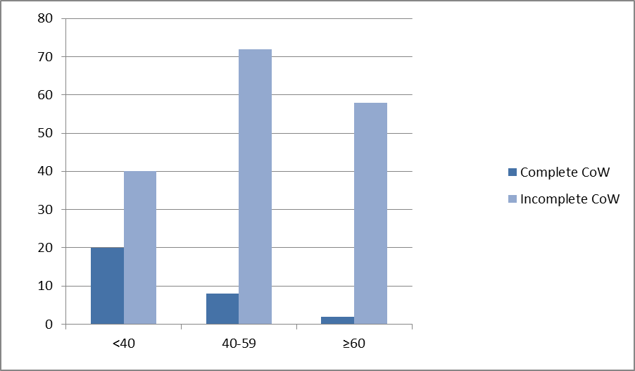
Figure 1a: Clustered column chart showing age-wise distribution of Circle of Willis configurations.
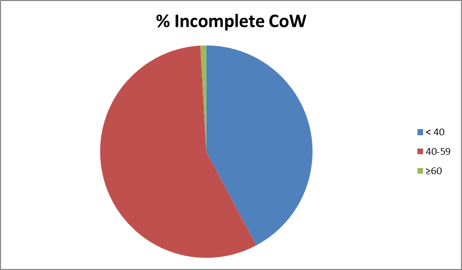
Figure 1b: Pie chart demonstrating the age-related prevalence of incomplete Circle of Willis configurations. The frequency of incomplete CoW increased from 66.7% in patients younger than 40 years to 90.0% in the 40–59-year age group and 96.7% in patients aged 60 years and above.
MRI Technique
Imaging Sequence
All examinations were performed using a three-dimensional time-of-flight (3D TOF) MRA sequence optimized for intracranial arterial visualization. TOF-MRA is a non-contrast technique that relies on flow-related enhancement, in which unsaturated blood entering the imaging volume produces high signal intensity, while stationary tissue is suppressed. This allows clear delineation of small arteries, including the A1, ACom, P1, and PCoA segments of the Circle of Willis.
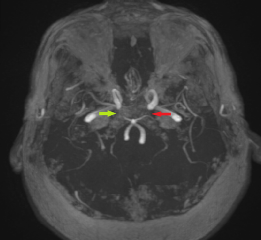
Image 1 : The 3D time-of-flight (TOF) MRA image shows bilateral hypoplastic posterior communicating arteries. (Green : Right , Red: Left ).
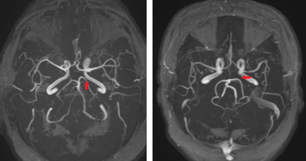
Image 2: The 3D time-of-flight (TOF) MRA images showing hypoplastic left posterior communicating artery
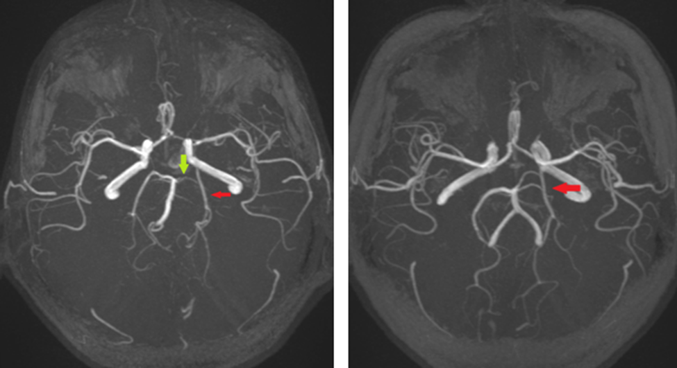
Image 3: The 3D time-of-flight (TOF) MRA images show 2 PCAs on the left (PCA duplication)—a dominant fetal-type PCA and a small-caliber classic PCA.
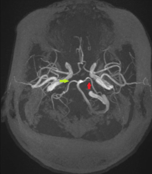
Image 4: The 3D time-of-flight (TOF) MRA image absent pcom on right side and hypoplastic left pcom artery.
Acquisition Parameters
The imaging protocol was standardized to ensure reproducibility and high spatial resolution. Typical parameters included:
· Repetition time (TR): 20–25 ms
· Echo time (TE): 3–7 ms
· Flip angle: 20–25°
· Field of view (FOV): 200–230 mm
· Matrix: high resolution (typically 256 × 512)
· Slice thickness: ≤1.0 mm, with overlapping thin slabs to improve signal uniformity and coverage of the entire CoW
· Number of excitations (NEX): 1–2, depending on patient cooperation and signal requirements
Images were reconstructed in multiple planes using maximum intensity projection (MIP) and multiplanar reconstruction (MPR) techniques. This allowed clear visualization of vessel continuity, detection of hypoplastic or absent segments, and assessment of variant anatomy such as fetal-type PCA. Thin-section reconstructions were particularly valuable for evaluating small communicating arteries, fenestrations, and segmental narrowing.
Variant Classification
For each patient, the Circle of Willis (CoW) was categorized as complete or incomplete based on the presence, caliber, and continuity of its arterial segments. A complete CoW required all principal arteries—the A1 segments of both anterior cerebral arteries, the anterior communicating artery (ACom), the P1 segments of both posterior cerebral arteries, and the posterior communicating arteries (PCoA)—to be patent, of normal caliber (≥1.0 mm), and continuous, forming a symmetric arterial ring capable of optimal collateral flow. An incomplete CoW was defined by hypoplastic (diameter <1.0 mm) or aplastic/absent segments, including PCoA, A1, ACom, or P1 variations, with fetal-type PCA considered a specific variant. The number, laterality, and type of affected segments were documented for each patient. This systematic classification using high-resolution 3D TOF-MRA allows reproducible assessment, facilitates age- and population-based analyses, and provides clinically relevant information regarding collateral capacity and cerebrovascular risk.
Image Analysis and Classification
Images were evaluated on high-resolution workstations using multiplanar reconstruction (MPR) and maximum intensity projection (MIP) to assess the Circle of Willis (CoW) in three dimensions, ensuring accurate visualization of both large and small communicating arteries.
Segmental Assessment
Each CoW component was systematically analyzed:
· A1 segments of the ACAs
· Anterior communicating artery (ACom)
· P1 segments of the PCAs
· Posterior communicating arteries (PCoA)
Segments were categorized as:
· Normal: clearly visualized, diameter ≥1.0 mm, continuous flow signal.
· Hypoplastic: diameter <1.0 mm, thin but patent, with preserved flow.
· Aplastic/Absent: not visualized on any projection, indicating congenital absence or extreme hypoplasia below detection threshold.
A1 Segments of the Anterior Cerebral Arteries (ACA)
The A1 segments arise from the internal carotid arteries and course medially to connect via the anterior communicating artery (ACom), forming the anterior portion of the Circle of Willis (CoW). They supply the medial frontal lobes and anterior corpus callosum. Common variants include hypoplasia, in which one A1 segment is narrow (<1.0 mm) but patent, and aplasia or complete absence, where the segment is not visualized. On 3D TOF-MRA, a normal A1 appears as a continuous, high-signal artery, while a hypoplastic A1 is thinner and may show slower flow-related enhancement. Complete absence manifests as a gap between the ICA and ACom, and multiplanar reconstructions help distinguish true aplasia from flow artifacts.
Anterior Communicating Artery (ACom)
The ACom connects the left and right ACAs, completing the anterior part of the CoW and allowing collateral flow between hemispheres in cases of ICA stenosis. Variants include hypoplasia or small diameter (<1.0 mm), which limits collateral capacity; aplasia or absence; and fenestration or duplication, where the segment divides into two parallel channels. On 3D TOF-MRA, the ACom is best visualized on axial and sagittal MIP reconstructions. Hypoplastic segments are narrow but continuous, fenestrations appear as dual channels, and absent ACom results in discontinuity between the A1 segments.
Posterior Communicating Arteries (PCoA)
The PCoAs connect the ICAs to the P1 segments of the posterior cerebral arteries, linking the anterior and posterior circulations and providing collateral flow in posterior circulation compromise. Variants include hypoplasia (diameter <1.0 mm), aplasia or absence, and fetal-type PCA (fPCA), where the PCoA predominates in supplying the PCA territory and the P1 segment is hypoplastic or absent. On 3D TOF-MRA, PCoAs are visualized as high-signal arteries connecting ICA to P1. Hypoplastic PCoAs may be subtle and require thin-slice MIP reconstructions, while fPCA is identified when P1 is thin or absent and the PCoA is dominant.
P1 Segments of the Posterior Cerebral Arteries (PCA)
The P1 segments originate from the basilar artery and connect to the posterior circulation, supplying the occipital and inferior temporal lobes. Common variants include hypoplasia or aplasia, often associated with fetal-type PCA where P1 is small or absent. On 3D TOF-MRA, P1 segments are best evaluated on coronal and axial MIP views. Normal P1 segments are symmetric and continuous, while hypoplastic segments appear thin with reduced flow-related signal, requiring careful multiplanar assessment to differentiate from imaging artifacts.
Fetal-Type Posterior Cerebral Artery (fPCA)
A fetal-type posterior cerebral artery (fPCA) occurs when the posterior communicating artery (PCoA) remains dominant in supplying the PCA territory, and the P1 segment of the PCA is hypoplastic or absent. This variant reflects persistence of the embryonic carotid–vertebrobasilar connection and can be unilateral or bilateral. Clinically, fPCA has implications for collateral circulation and stroke risk, as the PCA territory becomes dependent on the anterior circulation via the ICA. On 3D TOF-MRA, fPCA is identified when the PCoA is large and continuous, while the corresponding P1 segment is narrow or absent. Multiplanar reconstructions, particularly coronal and sagittal MIP views, are essential to distinguish fPCA from true P1 aplasia or hypoplasia, and to accurately document laterality and symmetry for clinical and research evaluation.
DISCUSSION:
Prevalence and Anatomical Variations of the Circle of Willis
The Circle of Willis (CoW) is the principal collateral arterial network of the brain, providing an important communication pathway between the carotid and vertebrobasilar circulations. Anatomical variations within this vascular ring are common and may significantly influence cerebral hemodynamics, collateral circulation, and susceptibility to cerebrovascular disease. In the present study, a complete classical CoW configuration was identified in only 15% of patients, while 85% demonstrated one or more anatomical variants. The most common abnormalities involved the posterior communicating arteries (PCoA), with bilateral PCoA hypoplasia/aplasia accounting for 29% of cases and unilateral PCoA hypoplasia/aplasia for 20%. In addition, fetal-type posterior cerebral artery (fPCA), anterior communicating artery (ACom) abnormalities, and A1 segment hypoplasia/aplasia were observed in smaller proportions. These findings confirm that the classical CoW configuration is relatively uncommon and that posterior circulation components are the most variable elements of the cerebral arterial network.
The prevalence of a complete CoW in our study is consistent with previously published anatomical and radiological investigations, which have reported complete configurations in approximately 10–30% of individuals. Krabbe-Hartkamp et al., in a landmark 3D TOF-MRA study, reported a complete CoW in approximately 21% of healthy adults, while Qiu et al. demonstrated similar findings in a large population-based cohort. Likewise, Chen et al. observed a predominance of posterior circulation variants, particularly involving the PCoA, supporting the observations of the present study. Anatomical studies conducted by Kapoor et al. also demonstrated that posterior communicating artery abnormalities represent the most frequent deviations from the classical configuration. Furthermore, Hoksbergen et al. emphasized the importance of these variants in determining collateral circulation capacity and cerebrovascular adaptability. The concordance of our findings with these established studies strengthens the validity of our observations and suggests that the pattern of CoW variation is remarkably consistent across different populations and imaging modalities.
The predominance of PCoA abnormalities can be explained by the embryological development of the cerebral circulation. During fetal life, the posterior cerebral arteries derive their blood supply predominantly from the internal carotid arteries through the primitive posterior communicating arteries. As the vertebrobasilar system matures, the P1 segments enlarge and gradually assume dominance. Variations in this developmental transition may result in persistent fetal-type circulation, hypoplastic communicating arteries, or absent segments, accounting for the high frequency of posterior circulation variants observed in both anatomical and radiological studies.
Clinical Significance and Neurointerventional Relevance
The clinical importance of CoW anatomy extends far beyond anatomical description. The completeness and symmetry of the CoW directly influence the effectiveness of collateral cerebral circulation during arterial stenosis, occlusion, or other hemodynamic disturbances. Patients with a complete CoW generally possess a greater capacity for redistribution of blood flow and may therefore demonstrate improved tolerance to carotid artery stenosis, intracranial arterial occlusion, and ischemic stroke. Conversely, incomplete configurations may compromise collateral reserve, increasing the likelihood of cerebral ischemia and potentially resulting in larger infarct volumes and poorer neurological outcomes.
From a territory-based perspective, specific variants demonstrate predictable perfusion consequences. A1 segment hypoplasia primarily affects the anterior cerebral artery (ACA) territory and may alter interhemispheric collateral flow through the anterior communicating artery. Fetal-type posterior cerebral artery (fPCA) results in posterior cerebral artery territory dependence on the internal carotid system, particularly affecting occipital lobe perfusion. Posterior communicating artery hypoplasia may impair collateral flow between the middle cerebral artery (MCA) and posterior circulation watershed zones, thereby reducing compensatory capacity in hypoperfusion states. Similarly, P1 segment hypoplasia limits vertebrobasilar contribution to the posterior cerebral artery (PCA) territory, increasing reliance on anterior circulation pathways.
The present study demonstrated a significant increase in CoW incompleteness with advancing age, with incomplete configurations identified in 66.7% of individuals younger than 40 years, 90.0% of those aged 40–59 years, and 96.7% of patients aged 60 years and above. This age-related trend was statistically significant (χ² = 23.95, p < 0.001). Similar observations have been reported by Hoksbergen et al., who demonstrated reduced collateral functionality in older individuals. Age-related vascular remodeling, atherosclerotic changes, reduced arterial compliance, and diminished flow velocities may contribute to the decreased visualization and functional capacity of communicating arteries. These findings may partly explain the increased susceptibility of elderly patients to cerebrovascular ischemic events and underscore the importance of careful vascular assessment in this population.
Knowledge of CoW anatomy has become increasingly important in modern neurointerventional practice. Mechanical thrombectomy has emerged as the standard of care for large-vessel occlusive stroke, and the adequacy of collateral circulation is now recognized as a major determinant of treatment outcome. Patients with well-developed communicating arteries often demonstrate slower infarct progression and better functional recovery, whereas those with incomplete CoW configurations may exhibit rapid infarct expansion due to limited collateral support. Consequently, pre-procedural evaluation of CoW anatomy may contribute valuable information regarding prognosis and treatment planning.
Similarly, detailed assessment of CoW variants is essential before carotid artery stenting, carotid endarterectomy, intracranial bypass procedures, balloon occlusion testing, and parent-vessel sacrifice. In these situations, communicating arteries may provide critical collateral pathways during temporary or permanent interruption of arterial flow. Recognition of absent or hypoplastic communicating vessels allows identification of patients at increased risk of peri-procedural ischemia and may influence therapeutic decision-making. Therefore, routine evaluation of the CoW should be considered an integral component of pre-interventional neurovascular imaging.
The fetal-type posterior cerebral artery observed in 10% of our cohort has additional clinical implications. In this configuration, the occipital and inferomedial temporal lobes are predominantly supplied by the internal carotid artery via the posterior communicating artery. Consequently, embolic events originating from carotid pathology may result in infarcts involving both anterior and posterior circulation territories, potentially leading to atypical stroke patterns. Failure to recognize this variant may therefore result in misinterpretation of stroke distribution and incomplete etiological evaluation.
Another clinically important aspect of CoW anatomy is its relationship to intracranial aneurysm formation. Variations such as A1 segment hypoplasia, absent communicating arteries, and fetal-type posterior circulation alter intracranial blood-flow patterns and wall shear stress. These hemodynamic changes may contribute to aneurysm development, particularly within the anterior communicating artery complex and posterior communicating artery region. Consequently, comprehensive evaluation of CoW anatomy is valuable not only for stroke assessment but also for aneurysm detection, risk stratification, and treatment planning.
Radiological Advances in Circle of Willis Evaluation
Advances in neurovascular imaging have significantly improved the ability to evaluate CoW anatomy non-invasively. Although digital subtraction angiography (DSA) remains the reference standard for vascular imaging because of its superior spatial and temporal resolution, its invasive nature limits routine use. Computed tomography angiography (CTA) provides excellent vascular detail and rapid acquisition but requires ionizing radiation and iodinated contrast administration.
Three-dimensional TOF-MRA has emerged as a highly effective alternative for evaluating intracranial arterial anatomy. The technique utilizes flow-related enhancement to produce high vascular contrast without the need for intravenous contrast agents, making it particularly suitable for screening and longitudinal follow-up. High-resolution thin-section acquisition combined with multiplanar reconstruction (MPR) and maximum-intensity projection (MIP) allows accurate visualization of small communicating arteries and variant configurations. In the present study, 3D TOF-MRA enabled reliable identification of CoW variants and demonstrated its value as a practical tool for routine neurovascular assessment.
Recent developments in MRI technology have further enhanced vascular imaging. The increasing availability of 3-Tesla and ultra-high-field 7-Tesla MRI systems has improved signal-to-noise ratio and spatial resolution, enabling better visualization of small arterial segments that may be difficult to assess on conventional systems. Advanced vessel wall imaging techniques now permit simultaneous evaluation of both luminal anatomy and arterial wall pathology, facilitating assessment of intracranial atherosclerosis, aneurysm instability, and inflammatory vascular disorders. In addition, artificial intelligence-based image analysis and automated vessel segmentation are emerging as promising tools for quantitative evaluation of cerebral vasculature, potentially improving diagnostic consistency and enabling large-scale population studies of CoW anatomy.
CONCLUSION:
In conclusion, the present study demonstrates that a complete classical Circle of Willis is uncommon, being present in only 15% of individuals. Posterior communicating artery abnormalities represent the most frequent anatomical variation, and incomplete CoW configurations increase significantly with advancing age. These findings are clinically important because CoW anatomy directly influences collateral cerebral circulation, stroke vulnerability, aneurysm hemodynamics, and neurointerventional planning. Modern 3D TOF-MRA provides a reliable, non-invasive, and high-resolution method for evaluating these vascular patterns and should be considered an essential component of contemporary neurovascular imaging.
Limitations
Several limitations should be acknowledged. First, this was a retrospective single-center study, which may limit the generalizability of the findings to other populations. Second, although the sample size was adequate for prevalence analysis, larger multicenter studies would provide more representative estimates of variant distribution. Third, TOF-MRA relies on flow-related enhancement and may underestimate vessel caliber in the presence of slow or turbulent flow, potentially resulting in overestimation of hypoplasia or aplasia. Fourth, direct comparison with CTA or DSA was not performed. Finally, clinical outcomes such as stroke severity, collateral performance, and procedural success were not assessed, limiting correlation between anatomical variants and patient prognosis.
REFERENCES:
1. Krabbe-Hartkamp MJ, van der Grond J, de Leeuw FE, et al. Circle of Willis: morphologic variation on three-dimensional time-of-flight MR angiograms. Radiology. 1998;207(1):103–111.
2. Hoksbergen AW, Majoie CB, Hulsmans FJ, et al. Collateral function of the circle of Willis: MR angiographic study. Radiology. 2003;226(2):511–516.
3. Schomer DF, Marks MP, Steinberg GK, et al. The anatomy of the posterior communicating artery. AJNR Am J Neuroradiol. 1994;15(1):57–64.
4. Kapoor K, Singh B, Dewan LI. Variations in the configuration of the circle of Willis. Anat Sci Int. 2008;83(2):96–106.
5. van Seeters T, Biessels GJ, van der Schaaf IC, et al. Completeness of the circle of Willis and risk of ischemic stroke. Stroke. 2015;46(9):2454–2459.
6. Qiu C, Zhang Y, Xue Y, et al. Variations of the circle of Willis in a large population study using MR angiography. Eur Radiol. 2015;25(7):2035–2043.
7. Zaninovich OA, et al. Circle of Willis completion varies with age and sex. World Neurosurg. 2017;106:953–963.
8. Jones JD, Castanho P, Bazira P, Sanders K. Anatomical variations of the circle of Willis and their prevalence: a literature review and meta-analysis. Clin Anat. 2021;34(7):978–990.
9. Ayre JR, Bazira PJ, Abumattar M, et al. A new classification system for the anatomical variations of the human circle of Willis: a systematic review. J Anat. 2022;240(6):1187–1204.
10. Feng L, Mao HJ, Zhang DD, et al. Anatomical variations in the circle of Willis and the formation and rupture of intracranial aneurysms: systematic review and meta-analysis. Front Neurol. 2023;13:1098950.
11. Shirin H, Roy DK, Bhoi S, et al. Anatomical variations of the circle of Willis: a 3D TOF-MRA study. Int J Sci Res. 2024;13(11):1505–1508.
12. Anderson FA, et al. Hemodynamic impact of circle of Willis variants. Stroke. 2005;36(2):214–220.
13. Wintermark M, Sanelli PC, Anzai Y, et al. Imaging of acute ischemic stroke. Lancet Neurol. 2013;12(2):173–186.
14. Zlokovic BV. Neurovascular pathways in stroke. Nat Rev Neurosci. 2011;12(12):723–738.
15. Bruneau M, et al. Anatomical variations of cerebral arteries: clinical relevance. Neurosurgery. 2013;72(3):602–613.
16. Schuette AJ, et al. Fetal posterior cerebral artery: clinical relevance and imaging features. J Neuroimaging. 2011;21(1):52–58.