Evaluating Diffuse Interstitial Lung Diseases Using High-Resolution CT: A Radiologic Perspective.
- Sathish Babu , Head of Department, Department of Radiodiagnosis, Sree Mookambika Institute of Medical Sciences.
- Rajashree , Junior resident, Department of Radiodiagnosis, Sree Mookambika Institute of Medical Sciences.
- Bharath Chandran , Assistant Professor, Department of Radiodiagnosis, Sree Mookambika Institute of Medical Sciences.
Article Information:
Abstract:
Background: Diffuse lung diseases (DLDs) encompass a diverse spectrum of pulmonary disorders primarily affecting the lower respiratory tract. These conditions are marked by inflammation and structural disruption of the pulmonary interstitium, leading to compromised alveolar integrity. The pathological process is not limited to the interstitium but extends to involve alveolar epithelium, capillary endothelium, and supporting mesenchymal tissues—resulting in widespread parenchymal involvement. This study aimed to assess the diagnostic utility of high-resolution computed tomography (HRCT) in the evaluation of diffuse lung diseases. Aims and Objectives: The primary aim of this study was to analyze the imaging characteristics of common interstitial lung diseases (ILDs), evaluate the severity of pulmonary involvement, and explore the potential for reversibility of parenchymal damage. Patients and Methods: This prospective observational study included 50 patients with clinical or radiological suspicion of interstitial lung disease. All patients underwent high-resolution computed tomography (HRCT) of the chest over a one-year period for detailed evaluation. Results: Among the 50 patients assessed, idiopathic pulmonary fibrosis (IPF) was the most frequently diagnosed condition (n=16), followed by ILD associated with rheumatoid arthritis (n=13), scleroderma (n=10), and systemic lupus erythematosus (SLE) (n=9). Less commonly observed cases included occupational lung disease (n=1) and pulmonary involvement in mycosis fungoides (n=1). Conclusion: HRCT serves as an essential non-invasive imaging modality in the evaluation of suspected ILD cases. It enables precise and early diagnosis, often reducing the need for invasive procedures like lung biopsy when correlated with clinical findings.
Keywords:
Article :
INTRODUCTION:
Interstitial lung diseases (ILDs), also referred to as diffuse infiltrative lung diseases, encompass a diverse group of disorders primarily affecting the lung parenchyma. These conditions are characterized by alveolar and septal thickening, fibroblast proliferation, and the development of pulmonary fibrosis. Although more than 100 distinct types of ILD have been identified, the majority of cases are attributed to idiopathic pulmonary fibrosis (IPF), sarcoidosis, and connective tissue disease-associated ILDs.
The prevalence of IPF varies depending on diagnostic criteria—ranging from approximately 54 per 100,000 adults using broad criteria to about 17 per 100,000 under stricter definitions. While ILDs are more commonly seen in adults, certain types—such as hypersensitivity pneumonitis and idiopathic interstitial pneumonias—can also occur in pediatric populations [1].
Clinically, patients with ILD often present with progressive exertional dyspnea, fatigue, generalized weakness, anorexia, weight loss, non-productive cough, and vague chest discomfort. Radiographically, these patients frequently exhibit diffuse infiltrative patterns on chest X-rays [2]. The purpose of this study is to examine the imaging features of commonly encountered ILDs, aiming for early diagnosis and prevention of irreversible pulmonary damage.
ILDs can be broadly categorized into those with identifiable causes and those without. The former includes ILDs related to connective tissue disorders, occupational exposures (pneumoconiosis), certain medications, smoking, radiation, and toxic inhalants. The idiopathic group includes conditions such as idiopathic pulmonary fibrosis, sarcoidosis, pulmonary lymphangioleiomyomatosis, and pulmonary alveolar proteinosis [1].
Imaging findings in ILDs may appear as reticular, nodular, or reticulonodular patterns. Interlobular septa, which contain lymphatic vessels, veins, and connective tissue, become thickened in conditions that involve these structures—such as IPF, lymphangitic carcinomatosis, and pulmonary edema—leading to irregular pleural surface appearances [3].
Historically, chest radiographs were the primary imaging modality for ILD diagnosis. The advent of computed tomography (CT), particularly with 8–10 mm collimation, offered better visualization of lung architecture. However, its diagnostic role remained limited until the introduction of high-resolution computed tomography (HRCT). By eliminating overlapping anatomical structures, HRCT enables detailed assessment of the type, distribution, and severity of lung parenchymal abnormalities [4].
Since its development in the 1980s, HRCT has become the preferred imaging technique for evaluating ILDs due to its superior ability to depict fine intrapulmonary detail. HRCT not only aids in detecting disease at an early stage—often when other diagnostic tests show minimal abnormalities—but also plays a pivotal role in assessing disease extent, identifying specific patterns, refining differential diagnoses, guiding biopsy locations, and evaluating treatment response and disease progression [5].
The main objectives of this study are to explore the role of HRCT in diagnosing ILD, especially in symptomatic individuals with inconclusive or normal chest radiographs; to assess disease patterns, severity, and distribution; to distinguish between reversible and irreversible parenchymal changes for prognostication; and to evaluate HRCT’s predictive value in treatment response.
For evaluating suspected idiopathic interstitial pneumonia or patients with restrictive pulmonary function test patterns, both prone and supine HRCT scans are typically employed. These positions help differentiate dependent atelectasis from fibrotic or inflammatory changes. Optimal HRCT image analysis requires specific window settings—usually with a window level of –600 to –700 Hounsfield Units (HU) and a window width of +1000 to +1500 HU [6].
Both inspiratory and expiratory HRCT scans are important in ILD evaluation. Expiratory images are particularly helpful for identifying air trapping and subtle ground-glass opacities that may not be visible during inspiration [7, 8]. While chest radiography remains a valuable initial imaging tool, HRCT has demonstrated significantly higher diagnostic accuracy in conditions like sarcoidosis, silicosis, and lymphangitic carcinomatosis. HRCT is especially recommended in cases where clinical, radiographic, and pulmonary function tests fail to yield a definitive diagnosis and should ideally be performed prior to biopsy [9].
MATERIALS AND METHODS:
Study Design and Setting:
This prospective observational study was conducted in the Department of Radiodiagnosis at Sree Mookambika Institute of medical sciences over a 6-month period, from January 2025 to June 2025.
Inclusion Criteria:
Participants were enrolled based on the following criteria:
1) Individuals with a suspected diagnosis of interstitial lung disease based on abnormalities detected on chest radiographs.
2) Patients presenting with clinical features suggestive of ILD but with normal or inconclusive chest X-ray findings.
3) Previously diagnosed cases of ILD undergoing HRCT evaluation for quantification of disease extent and to monitor therapeutic response.
Exclusion Criteria:
The following individuals were excluded from the study:
1) Pregnant women.
2) Adolescent females (due to radiation safety concerns).
3) Children under the age of 15 years.
Study Protocol
High-resolution computed tomography (HRCT) of the chest was carried out using a dual-slice CT scanner (Somatom Spirit, Siemens Healthcare, Germany). Imaging parameters were optimized to achieve maximum spatial resolution. Thin slices of 1 mm thickness were acquired at 10 mm intervals from the lung apices to the bases, providing representative cross-sectional images of the entire lungs.
A high-resolution image reconstruction algorithm was employed, utilizing a minimal field of view to reduce pixel size and enhance image clarity. For accurate interpretation, images were evaluated using a window level of -750 Hounsfield Units (HU) and a window width of +1500 HU.
Scans were obtained during full inspiratory breath-hold in the supine position. In select cases, prone imaging was performed to differentiate gravity-dependent atelectasis from early fibrotic changes suggestive of idiopathic pulmonary fibrosis. For patients with suspected airway obstruction, additional expiratory-phase scans were acquired to assess for air trapping.
Complementary laboratory investigations were conducted to support the diagnosis of connective tissue disease-related ILD. These included serological tests such as erythrocyte sedimentation rate (ESR), rheumatoid factor (RF), antinuclear antibody (ANA), and anticentromere antibody testing.
Data Sources, Variables, and Statistical Analysis
Data for this study were derived from the clinical history, radiological findings, and laboratory results of the 50 patients included. Relevant statistical tools were applied to analyze the collected data, as described in the results section. The study design ensured consistency in image interpretation, and efforts were made to eliminate inter-observer variability.
RESULTS:
In the current study, patient ages ranged from 22 to 85 years, with a mean age of 53.5 years. The highest percentage of participants (38%) belonged to the 21–40 year age group (Table 2). Gender distribution showed a predominance of female patients, comprising 56% of the cohort, while males accounted for the remaining 44% (Table 3).
The most common clinical symptom reported was shortness of breath on exertion, present in 64% of patients. This was followed by cough, observed in 60% of the cases. Fever was noted in 24% of individuals. Both skin thickening and joint pain (arthralgia) were reported in 8% of patients each. Additionally, 6% of the participants experienced weight loss and Raynaud’s phenomenon (Table 1).
Clinical Features
Table 1 summarizes the clinical presentations observed in patients diagnosed with interstitial lung disease (ILD). The most frequent symptom was exertional dyspnea, noted in 64% of the study population, followed closely by cough, present in 60% of patients. Fever was documented in 24% of cases. Arthralgia and skin thickening were seen in 8% each, while Raynaud’s phenomenon and weight loss were reported by 6% of patients, respectively. Less commonly reported symptoms included chest pain (4%) and hemoptysis (8%).
Table 1: Frequency of Clinical Features in ILD Patients
|
S. No |
Clinical Feature |
No. of Patients |
Percentage (%) |
|
1 |
Dyspnea on exertion |
32 |
64 |
|
2 |
Cough |
30 |
60 |
|
3 |
Fever |
12 |
24 |
|
4 |
Arthralgia |
4 |
8 |
|
5 |
Skin thickening |
4 |
8 |
|
6 |
Raynaud’s phenomenon |
3 |
6 |
|
7 |
Weight loss |
3 |
6 |
|
8 |
Chest pain |
2 |
4 |
|
9 |
Hemoptysis |
4 |
8 |
Age and Gender Distribution
Patients ranged in age from 22 to 85 years, with a mean age of 53.5 years. The largest group (38%) was aged between 21 and 40 years, followed by 36% in the 41–60 age group. Only one patient was over 80 years of age (Table 2).
Table 2: Age-wise Distribution of Patients
|
Age Group (Years) |
No. of Patients |
Percentage (%) |
|
0–20 |
0 |
0 |
|
21–40 |
19 |
38 |
|
41–60 |
18 |
36 |
|
61–80 |
12 |
24 |
|
81–100 |
1 |
2 |
Female patients comprised a slightly higher percentage of the study population (56%), while males accounted for 44% (Table 3).
Table 3: Gender Distribution of Patients
|
Gender |
No. of Patients |
Percentage (%) |
|
Male |
22 |
44 |
|
Female |
28 |
56 |
|
Total |
50 |
100 |
Indications for HRCT
The most common clinical indication for performing HRCT chest was idiopathic pulmonary fibrosis (IPF), accounting for 32% of cases. Rheumatoid arthritis-associated ILD followed at 26%, and scleroderma at 20%. Systemic lupus erythematosus (SLE) accounted for 18%, while occupational lung disease and mycosis fungoides were each responsible for 2% of cases (Figure 4). Only 42% (n=21) of patients demonstrated abnormalities on chest X-ray. Serum markers were positive in 70% (n=35) of patients.
HRCT Findings
The most frequent HRCT feature observed across ILD cases was the presence of interlobular septal thickening, seen in 42% of patients. Bronchiectasis appeared in 40%, while ground-glass opacity was noted in 32% of cases. Honeycombing was observed in 20%, and subpleural nodules in 18%. Less frequent findings included parenchymal bands (12%), consolidation (4%), subpleural cysts (4%), and esophageal dilatation (4%). Pleural effusion and suspected scar carcinoma were each noted in 2% of patients.
Table 4: HRCT Features by ILD Type
|
Finding |
RA |
Scleroderma |
IPF |
SLE |
|
Ground-glass haze |
✓ |
✓ |
✓ |
|
|
Septal lines |
✓ |
✓ |
✓ |
✓ |
|
Bronchiectasis |
✓ |
✓ |
✓ |
✓ |
|
Honeycombing |
✓ |
✓ |
✓ |
|
|
Subpleural nodules |
✓ |
✓ |
||
|
Parenchymal bands |
✓ |
|||
|
Subpleural cysts |
✓ |
|||
|
Irregular interface |
✓ |
|||
|
Consolidation |
✓ |
|||
|
Pleural effusion |
✓ |
|||
|
Dilated esophagus |
✓ |
Rheumatoid Arthritis:
The most frequently observed HRCT abnormality in patients with rheumatoid arthritis (RA) was ground-glass opacity, identified in 7 patients. This was followed by bronchiectasis seen in 5 patients, reticulations in 3 cases, honeycombing in 2 patients, and pleural effusion in 1 patient (7.6%) . HRCT detected RA-related lung changes in 10 cases (76.9%), whereas chest X-rays were positive in only 6 patients (46.2%). Regarding serological markers, all patients showed elevated ESR, rheumatoid factor (RF) was positive in 10 patients (76.9%), and antinuclear antibodies (ANA) were present in 1 patient (7.6%).
Scleroderma:
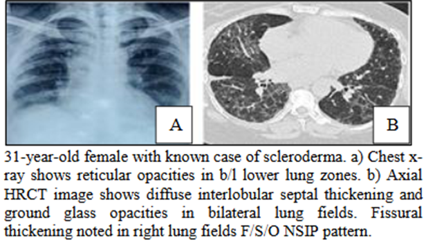
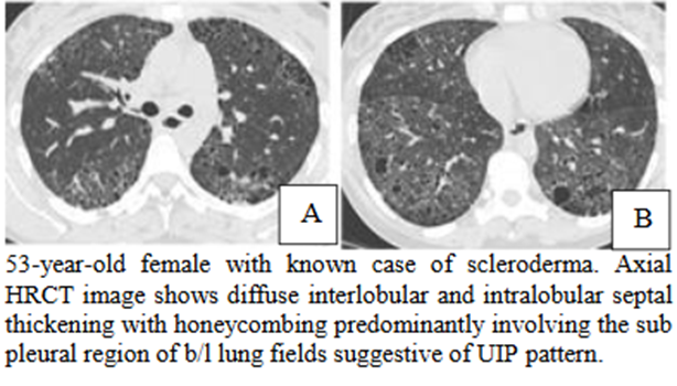
In scleroderma patients, the predominant HRCT findings were septal lines and parenchymal bands, each present in 5 cases. Subpleural nodules and bronchiectasis were noted in 4 patients each, while honeycombing, ground-glass opacities, and dilated esophagus appeared in 2 patients respectively. Scar carcinoma was observed in 1 patient . HRCT revealed abnormalities in 8 cases (80%), compared to 5 cases (50%) on chest X-ray. The most common serum marker was ANA, found in 6 patients (60%), while Scl-70 antibody was present in 1 patient (10%).
Systemic Lupus Erythematosus (SLE):
Septal lines were the most common HRCT feature seen in 4 patients with SLE, followed by subpleural nodules and bronchiectasis in 3 patients each. Ground-glass haze was identified in 2 patients, and irregular interface and consolidation were each seen in 1 patient (Figure 1). HRCT detected lung involvement in 7 patients (77.8%), whereas only 2 patients (22.2%) had abnormalities on chest X-ray.
Idiopathic Pulmonary Fibrosis (IPF):
The predominant HRCT findings in IPF patients were septal lines and honeycombing, each observed in 8 patients. Bronchiectasis was present in 7 cases, ground-glass haze in 4, and subpleural cysts in 2 cases . HRCT identified abnormalities in all 16 IPF patients (100%), while chest X-rays were positive in 7 patients (43.7%). Raised ESR was the only serum marker noted in 8 patients (50%). Diagnosis was based on clinical and radiological evaluation with exclusion of other causes. Lung biopsy was recommended for confirmation; however, only 3 patients (19%) underwent the procedure, which confirmed HRCT findings. The remaining 13 patients (81%) were followed clinically and showed improvement with treatment.
Mycosis Fungoides:
In patients with mycosis fungoides, HRCT demonstrated septal lines, ground-glass haze, bronchiectasis, and miliary nodules. Both HRCT and chest X-ray revealed these findings. Additional associated features included lytic bone lesions and liver cysts.
Occupational Lung Disease:
HRCT findings in pneumoconiosis included subpleural consolidation, centrilobular nodules, and parenchymal bands. These abnormalities were detected on both chest X-ray and HRCT.
DISCUSSION:
Interstitial lung diseases (ILDs), also referred to as diffuse parenchymal lung diseases, represent a heterogeneous group of pulmonary disorders that share common clinical, radiographic, physiological, or pathological characteristics. Patients suspected of having diffuse ILD typically undergo chest radiography as the first imaging modality. Although most patients demonstrate abnormalities on chest X-rays, the findings are often nonspecific. In a smaller subset, the chest radiograph may even appear normal [5]. The limitations of plain chest radiographs in accurately evaluating lung diseases, particularly diffuse ILDs, became more apparent with the introduction of high-resolution computed tomography (HRCT), which offers superior lung morphology characterization [10]. Key HRCT features valuable for diagnosing ILD include the pattern of parenchymal abnormalities (such as consolidation or reticulation), anatomical distribution (upper vs. lower lung zones, central vs. peripheral), and associated findings like mediastinal lymphadenopathy. Idiopathic pulmonary fibrosis (IPF) is recognized as the most common cause of ILD. In this study, connective tissue diseases accounted for the majority of cases (32), followed by IPF (16 cases).
Rheumatoid Arthritis (RA):
RA is a connective tissue disorder marked by symmetrical inflammatory arthritis and is the most prevalent connective tissue disease. Extra-articular manifestations are common, and pulmonary involvement presents with a broad range of pleural and parenchymal abnormalities. Most patients with pulmonary involvement also have systemic signs of RA [11]. HRCT is the most sensitive tool for early detection of interstitial changes in RA, often identifying abnormalities before clinical symptoms or pulmonary function tests become abnormal. Compared to chest X-rays, HRCT is far superior in detecting early lung involvement [12]. In our study, HRCT findings in RA included ground-glass opacities (7 cases), bronchiectasis (5 cases), reticulations (3 cases), honeycombing (2 cases), and architectural distortion (1 case). No nodules were observed, but pleural effusion was present in one case. These findings are consistent with previous research [13,14].
Scleroderma:
Progressive systemic sclerosis (scleroderma) predominantly affects women aged 30-50 years and is characterized by excessive collagen deposition causing fibrosis in skin, lungs, and other organs. Clinically, patients present with skin thickening, Raynaud’s phenomenon, and visceral fibrosis [15]. HRCT is more sensitive than chest radiography for detecting subtle lung involvement in scleroderma and is considered the preferred imaging method for assessing lung damage. While 35% of scleroderma patients may have normal chest X-rays, HRCT detects abnormalities in over 90% [8]. Our study similarly showed predominant lower lobe involvement, with septal lines and parenchymal bands present in 5 cases each. Subpleural nodules and bronchiectasis were seen in 4 cases each; honeycombing, ground-glass haze, and dilated esophagus were noted in 2 cases each. One patient developed carcinoma on fibrotic lung tissue. These results align with prior studies [16,17].
Systemic Lupus Erythematosus (SLE):
SLE is a systemic autoimmune disorder characterized by immune complex deposition causing widespread tissue damage. It predominantly affects women aged 20-50 years [18]. In this study, 9 patients with SLE showed HRCT findings of interlobular septal thickening (4 cases), irregular interfaces (1 case), subpleural nodules and bronchiectasis (3 cases each), ground-glass haze (2 cases), and consolidation (1 case). No pleural thickening or effusion was observed. Notably, HRCT identified lung involvement in cases where chest X-rays were normal, confirming its superiority in detecting ILD changes [19,20].
Idiopathic Pulmonary Fibrosis (IPF):
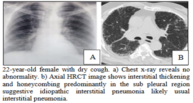
IPF is a chronic fibrosing interstitial pneumonia of unknown cause, typically affecting men over 50 years. It presents with gradual onset of dry cough and breathlessness, and has a poor prognosis with median survival of 2.5 to 3.5 years [5]. Among 16 IPF cases in this study, bilateral parenchymal abnormalities were seen in 15, with basal and subpleural distribution in 13. Honeycombing and septal lines were noted in 8 cases each, bronchiectasis in 7, ground-glass haze in 4, and subpleural cysts in 2. HRCT detected abnormalities in all patients, while chest X-rays were positive in only 7. Only 3 patients underwent lung biopsy confirming HRCT findings; the rest showed clinical and radiological improvement after treatment. Diagnosis relies on clinical evaluation combined with HRCT patterns [21,22].
Mycosis Fungoides:
Mycosis fungoides is a malignant lymphoma-like skin condition that may progress systemically involving lymph nodes, liver, lungs, and other organs [23]. HRCT lung findings include ground-glass opacities, bronchiectasis, septal lines, and small nodules [24]. Our study corroborated these observations, with associated systemic findings such as liver cysts and lytic bone lesions, consistent with earlier reports [25].
Occupational Lung Diseases:
Exposure to dust, chemicals, and toxins in various occupations can cause lung diseases such as silicosis, asbestosis, and coal-worker’s pneumoconiosis. Silicosis, commonly seen in miners and construction workers, involves upper lobe predominant fibrosis. HRCT in our patients showed bilateral subpleural consolidations, centrilobular nodules, septal thickening, and parenchymal bands with upper lobe predominance, matching previous findings [26,27]. HRCT often enables confident diagnosis in ILD, reducing the need for invasive lung biopsies [28].
HRCT plays a crucial role in evaluating diffuse lung diseases by eliminating overlapping structures seen in plain radiographs and providing detailed information on type, distribution, and severity of lung abnormalities. Its superior sensitivity and specificity aid in accurate diagnosis and management planning, especially in cases with normal or inconclusive chest X-rays [4].
CONCLUSION:
In summary, HRCT is an indispensable tool for assessing the extent of lung involvement in various interstitial lung diseases, even when chest radiographs are normal. It offers excellent spatial resolution and detailed anatomical visualization, enabling specific diagnosis and guiding patient management. When combined with clinical assessment, HRCT can often eliminate the need for invasive lung biopsy.
REFERENCES:
1. Ryu JH, Daniels CE, Hartman TE, Yi ES. Diagnosis of interstitial lung diseases. Mayo Clin Proc. 2007 Aug;82(8):976-86.
2. Fulmer JD. The interstitial lung diseases. Chest. 1982 Aug;82(2):172-8.
3. Meziane MA, Hruban RH, Zerhouri EA, Wheeler EA, Khouri NF, Fishman EK, et al. High resolution computed tomography of the lung parenchyma with pathological correlation. Radiology. 1988;8(1):27-54.
4. Müller NL. Computed tomography and magnetic resonance imaging: past, present and future. Eur Respir J Suppl. 2002 Feb;35:3s-12s.
5. King TE Jr. Clinical advances in the diagnosis and therapy of the interstitial lung diseases. Am J Respir Crit Care Med. 2005 Aug 1;172(3):268-79. Epub 2005 May 5.
6. Gotway MB, Reddy GP, Webb WR, Elicker BM, Leung JW. High-resolution CT of the lung: patterns of disease and differential diagnoses. Radiol Clin North Am. 2005 May;43(3):513-42, viii.
7. Brody AS. Imaging considerations: interstitial lung disease in children. Radiol Clin North Am. 2005 Mar;43(2):391-403.
8. Schurawitzki H, Stiglbauer R, Graninger W, Herold C, Polzleitner D, Burghuber OC, et al. Interstitial lung disease in progressive systemic sclerosis: high resolution CT versus radiography. Radiology. 1990;176(3):755-9.
9. Mathieson JR, Mayo JR, Chatherine AS, Muller NL. Chronic diffuse infiltrative lung disease: comparison of diagnostic accuracy of CT and chest radiography. Radiology. 1989;171(1):111-6.
10. Müller NL. Clinical value of high-resolution CT in chronic diffuse lung disease. AJR Am J Roentgenol. 1991 Dec;157(6):1163-70.
11. Lamblin C, Bergoin C, Saelens T, Wallaert B. Interstitial lung diseases in collagen vascular diseases. Eur Respir J Suppl. 2001 Sep;32:69s-80s.
12. Raniga S, Sharma P, Kaur G, Arora A, Khalasi Y, Vohra PV. Interstitial lung disease in rheumatoid arthritis: study of thirty cases. Indian J Radiol Imaging. 2006;16(4):835-9.
13. Remy-Jardin M, Giraud F, Remy J, Copin MC, Gosselin B, Duhamel A. Importance of ground-glass attenuation in chronic diffuse infiltrative lung disease: pathologic-CT correlation. Radiology. 1993 Dec;189(3):693-8.
14. Tanaka N, Kim JS, Newell JD, Brown KK, Cool CD, Meehan R, et al. Rheumatoid arthritis-related lung diseases: CT findings. Radiology. 2004 Jul;232(1):81-91. Epub 2004 May 27.
15. Mayberry JP, Primack SL, Müller NL. Thoracic manifestations of systemic autoimmune diseases: radiographic and high-resolution CT findings. Radiographics. 2000 Nov-Dec;20(6):1623-35.
16. Goldin JG, Lynch DA, Strollo DC, Suh RD, Schraufnagel DE, Clements PJ, et al.; Scleroderma Lung Study Research Group. High-resolution CT scan findings in patients with symptomatic scleroderma-related interstitial lung disease. Chest. 2008 Aug;134(2):358-67. doi:10.1378/chest.07-2444. Epub 2008 Jul 18.
17. Strollo D, Goldin J. Imaging lung disease in systemic sclerosis. Curr Rheumatol Rep. 2010 Apr;12(2):156-61. doi:10.1007/s11926-010-0095-0.
18. Strange C, Highland KB. Interstitial lung disease in the patient who has connective tissue disease. Clin Chest Med. 2004 Sep;25(3):549-59, vii.
19. Fenlon HM, Doran M, Sant SM. HRCT in systemic lupus erythematosus. AJR Am J Roentgenol. 1996;166(2):301-7.
20. Kakati S, Doley B, Pal S, Deka UJ. Pulmonary manifestations in systemic lupus erythematosus (SLE) with special reference to HRCT. J Assoc Physicians India. 2007 Dec;55:839-41.
21. Müller NL, Staples CA, Miller RR, Vedal S, Thurlbeck WM, Ostrow DN. Disease activity in idiopathic pulmonary fibrosis: CT and pathologic correlation. Radiology. 1987 Dec;165(3):731-4.
22. Souza CA, Müller NL, Flint J, Wright JL, Churg A. Idiopathic pulmonary fibrosis: spectrum of high-resolution CT findings. AJR Am J Roentgenol. 2005 Dec;185(6):1531-9.
23. Shapeero LG, Young SW. Mycosis fungoides: manifestations on computed tomography. Radiology. 1983 Jul;148(1):202.
24. Ueda T, Hosoki N, Isobe K, Yamamoto S, Motoori K, Shinkai H. Diffuse pulmonary involvement by mycosis fungoides: HRCT and pathological findings. J Thorac Imaging. 2002;17(2):157-9.
25. Byun JH, Ha HK, Kim AY, Kim TK, Ko EY, Lee JK, et al. CT findings in peripheral T-cell lymphoma involving the gastrointestinal tract. Radiology. 2003 Apr;227(1):59-67. Epub 2003 Feb 11.
26. Kim KI, Kim CW, Lee MK, Lee KS, Park CK, Choi SJ, Kim JG. Imaging of occupational lung disease. Radiographics. 2001 Nov-Dec;21(6):1371-91.
27. Sharifian SA, Mehrparvar AH, Mohammadi S. An unusual form of silicosis. Acta Med Iran. 2007;45(2):158-60.
28. Swensen SJ, Aughenbaugh GL, Myers JL. Diffuse lung disease: diagnostic accuracy of CT in patients undergoing surgical biopsy of the lung. Radiology. 1997 Oct;205(1):229-34.