Role of antenatal multivessel doppler assessment for prediction of adverse perinatal outcomes.
- Vinod , Associate Professor, Department of Radiodiagnosis, Sree Mookambika Institute of Medical Sciences.
- Dharshika , Junior Resident, Department of Radiodiagnosis, Sree Mookambika Institute of Medical Sciences.
- Najila , Senior Resident, Department of Radiodiagnosis, Sree Mookambika Institute of Medical Sciences.
Article Information:
Abstract:
Background: Adverse perinatal outcomes remain a major cause of neonatal morbidity and mortality, particularly in pregnancies complicated by placental insufficiency. Doppler ultrasonography is a non-invasive imaging modality that evaluates uteroplacental and fetoplacental circulation and facilitates early detection of fetal hemodynamic compromise. Multivessel Doppler assessment may improve the prediction of adverse perinatal outcomes by evaluating both arterial and venous circulations. Aim: To evaluate the role of antenatal multivessel Doppler ultrasonography in predicting adverse perinatal outcomes in high-risk pregnancies. Materials and Methods: A prospective descriptive observational study was conducted in the Department of Radiodiagnosis, Sree Mookambika Institute of Medical Sciences, Kulasekharam, over a period of 10 months. Sixty-five high-risk pregnant women with a gestational age of 26 weeks or more underwent detailed obstetric ultrasonography followed by multivessel Doppler assessment of the uterine artery, umbilical artery, middle cerebral artery, ductus venosus, and umbilical vein. Doppler indices including pulsatility index (PI), resistance index (RI), systolic-to-diastolic (S/D) ratio, and cerebroplacental ratio (CPR) were recorded. Perinatal outcomes were evaluated using birth weight, mode of delivery, Apgar score at 5 minutes, NICU admission, preterm birth, stillbirth, and neonatal death. Statistical analysis was performed using IBM SPSS Statistics version 26.0, and a p-value <0.05 was considered statistically significant. Results: Among the 65 study participants, the majority were aged 26–30 years (43.1%), and 60% were primigravida. Hypertensive disorders of pregnancy were the most common high-risk condition (29.2%). Abnormal uterine artery Doppler findings were observed in 36.9% of cases, while abnormal umbilical artery, middle cerebral artery, ductus venosus, and umbilical vein Doppler findings were noted in 38.5%, 32.3%, 20.0%, and 13.9% of pregnancies, respectively. An abnormal cerebroplacental ratio (CPR <1.08) was present in 27 (41.5%) patients, of whom 21 (77.8%) developed adverse perinatal outcomes. The CPR demonstrated the highest diagnostic performance with a sensitivity of 78.6%, specificity of 86.5%, positive predictive value of 81.5%, and negative predictive value of 84.2%. Low birth weight (27.7%) was the most common adverse neonatal outcome, followed by NICU admission (21.5%), preterm birth (16.9%), low Apgar score (15.4%), stillbirth (4.6%), and neonatal death (15.4%). Overall, pregnancies with abnormal multivessel Doppler findings had a markedly higher incidence of adverse perinatal outcomes compared with those with normal Doppler studies (78.6% vs. 18.9%). Conclusion: Antenatal multivessel Doppler ultrasonography is an effective, non-invasive imaging technique for predicting adverse perinatal outcomes in high-risk pregnancies. Combined assessment of the uterine artery, umbilical artery, middle cerebral artery, ductus venosus, and umbilical vein provides comprehensive evaluation of placental function and fetal circulatory adaptation. Among the Doppler parameters studied, the cerebroplacental ratio demonstrated the highest diagnostic accuracy and may serve as a valuable marker for identifying fetuses at increased risk, thereby facilitating timely obstetric intervention and improving perinatal outcomes.
Keywords:
Article :
INTRODUCTION:
Adverse perinatal outcomes remain a significant contributor to neonatal morbidity and mortality worldwide, particularly in pregnancies complicated by placental insufficiency. Inadequate trophoblastic invasion of the maternal spiral arteries results in impaired placentation, leading to increased uteroplacental vascular resistance and reduced placental perfusion. These hemodynamic alterations predispose the fetus to chronic hypoxia, fetal growth restriction (FGR), pre-eclampsia, placental abruption, preterm birth, and, in severe cases, intrauterine fetal demise.
Early recognition of fetal compromise is essential for optimizing the timing of delivery and improving neonatal survival. Conventional fetal surveillance techniques, including the non-stress test (NST), biophysical profile (BPP), and electronic fetal heart rate monitoring, remain important components of antenatal care. However, these methods primarily detect late manifestations of fetal hypoxia and may demonstrate relatively high false-positive rates, limiting their ability to accurately predict adverse perinatal outcomes.
Doppler ultrasonography has emerged as a reliable, non-invasive imaging modality for evaluating maternal and fetal circulation. By assessing blood flow velocity waveforms in the uteroplacental and fetoplacental circulations, Doppler examination provides valuable information regarding fetal hemodynamic adaptation to placental insufficiency. Progressive trophoblastic invasion during normal pregnancy leads to a gradual reduction in uterine artery vascular resistance. Failure of this physiological process results in persistent high-resistance flow, reflecting impaired placental development and increased risk of hypertensive disorders of pregnancy and fetal growth restriction.
Among fetal vessels, the umbilical artery (UA) is the most widely utilized Doppler parameter for monitoring high-risk pregnancies. Increased pulsatility index (PI), absent end-diastolic flow, or reversed end-diastolic flow in the umbilical artery reflects elevated placental vascular resistance and is associated with poor perinatal outcome. Evidence from randomized trials and systematic reviews has demonstrated that the use of umbilical artery Doppler surveillance in high-risk pregnancies reduces perinatal mortality and assists in appropriate obstetric management.
As placental insufficiency progresses, the fetus undergoes circulatory redistribution to preserve oxygen delivery to vital organs, a compensatory mechanism known as the brain-sparing effect. This adaptation is characterized by cerebral vasodilatation and a reduction in the pulsatility index of the middle cerebral artery (MCA). The cerebroplacental ratio (CPR), calculated from the MCA and UA pulsatility indices, integrates information regarding both placental resistance and fetal cerebral adaptation and has been shown to improve the prediction of fetal compromise compared with assessment of either vessel alone. Recent studies have demonstrated that abnormal UA Doppler and reduced CPR are associated with increased risks of fetal distress, operative delivery, low Apgar scores, neonatal intensive care unit (NICU) admission, and low birth weight.
Venous Doppler evaluation provides additional insight into fetal cardiovascular status during advanced stages of compromise. The ductus venosus (DV), which directly connects the umbilical vein to the inferior vena cava, reflects fetal cardiac preload and myocardial function. Abnormal DV waveforms, particularly absent or reversed A-wave flow, together with pulsatile flow in the umbilical vein, indicate severe fetal deterioration and are associated with increased risks of perinatal morbidity and mortality. These venous Doppler abnormalities generally occur after arterial Doppler changes and therefore represent late manifestations of fetal compromise.
Recent evidence suggests that assessment of multiple fetal vessels provides a more comprehensive evaluation of fetal well-being than individual Doppler parameters alone. Simultaneous analysis of uterine artery, umbilical artery, middle cerebral artery, ductus venosus, and umbilical vein Doppler allows assessment of placental function, fetal adaptive responses, and cardiovascular decompensation across different stages of disease. Such a multivessel approach has shown promising value in identifying pregnancies at increased risk of adverse perinatal outcomes and may facilitate timely intervention before irreversible fetal compromise occurs.
In view of the growing evidence supporting multivessel Doppler assessment in high-risk pregnancies, the present study was undertaken to evaluate the role of multivessel Doppler ultrasonography in predicting adverse perinatal outcomes. Doppler parameters obtained from the uterine artery, umbilical artery, middle cerebral artery, ductus venosus, and umbilical vein were correlated with neonatal outcomes to determine their usefulness in identifying fetuses at risk and guiding timely obstetric management.
MATERIALS AND METHODS:
Study Design and Setting
A prospective descriptive observational study was conducted in the Department of Radiodiagnosis, SreeMookambika Institute of Medical Sciences, Kulasekharam, over a period of 10 months. Ethical clearance was obtained from the Institutional Ethics Committee before commencement of the study. Written informed consent was obtained from all participants prior to enrollment. Pregnant women attending the Department of Obstetrics and Gynaecology, including both outpatient and inpatient services, who were referred for fetal Doppler evaluation during the study period were considered for inclusion. A total of 65 consecutive eligible high-risk pregnant women were enrolled during the study period.
Inclusion Criteria
The study included pregnant women with a gestational age of 26 weeks or more who had one or more high-risk factors, including:
· Maternal anemia
· Thyroid disorders
· Diabetes mellitus
· Chronic hypertension
· Renal disease
· Elderly primigravida (age >30 years)
· Previous pre-eclampsia requiring delivery before 34 weeks
· Previous pre-eclampsia or gestational hypertension with delivery after 34 weeks
· Previous spontaneous preterm delivery
· Previous low birth weight infant
· Placental abruption
· Previous stillbirth or early neonatal death
· History of two or more miscarriages or induced abortions
Exclusion Criteria
Patients were excluded if they had:
· Multiple gestation
· Known fetal structural or chromosomal abnormalities diagnosed antenatally or postnatally
· Congenital uterine malformations
· Pelvic malignancy
Ultrasound Equipment
All examinations were performed using a Siemens Acuson 300 ultrasound system (Siemens Medical Solutions, USA) equipped with a 2–5 MHz curvilinear transducer. Gray-scale ultrasound, colour Doppler, and pulsed-wave Doppler examinations were performed in all cases using standard imaging protocols.
Study method
Each participant initially underwent a detailed gray-scale obstetric ultrasound examination. Standard fetal biometric parameters, including biparietal diameter (BPD), head circumference (HC), abdominal circumference (AC), and femur length (FL), were recorded. Estimated fetal weight was calculated using standard biometric formulae. Amniotic fluid volume and placental maturity were also assessed.
Subsequently, multivessel Doppler assessment was performed using colour and pulsed Doppler techniques. All Doppler measurements were obtained during periods of fetal quiescence in the absence of fetal breathing movements.
Middle Cerebral Artery (MCA)
The fetal head was examined in the transverse plane at the level used for biparietal diameter measurement. The transducer was then adjusted to visualize the circle of Willis at the level of the sphenoid bone. After identifying the proximal middle cerebral artery using colour Doppler, pulsed Doppler was used to obtain waveforms. Peak systolic velocity (PSV) and pulsatility index (PI) were recorded while maintaining an angle of insonation less than 15°
Uterine Artery (UtA)
The uterine arteries were identified transabdominally at the apparent crossover with the external iliac artery using colour Doppler imaging. Pulsed-wave Doppler was then applied to obtain at least three consecutive uniform waveforms from both the right and left uterine arteries. The pulsatility index (PI) of each uterine artery was recorded, and the mean uterine artery PI was calculated. The presence or absence of an early diastolic notch was also documented as an indicator of impaired placentation.
Umbilical Artery (UA)
Umbilical artery Doppler interrogation was performed on a free-floating loop of the umbilical cord. Pulsatility index (PI), resistance index (RI), and systolic-to-diastolic (S/D) ratio were measured from at least three consecutive uniform waveforms.
Umbilical Vein (UV)
Umbilical venous flow was evaluated from the free-floating portion of the umbilical cord. Flow characteristics were examined for venous pulsations or reversal. Umbilical venous pulsation was considered significant when a reduction in diastolic flow velocity exceeded 15% of the baseline maximum velocity.
Ductus Venosus (DV)
The ductus venosus was identified in the fetal abdomen using either a midsagittal or an oblique transverse plane. Colour Doppler was used to localize the vessel, followed by pulsed Doppler assessment at its isthmic portion. Doppler waveforms from at least four cardiac cycles were recorded. The Peak Velocity Index for Veins (PVIV) was automatically calculated using the formula:
PVIV = (S − A) / D
where S represents peak systolic velocity, D represents peak early diastolic velocity, and A represents the lowest forward velocity during atrial contraction. Any absent or reversed A-wave was documented.
Outcome Assessment
Pregnancy outcomes were recorded after delivery by reviewing labour room records, neonatal records, and neonatal intensive care unit (NICU) records.
The following adverse perinatal outcome measures were evaluated:
· Birth weight
· Mode of delivery
· Fetal distress requiring emergency caesarean section
· Five-minute APGAR score
· NICU admission
· Requirement of ventilatory support
· Metabolic complications
· Perinatal mortality
· Neonatal mortality
Small-for-gestational-age (SGA) neonates were defined as those with an estimated fetal weight below the 10th percentile for gestational age.
An adverse perinatal outcome was considered when one or more of the following were present:
· Perinatal death
· Emergency caesarean section for fetal distress
· Five-minute APGAR score less than 7
· NICU admission for complications related to low birth weight
Pregnancies without these complications were categorized as having a favourable perinatal outcome.
Doppler Interpretation
Uterine artery Doppler was considered abnormal when the mean uterine artery pulsatility index (PI) was above the 95th percentile for gestational age and/or when a persistent early diastolic notch was present. Umbilical artery Doppler was considered abnormal when the PI was above the 95th percentile for gestational age or when absent or reversed end-diastolic flow was observed. Middle cerebral artery PI values below the 5th percentile for gestational age were considered abnormal. The cerebroplacental ratio (CPR), calculated as the ratio of MCA PI to UA PI, was also assessed, and a CPR value below 1.08 was considered abnormal and suggestive of fetal circulatory redistribution in accordance with published reference standards. Ductus venosus Doppler was considered abnormal in the presence of an absent or reversed A-wave, while significant pulsations in the umbilical vein were considered indicative of advanced fetal compromise.
Statistical Analysis
Clinical, Doppler, and neonatal outcome data were analyzed
Statistical analysis was performed using IBM SPSS Statistics version 26.0 (IBM Corp., Armonk, NY, USA).Associations between Doppler abnormalities and adverse perinatal outcome were evaluated using Chi-square test or Fisher's exact test.Diagnostic performance was assessed by calculating sensitivity, specificity, positive predictive value (PPV), negative predictive value (NPV), and overall diagnostic accuracy. A p-value <0.05 was considered statistically significant.
RESULTS:
A total of 65 high-risk pregnant women underwent antenatal multivessel Doppler assessment during the study period. The majority of the participants were aged 26–30 years (43.1%), followed by 21–25 years and 31–35 years (24.6% each). Primigravida constituted 60.0% of the study population, while 40.0% were multigravida. Most Doppler examinations were performed between 31 and 34 weeks of gestation (43.1%). Hypertensive disorders of pregnancy (pregnancy-induced hypertension/preeclampsia) were the most common high-risk condition (29.2%), followed by maternal anemia (15.4%) and gestational diabetes mellitus (13.8%). Abnormal uterine artery Doppler findings were observed in 24 (36.9%) patients. Umbilical artery Doppler demonstrated normal flow in 40 (61.5%) patients, while 12 (18.5%) had decreased end-diastolic flow, 8 (12.3%) had absent end-diastolic flow, and 5 (7.7%) had reversed end-diastolic flow. Abnormal middle cerebral artery Doppler suggestive of the brain-sparing effect was identified in 21 (32.3%) patients. Abnormal ductus venosus waveforms were observed in 13 (20.0%), and abnormal umbilical vein flow was noted in 9 (13.9%) patients. The mean umbilical artery pulsatility index, resistance index, and S/D ratio were 1.28 ± 0.42, 0.74 ± 0.15, and 3.10 ± 1.05, respectively, whereas the corresponding middle cerebral artery values were 1.62 ± 0.48, 0.78 ± 0.12, and 3.55 ± 1.10.
An abnormal cerebroplacental ratio (CPR <1.08) was identified in 27 (41.5%) patients and was associated with adverse perinatal outcomes in 21 (77.8%) cases, compared with 7 (18.4%) among those with a normal CPR. Among the Doppler parameters evaluated, the cerebroplacental ratio demonstrated the highest diagnostic performance, with a sensitivity of 78.6%, specificity of 86.5%, positive predictive value of 81.5%, and negative predictive value of 84.2%. Neonatal outcomes included low birth weight in 18 (27.7%), NICU admission in 14 (21.5%), preterm birth in 11 (16.9%), low Apgar score (<7 at 5 minutes) in 10 (15.4%), and stillbirth in 3 (4.6%), while 9 (13.8%) neonates had no adverse outcome. Elective lower segment caesarean section was the most common mode of delivery (41.5%), followed by emergency caesarean section (30.8%) and normal vaginal delivery (27.7%). Overall, 52 (80.0%) pregnancies resulted in live births, 3 (4.6%) resulted in stillbirth, and 10 (15.4%) were associated with neonatal death.
Adverse perinatal outcomes occurred significantly more frequently in pregnancies with abnormal multivessel Doppler findings (78.6%) than in those with normal Doppler studies (18.9%), demonstrating the clinical value of multivessel Doppler ultrasonography in identifying fetuses at increased risk of adverse perinatal outcomes.
Table 1. Age Distribution of Study Participants (n = 65)
|
Age (years) |
Frequency (n) |
Percentage (%) |
|
<20 |
2 |
3.1 |
|
21–25 |
16 |
24.6 |
|
26–30 |
28 |
43.1 |
|
31–35 |
16 |
24.6 |
|
>35 |
3 |
4.6 |
Table 2. Distribution According to Gravida
|
Gravida |
Frequency (n) |
Percentage (%) |
|
Primigravida |
39 |
60.0 |
|
Multigravida |
26 |
40.0 |
Table 3. Gestational Age at the Time of Doppler Examination
|
Gestational Age (weeks) |
Frequency (n) |
Percentage (%) |
|
26–30 |
15 |
23.1 |
|
31–34 |
28 |
43.1 |
|
35–37 |
17 |
26.2 |
|
>37 |
5 |
7.7 |
Table 4. Distribution of Study Participants According to Primary High-Risk Obstetric Condition
|
High-Risk Condition |
Frequency (n) |
Percentage (%) |
|
Hypertensive disorders of pregnancy (PIH / Preeclampsia) |
19 |
29.2 |
|
Gestational diabetes mellitus |
9 |
13.8 |
|
Maternal anemia |
10 |
15.4 |
|
Thyroid disorders |
6 |
9.2 |
|
Renal disease |
4 |
6.2 |
|
Obesity |
5 |
7.7 |
|
Oligohydramnios |
6 |
9.2 |
|
Intrauterine growth restriction (IUGR) |
6 |
9.2 |
UTERINE ARTERY
Table.5: Percentage and no of patients of Uterine Artery Doppler Waveform Pattern
|
Uterine Artery Doppler Pattern |
Frequency (n) |
Percentage (%) |
|
Normal waveform (normal PI, no notch) |
41 |
63.1 |
|
Abnormal waveform (increased PI and/or diastolic notch present) |
24 |
36.9 |
UMBILICAL ARTERY
Table.6: Percentage and no of patients of umbilical Artery Doppler Waveform Pattern
|
Umbilical Artery Doppler Pattern |
Frequency (n) |
Percentage (%) |
|
Normal waveform |
40 |
61.5 |
|
Decreased end-diastolic flow |
12 |
18.5 |
|
Absent end-diastolic flow |
8 |
12.3 |
|
Reversed end-diastolic flow |
5 |
7.7 |
Table.7: mean values of Doppler velocimetry of Umbilical Artery
|
Parameter |
Mean |
Standard Deviation (SD) |
|
Pulsatility Index (PI) |
1.28 |
0.42 |
|
Resistance Index (RI) |
0.74 |
0.15 |
|
Umbilical Artery S/D Ratio |
3.10 |
1.05 |
MIDDLE CEREBRAL ARTERY (MCA)
Table.8: Percentage and no of patients of MCA Doppler Waveform Pattern
|
MCA Doppler Pattern |
Frequency (n) |
Percentage (%) |
|
Normal waveform (PI ≥ 5th percentile) |
44 |
67.7 |
|
Abnormal waveform (PI < 5th percentile – brain sparing effect) |
21 |
32.3 |
Table.9: mean values of Doppler velocimetry of MCA Artery
|
Parameter |
Mean |
Standard Deviation (SD) |
|
MCA Pulsatility Index (PI) |
1.62 |
0.48 |
|
MCA Resistance Index (RI) |
0.78 |
0.12 |
|
MCA S/D Ratio |
3.55 |
1.10 |
UMBILICAL VEIN
Table.7: Percentage and no of patients of Umbilical Vein Doppler Waveform Pattern
|
Umbilical Vein Doppler Pattern |
Frequency (n) |
Percentage (%) |
|
Normal flow (continuous non-pulsatile flow) |
56 |
86.2 |
|
Pulsatile flow |
7 |
10.8 |
|
Reversed flow |
2 |
3.1 |
DUCTUS VENOSUS
Table.8: Percentage and no of patients of Ductus Venosus Doppler Waveform Pattern
|
Ductus Venosus Doppler Pattern |
Frequency (n) |
Percentage (%) |
|
Normal waveform |
52 |
80.0 |
|
Decreased flow in A-wave |
9 |
13.8 |
|
Reversal of flow in A-wave |
4 |
6.2 |
Table 9: Fetal Blood Flow Redistribution (CPR: MCA/UA PI) and Outcome
|
CPR (MCA/UA PI) |
n |
Adverse Outcome (n) |
% Adverse |
|
< 1.08 |
27 |
21 |
77.8 |
|
≥ 1.08 |
38 |
7 |
18.4 |
|
Total |
65 |
28 |
43.1 |
Table 10: Diagnostic Performance of Doppler Parameters in Predicting Adverse Perinatal Outcome
|
Doppler Parameter |
Cut-off |
TP |
TN |
FP |
FN |
Sensitivity (%) |
Specificity (%) |
PPV (%) |
NPV (%) |
|
Umbilical Artery RI |
>0.72 |
18 |
33 |
4 |
10 |
64.3 |
89.2 |
81.8 |
76.7 |
|
Umbilical Artery PI |
>1.20 |
20 |
32 |
5 |
8 |
71.4 |
86.5 |
80.0 |
80.0 |
|
Umbilical Artery S/D |
>3.0 |
17 |
34 |
3 |
11 |
60.7 |
91.9 |
85.0 |
75.6 |
|
Middle Cerebral Artery PI |
<1.0 |
21 |
31 |
6 |
7 |
75.0 |
83.8 |
77.8 |
81.6 |
|
MCA/UA PI Ratio (CPR) |
<1.08 |
22 |
32 |
5 |
6 |
78.6 |
86.5 |
81.5 |
84.2 |
Table.11: Percentage and no of patients of neonatal complications
|
Neonatal Outcome |
Number (n) |
Percentage (%) |
|
NICU admission |
14 |
21.5 |
|
Low birth weight |
18 |
27.7 |
|
Preterm birth |
11 |
16.9 |
|
Low Apgar score (<7 at 5 min) |
10 |
15.4 |
|
Stillbirth |
3 |
4.6 |
|
No adverse outcome (normal neonates) |
9 |
13.8 |
Table.12: Percentage and number of mode of delivery
|
Mode of Delivery |
Number (n) |
Percentage (%) |
|
Normal vaginal delivery |
18 |
27.7 |
|
Emergency LSCS |
20 |
30.8 |
|
Elective LSCS |
27 |
41.5 |
|
Total |
65 |
100.0 |
Table 13. Status of Birth in Study Participants
|
Status of Birth |
Number (n) |
Percentage (%) |
|
Live birth |
52 |
80.0 |
|
Stillbirth |
3 |
4.6 |
|
Neonatal death |
10 |
15.4 |
|
Total |
65 |
100.0 |
Table 14 : Doppler Abnormality vs Adverse Perinatal Outcome
|
Doppler Status |
Number of Patients (n) |
Adverse Outcome (n) |
No Adverse Outcome (n) |
Adverse Outcome (%) |
|
Normal Doppler (no vessel abnormality) |
37 |
7 |
30 |
18.9 |
|
Any Doppler abnormality (≥1 vessel abnormal) |
28 |
22 |
6 |
78.6 |
|
Total |
65 |
29 |
36 |
44.6 |
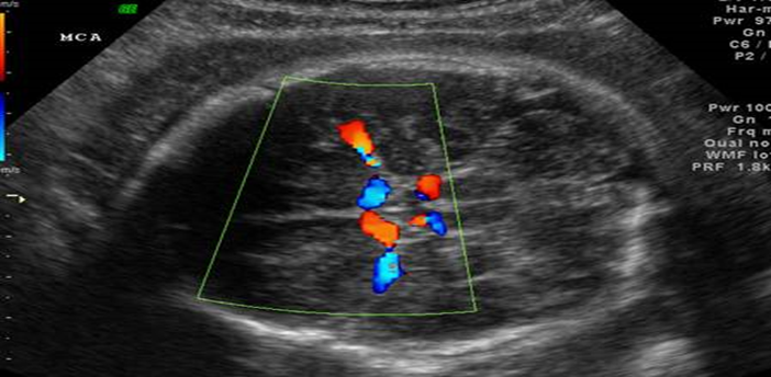
FIGURE 1: MIDDLE CEREBRAL ARTERY.
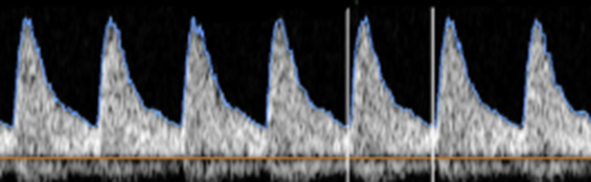
FIGURE IB: NORMAL DOPPLER WAVEFORM OF MIDDLE CEREBRAL ARTERY

FIGURE IC:ABSENT DIASTOLIC FLOW IN MIDDLE CEREBRAL ARTERY DOPPLER
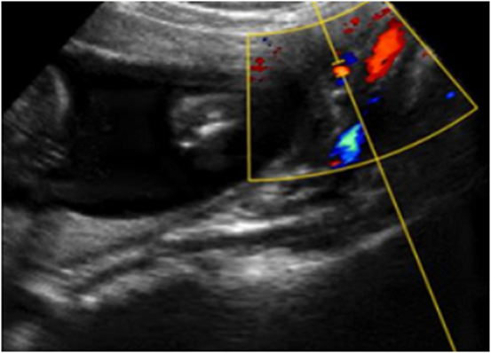
FIGURE II : UTERINE ARTERY
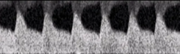
FIGURE IIA: Colour Doppler image of the right uterine artery.
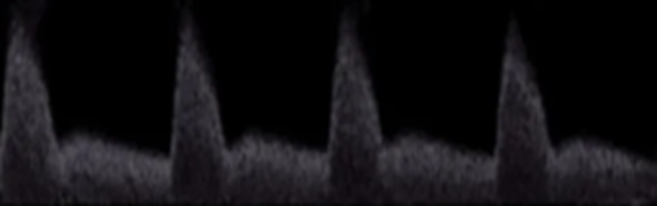
FIGURE IIB: NORMAL UTERINE ARTERY WAVEFORM
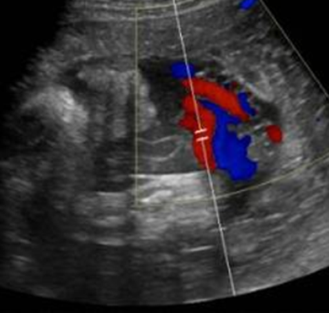
FIGURE IIC: ABNORMAL UTERINE ARTERY SPECTRAL WAVEFORM INDICATING THE PRESENCE OF A DIASTOLIC NOTCH.
FIGURE III : UMBILICAL ARTERY
FIGURE III A : Colour Doppler image of the fetal umbilical artery
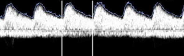
FIGURE III B : NORMAL UMBILICAL ARTERY DOPPLER
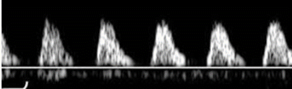
FIGURE III C : UMBILICAL ARTERY DOPPLER WITH ABSENT DIASTOLIC FLOW
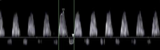
FIGURE III D: UMBILICAL ARTERY DOPPLER WITH REVERSE END DIASTOLIC FLOW
FIGURE IV : DUCTUS VENOSUS
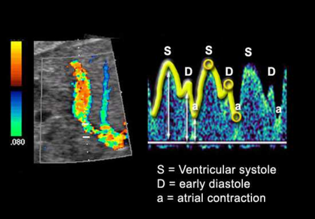
FIGURE IV A : Colour Doppler image and waveform of ductus venosus.
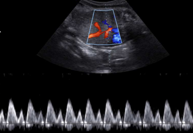
FIGURE IV B :NORMAL DOPPLER WAVEFORM OF DUCTUS VENOSUS.
CONCLUSION:
Antenatal multivessel Doppler ultrasonography is a valuable, non-invasive imaging modality for the assessment of fetal well-being in high-risk pregnancies. Simultaneous evaluation of the uterine artery, umbilical artery, middle cerebral artery, ductus venosus, and umbilical vein provides comprehensive information regarding placental function, fetal circulatory adaptation, and the progression of fetal compromise.
Among the Doppler parameters evaluated, the cerebroplacental ratio (CPR) demonstrated the highest diagnostic accuracy for predicting adverse perinatal outcomes, followed by abnormalities in the umbilical artery and middle cerebral artery Doppler indices. Abnormal multivessel Doppler findings were strongly associated with increased rates of low birth weight, NICU admission, low Apgar scores, preterm birth, stillbirth, and neonatal death.
The findings of this study support the incorporation of multivessel Doppler assessment as a routine component of antenatal surveillance in high-risk pregnancies, as it facilitates early identification of fetal compromise, enables timely obstetric intervention, and has the potential to improve perinatal outcomes while reducing neonatal morbidity and mortality.
Multivessel Doppler ultrasonography, including assessment of the uterine artery, umbilical artery, middle cerebral artery, ductus venosus, and umbilical vein, should be incorporated into routine antenatal surveillance of high-risk pregnancies for early detection of fetal compromise. Particular emphasis should be given to the cerebroplacental ratio (MCA/UA PI), which demonstrated the highest predictive accuracy for adverse perinatal outcomes in this study and may be used as a key screening parameter in clinical practice. Pregnancies showing abnormal Doppler findings should undergo closer monitoring with timely obstetric intervention to reduce perinatal morbidity and mortality. Standardized Doppler measurement protocols should be adopted to minimize inter-observer variability and improve diagnostic reliability. Further large-scale multicentric studies with long-term neonatal follow-up are recommended to validate these findings and strengthen the clinical utility of multivessel Doppler assessment in high-risk pregnancies.
REFERENCES:
1. Fong KW, Ohlsson A, Hannah ME, Grisaru S, Kingdom J, Cohen H, et al. Prediction of perinatal outcome in fetuses suspected to have intrauterine growth restriction: Doppler US study of fetal cerebral, renal, and umbilical arteries. Radiology. 1999;213(3):681-689.
2. Arduini D, Rizzo G. Normal values of pulsatility index from fetal vessels: a cross-sectional study on 1556 healthy fetuses. Ultrasound Med Biol. 1990;16(7):637-643.
3. Acharya G, Wilsgaard T, Berntsen GKR, Maltau JM, Kiserud T. Reference ranges for serial measurements of umbilical artery Doppler indices in normal pregnancy. Am J Obstet Gynecol. 2005;192(3):937-944.
4. Kiserud T, Eik-Nes SH, Blaas HGK, Hellevik LR. Ductus venosus blood velocity and the umbilical circulation in the severely growth-restricted fetus. Ultrasound Obstet Gynecol. 1994;4(2):109-114.
5. Ferrazzi E, Bozzo M, Rigano S, Bellotti M, Galan H, Battaglia FC, et al. Temporal sequence of abnormal Doppler changes in the peripheral and central circulatory systems of severely growth-restricted fetuses. Ultrasound Obstet Gynecol. 2002;19(2):140-146.
6. Hecher K, Bilardo CM, Stigter RH, Ville Y, Hackelöer BJ, Kok HJ, et al. Monitoring of fetuses with intrauterine growth restriction using arterial and venous Doppler velocimetry. Ultrasound Obstet Gynecol. 2001;18(6):564-570.
7. Bilardo CM, Wolf H, Stigter RH, Ville Y, Baez E, Visser GHA, et al. Relationship between Doppler monitoring parameters and perinatal outcome in severe fetal growth restriction. Ultrasound Obstet Gynecol. 2004;23(2):119-125.
8. Baschat AA, Gembruch U. The cerebroplacental Doppler ratio revisited. Ultrasound Obstet Gynecol. 2003;21(2):124-127.
9. Gramellini D, Folli MC, Raboni S, Vadora E, Merialdi A. Cerebral-umbilical Doppler ratio as a predictor of adverse perinatal outcome. Obstet Gynecol. 1992;79(3):416-420.
10. Mari G, Deter RL. Middle cerebral artery flow velocity waveforms in normal and growth-restricted fetuses. Am J Obstet Gynecol. 1992;166(4):1262-1270.
11. Harrington K, Carpenter RG, Nguyen M, Campbell S. Changes observed in Doppler studies of the maternal circulation in pregnancies complicated by pre-eclampsia and fetal growth restriction. Br J Obstet Gynaecol. 1995;102(10):789-796.
12. Kiserud T. Physiology of the fetal circulation. Semin Fetal Neonatal Med. 2005;10(6):493-503.
13. Maulik D. Doppler ultrasound in obstetrics: from waveform analysis to hemodynamic assessment. J Ultrasound Med. 2005;24(5):643-656.
14. Pretorius DH, Nelson TR. Fetal Doppler imaging: principles and clinical applications. J Ultrasound Med. 1995;14(8):575-586.
15. Abu-Yousef MM, Bleicher JJ, Williamson RA, Weiner CP. Uterine artery Doppler waveform analysis in normal and high-risk pregnancies. AJR Am J Roentgenol. 1987;149(5):1047-1050.
16. Taylor KJW, Burns PN, Wells PNT. Clinical Applications of Doppler Ultrasound. 2nd ed. New York: Raven Press; 1995.
17. Rumack CM, Wilson SR, Charboneau JW, Levine D. Diagnostic Ultrasound. 5th ed. Philadelphia: Elsevier; 2018.
18. Nyberg DA, McGahan JP, Pretorius DH, Pilu G. Diagnostic Imaging: Ultrasound. 3rd ed. Philadelphia: Elsevier; 2014.
19. Ali S, Heuving S, Kawooya MG, Byamugisha J, Grobbee DE, Papageorghiou AT, et al. Prognostic accuracy of antenatal Doppler ultrasound for adverse perinatal outcomes: a systematic review. BMJ Open. 2021;11:e055122.
20. Vollgraff Heidweiller-Schreurs CA, De Boer MA, Heymans MW, Schoonmade LJ, Bossuyt PM, Mol BW, et al. Prognostic accuracy of cerebroplacental ratio and middle cerebral artery Doppler for adverse perinatal outcome: systematic review and meta-analysis. Ultrasound Obstet Gynecol. 2018;51(3):313-322.