CROSS SECTIONAL STUDY ON THE MORPHOMETRY OF ACETABULUM IN HIP BONE.
- Girija Kumari K , Professor, Department of Anatomy, Sree Mookambika Institute of Medical Sciences Kulasekharam, Tamil Nadu, India.
- Rathisha S , Postgraduate, Department of Anatomy, Sree Mookambika Institute of Medical Sciences Kulasekharam, Tamil Nadu, India.
Article Information:
Abstract:
Background: The acetabulum is a deep hemispherical cavity of the hip bone forming the socket of the hip joint. Its morphometric evaluation is important for orthopedic procedures, prosthesis design, and forensic identification. Variations in its dimensions influence joint stability and surgical outcomes. Understanding its anatomical parameters helps in planning reconstructive surgeries and improving implant fitting accuracy in hip-related interventions. Aims: To study the morphometric parameters of the acetabulum of dry hip bones. Materials and Methods: This cross-sectional study was conducted on 55 dry adult human hip bones obtained from the department of Anatomy over a period of 10 months. Acetabular morphometric measurements were recorded using digital vernier calipers with an accuracy of 0.01 mm. Parameters assessed included acetabular diameter, depth, rim diameter, and acetabular notch width. Each measurement was taken twice by two independent observers and the mean value was recorded to reduce inter-observer error. Data were tabulated and statistically analyzed using descriptive statistics. Mean, standard deviation, and range were calculated. Comparison between right and left hip bones was performed where applicable. Ethical clearance was obtained prior to commencement of the study ensuring standardization and reproducibility of measurements throughout study period conducted. Results: The mean acetabular diameter was found to be 52.6 ± 3.4 mm, while the mean depth was 28.2 ± 2.1 mm. The rim diameter showed a mean value of 54.1 ± 3.2 mm. The acetabular notch width demonstrated a mean of 19.5 ± 1.8 mm. Slight variations were observed between right and left hip bones, though differences were not statistically significant (p > 0.05). Male bones demonstrated marginally higher morphometric values compared to presumed female bones based on morphological features. Strong correlation was observed between acetabular diameter and depth. Conclusion: The acetabulum shows measurable morphometric variation with clinical and forensic significance, supporting its role in implant design and orthopedic surgical planning applications in practice.
Keywords:
Article :
INTRODUCTION:
The hip bone is a crucial component of the pelvic girdle that plays a significant role in weight transmission, locomotion, and stability of the human body.1 One of its most important structural components is the acetabulum, a deep, cup-shaped cavity located on the lateral aspect of the hip bone.2 It articulates with the head of the femur to form the hip joint, which is a ball-and-socket type synovial joint. This joint allows a wide range of movements, including flexion, extension, abduction, adduction, circumduction, and rotation, thereby contributing to functional mobility.3
The acetabulum is formed by the fusion of three bones: ilium, ischium, and pubis. Its morphology varies with age, sex, ethnicity, and functional demands. The structural integrity and precise anatomical configuration of the acetabulum are essential for maintaining hip joint stability and biomechanics.4 Any alteration in its shape or dimensions may predispose individuals to degenerative joint diseases, dislocations, or impaired mobility. Therefore, accurate morphometric analysis of the acetabulum is of paramount importance in both anatomical and clinical contexts.5
In orthopedic practice, acetabular morphometry is especially relevant in procedures such as total hip arthroplasty, fracture fixation, and reconstructive surgeries. Proper knowledge of acetabular dimensions aids in selecting appropriate prosthetic components, ensuring optimal fit, alignment, and longevity of implants. Inadequate sizing may lead to complications such as implant loosening, dislocation, and abnormal wear patterns.6 Similarly, in forensic science, acetabular measurements contribute to sex estimation and identification of skeletal remains, as pelvic bones exhibit significant sexual dimorphism.7
Despite its clinical importance, variations in acetabular dimensions across different populations remain underreported, particularly in regional anatomical studies. Morphometric data derived from specific populations are essential for developing population-specific prosthetic designs and improving surgical outcomes. Global implant designs are often based on Western anatomical data, which may not accurately represent the anatomical variations seen in other populations. This highlights the need for localized morphometric studies.8
The acetabulum also demonstrates variability influenced by biomechanical stress and genetic factors. Weight-bearing patterns and locomotor activity can affect its depth, diameter, and orientation.9 Understanding these variations provides insights into functional anatomy and evolutionary adaptations of the human pelvis. Additionally, precise anatomical data are useful in anthropological research for reconstructing population characteristics and evolutionary trends.
The present study focuses on the detailed morphometric analysis of the acetabulum in dry human hip bones. Parameters such as acetabular diameter, depth, rim diameter, and acetabular notch width are measured to establish baseline anatomical data. These parameters are essential for understanding structural variability and its implications in clinical practice. The findings are expected to contribute to anatomical databases and assist orthopedic surgeons, prosthetists, and forensic experts in their respective fields.
AIMS AND OBJECTIVES
• To study the morphometric parameters of the acetabulum of dry hip bones.
MATERIALS AND METHODS:
The present cross-sectional study was conducted in the Department of Anatomy at Sree Mookambika Institute of Medical Sciences over a period of 10 months from July 2025 to April 2026. A total of 55 dry adult human hip bones were collected and included for morphometric analysis of the acetabulum.
The inclusion criteria comprised well-preserved, fully ossified adult human hip bones with clearly visible acetabular margins and intact anatomical landmarks. Only bones that were complete and suitable for accurate morphometric assessment were included in the study. Hip bones of unknown sex were also included provided they were free from deformities and suitable for measurement. The acetabular region had to be structurally intact to allow proper identification of landmarks for measurement of diameter, depth, rim diameter, and acetabular notch width.
The exclusion criteria included damaged, broken, eroded, or incomplete hip bones where acetabular landmarks were not clearly identifiable. Bones showing pathological changes, congenital deformities, or post-mortem damage affecting the acetabulum were excluded. Juvenile or partially ossified bones were also excluded to maintain uniformity and ensure that only adult morphometric data were analyzed.
All measurements were carried out in the anatomy laboratory under standardized conditions using a digital vernier caliper with an accuracy of 0.01 mm to ensure precision. To minimize observer bias and improve reliability, each measurement was taken twice independently by two observers, and the mean value was recorded for final analysis.
Morphometric parameters assessed included acetabular diameter (Fig 1), defined as the maximum transverse distance across the acetabular cavity; acetabular depth (Fig 2), measured as the maximum vertical distance from the rim to the deepest point of the socket; rim diameter; and acetabular notch width (Fig 3). All measurements were carefully recorded in a structured data collection sheet immediately after observation to avoid transcription errors.
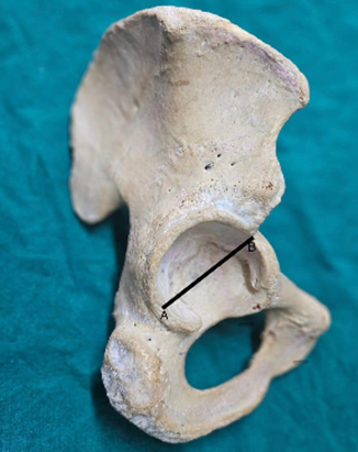
Fig 1: Acetabulum diameter
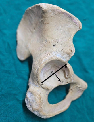
Fig 2: Acetabulum depth
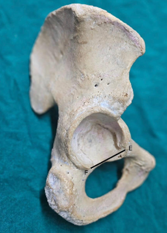
Fig 3: Acetabulum notch width
Statistical analysis was performed using standard statistical software. Data were expressed as mean, standard deviation, minimum, and maximum values. Comparison between right and left hip bones was done where applicable using appropriate tests. Correlation between different acetabular parameters was assessed using Pearson’s correlation coefficient. A p-value of less than 0.05 was considered statistically significant. The results were interpreted in relation to anatomical variability and clinical relevance in orthopedic and forensic applications.
RESULTS:
A total of 55 dry adult human hip bones were studied for morphometric analysis of the acetabulum. All parameters were measured using a digital vernier caliper and expressed as mean ± standard deviation. The rim diameter was slightly higher than acetabular diameter, suggesting marginal bony overhang at the acetabular margin. Depth showed relatively low variability, indicating stable structural anatomy across specimens. (Table 1)
Table 1: Overall acetabular morphometric parameters
|
Parameter |
Mean ± SD (mm) |
Minimum |
Maximum |
|
Acetabular diameter |
52.6 ± 3.4 |
46.2 |
60.8 |
|
Acetabular depth |
28.2 ± 2.1 |
24.0 |
33.5 |
|
Rim diameter |
54.1 ± 3.2 |
47.5 |
62.0 |
|
Acetabular notch width |
19.5 ± 1.8 |
16.0 |
23.2 |
No statistically significant differences were observed between right and left acetabular measurements for all parameters (p > 0.05). This indicates symmetrical development of the acetabulum on both sides of the pelvis. The slight variations in mean values are within normal anatomical limits and do not suggest any structural asymmetry. (Table 2).
Table 2: Side-wise Comparison of Acetabular Morphometric Parameters
|
Parameter |
Category / Side |
n (%) |
Mean ± SD (mm) |
p-value |
|
Acetabular diameter |
Right |
28 (50.9%) |
53.1 ± 3.2 |
0.42 |
|
Left |
27 (49.1%) |
52.0 ± 3.5 |
||
|
Acetabular depth |
Right |
28 (50.9%) |
28.5 ± 2.0 |
0.38 |
|
Left |
27 (49.1%) |
27.9 ± 2.2 |
||
|
Acetabular notch width |
Right |
28 (50.9%) |
19.7 ± 1.7 |
0.44 |
|
Left |
27 (49.1%) |
19.3 ± 1.9 |
||
|
Rim diameter |
Right |
28 (50.9%) |
54.4 ± 3.1 |
0.36 |
|
Left |
27 (49.1%) |
53.8 ± 3.3 |
The derived indices demonstrate proportional relationships between acetabular dimensions, indicating balanced structural geometry. The depth-to-diameter ratio suggests adequate coverage of the femoral head, reflecting functional stability of the hip joint. (Table 3).
Table 3: Derived Acetabular Indices
|
Index |
Mean ± SD |
|
Depth-to-diameter ratio |
0.54 ± 0.03 |
|
Notch index (%) |
36.9 ± 2.8 |
|
Rim-to-diameter ratio |
1.03 ± 0.02 |
Most acetabula (50.9%) fall within the 50–55 mm range, indicating a predominant mid-sized morphological pattern. More than half of the specimens (54.5%) exhibit moderate acetabular depth (26–30 mm), indicating a stable and consistent anatomical configuration. (Table 4).
Table 4: Distribution of Acetabular Diameter and depth
|
Parameter |
N(%) |
|
|
Acetabular diameter distribution |
<50 mm |
14 (25.5%) |
|
50–55 mm |
28 (50.9%) |
|
|
>55 mm |
13 (23.6%) |
|
|
Acetabular depth distribution |
<26 mm |
12 (21.8%) |
|
26–30 mm |
30 (54.5%) |
|
|
>30 mm |
13 (23.6%) |
|
DISCUSSION:
A total of 55 dry adult human hip bones were analyzed for morphometric evaluation of the acetabulum. The present study demonstrated that the mean acetabular diameter was 52.6 ± 3.4 mm, acetabular depth was 28.2 ± 2.1 mm, rim diameter was 54.1 ± 3.2 mm, and acetabular notch width was 19.5 ± 1.8 mm. The findings showed relatively uniform acetabular morphology with no significant side-wise differences. Similar absence of lateral variation was reported by Kumar VS et al.10 who observed no statistically significant differences between right and left acetabular parameters, and by Akhlaghi M et al.11 who also noted comparable morphometric symmetry between sides, reinforcing the bilateral uniformity observed in the present study.
Side-wise analysis in the present study revealed no significant differences between right and left hip bones across all parameters (p > 0.05). A similar pattern was observed by Tripathi M et al.12 who reported no statistically significant side differences in acetabular dimensions, although gender-based differences were noted. Likewise, Arunkumar KR et al.13 also found slightly higher left-sided values that were not statistically significant, supporting the present study’s observation of symmetrical pelvic architecture.
The present study identified a predominance of mid-range acetabular diameter (50–55 mm in 50.9%) and moderate acetabular depth (26–30 mm in 54.5%), indicating a relatively uniform distribution of acetabular morphology. Comparable morphometric averages were reported by Indurjeeth K et al.14 who documented an acetabular diameter of 54.84 ± 4.18 mm and depth of 31.30 ± 3.18 mm, values closely aligning with the present findings, suggesting consistency across populations despite minor variations in mean values.
Morphological variability of the acetabulum, particularly in relation to shape and structural configuration, has been widely documented. In the present study, derived indices such as depth-to-diameter ratio (0.54 ± 0.03) and rim-to-diameter ratio (1.03 ± 0.02) indicated proportional structural relationships. Similar morphometric consistency was observed by Singh A et al.15 who reported comparable acetabular dimensions and highlighted variability in acetabular ridge morphology. Additionally, Ulkir M et al.16 identified multiple acetabular ridge shapes (straight, curved, angular, and irregular) and reported significant associations between shape and depth, emphasizing structural variability within otherwise symmetrical acetabula.
The present study also demonstrated that most specimens clustered within mid-range morphometric categories, reflecting a stable anatomical pattern. Similar distribution trends were reported by Narasipuram A et al.17 who observed curved and straight acetabular shapes as most common, along with moderate mean diameters. Taşci M et al.18 further reported that curved anterior acetabular ridges were predominant, followed by angular and irregular types, supporting the concept of dominant morphological patterns with minor variations.
Correlation between acetabular parameters has been highlighted in previous literature. In the present study, proportional indices suggested coordinated structural relationships between diameter, depth, and rim dimensions. Similarly, Arunkumar KR et al.13 reported a significant positive correlation between acetabular diameter and depth (r = 0.388, p < 0.001), supporting the interdependence of acetabular morphometric parameters. This indicates that larger acetabula tend to be deeper, reflecting proportional developmental anatomy.
Gender and functional implications of acetabular morphology have also been explored in literature. Tripathi M et al.12 reported significant sexual dimorphism, with males showing larger and deeper acetabula compared to females, while notch width was greater in females. Although gender analysis was not included in the present study, their findings highlight the forensic importance of acetabular morphometry.
Advanced morphometric modeling approaches have also been described. Acikoz AK et al.19 developed regression models for estimating acetabular measurements from hip bone parameters, demonstrating low standard error of estimate values, suggesting strong predictive potential. Such approaches emphasize the clinical relevance of precise acetabular measurements in reconstructive and prosthetic applications.
Morphological variation of acetabular margins has been widely reported. Singh A et al.15 documented curved anterior acetabular ridges in 45.7% of specimens, while Uzun GB et al.20 and Tasci M et al.18 also described curved and angular patterns as most common. These findings collectively support the variability of acetabular morphology despite overall structural consistency.
CONCLUSION:
The study provides detailed morphometric data of the acetabulum in dry adult human hip bones. The findings demonstrate that acetabular diameter, depth, rim diameter, and notch width exhibit relatively consistent anatomical patterns with no significant side-wise differences. The derived indices indicate proportional structural relationships, reflecting balanced acetabular morphology. The majority of measurements fall within mid-range values, suggesting uniform pelvic architecture in the studied population. These results offer valuable baseline data for orthopedic procedures, particularly total hip arthroplasty, implant sizing, and reconstructive surgeries, as well as forensic applications in skeletal identification and sex estimation in clinical and anatomical practice settings.
REFERENCES:
1. Rybski MF. The Hip and Pelvis. InKinesiology for Occupational Therapy 2024 Jun 1 (pp. 291-303). Routledge.
2. Hazari A, Maiya AG, Nagda TV. Kinetics and kinematics of hip and pelvis. InConceptual Biomechanics and Kinesiology 2021 Nov 2 (pp. 125-144). Singapore: Springer Singapore.
3. Fisher MA, Bordoni B. Anatomy, Bony Pelvis and Lower Limb: Pelvic Joints. InStatPearls [Internet] 2023 Mar 6. StatPearls Publishing.
4. PELVIS B. Sacrum, Coccyx, and Hip Bones (Innominate Bones). Sectional Anatomy for Imaging Professionals-E-Book. 2025 Nov 28:409.
5. Prescher A, Drescher WR. Surgical anatomy of the hip. InAdvances in Specialist Hip Surgery 2021 Jan 30 (pp. 3-29). Cham: Springer International Publishing.
6. Thorp LE. Hip anatomy. Hip arthroscopy and hip joint preservation surgery. 2022 Aug 2:3-15.
7. Zhang M. The application of forensic imaging to sex estimation: focus on skull and pelvic structures. Perspectives in Legal and Forensic Sciences. 2024 May;1(1):10005.
8. Johnson LG, Bortolussi-Courval S, Chehil A, Schaeffer EK, Pawliuk C, Wilson DR et al. Application of statistical shape modeling to the human hip joint: a scoping review. JBI Evidence Synthesis. 2023 Mar 1;21(3):533-83.
9. Ferrante H, Schemitsch EH, Zdero R, Bagheri ZS. Biomechanical analysis of fixation methods for acetabular fractures: A review. Medical Engineering & Physics. 2021 Mar 1;89(1):51-62.
10. Kumar VS, Laxmi V, Kumar M, Rani S, Madhukar PK, Bharti JP. Morphometric Study of the Acetabulum and Its Clinical Correlation in Total Hip Arthroplasty. European Journal of Cardiovascular Medicine. 2023 Jul 1;13(3).
11. Akhlaghi M, Azizian A, Sadeghian MH, Forghani T, Tavakoli SA, Rafiee S et al. Comparing the Accuracy of Morphometric and Morphological Criteria of Hip Bone in Gender Determination. International Journal of Medical Toxicology and Forensic Medicine. 2019 Apr 1;9(2):57-64.
12. Tripathi M, Gajbhiye V. Morphometric and morphology study of acetabulum of human hip bone in central Indian population. International Journal of Health Sciences. 2022(I):1064-72.
13. Arunkumar KR, Delhiraj U, Kumar SS. Morphologic and Morphometric Study of Human Acetabulum and its Clinical Significance. Journal of Clinical & Diagnostic Research. 2021 Feb 1;15(2).
14. Indurjeeth K, Ishwarkumar S, De Gama BZ, Ndlazi Z, Pillay P, Indurjeeth K et al. Morphometry and morphology of the acetabulum within the black African population of South Africa. Int j morphol. 2019 Sep 1;37(3):971-6.
15. Ülkir M, Ilgaz HB. Morphologic and morphometric evaluation of the acetabulum. Online Turkish Journal of Health Sciences. 2023 Jun 6;8(2):155-60.
16. Narasipuram A, sheikh Vajir J. Morphometric study of the Acetabulum in Human hip bones: An anatomical analysis. Cuestiones de Fisioterapia. 2025 Jan 10;54(2):3409-14.
17. Taşci M, Pirinç B, Sevindik B, Doğan NÜ. Evaluation of the morphological and morphometric characteristics of the acetabulum in the Anatolian population. Anatomy. 2023 Apr 4;17(1):34-40.
18. Acikoz AK, Bozkır MG. The Importance of Morphometric Measurements of Adult Human Dry Hip Bone in Acetabular Reconstruction. Journal of Contemporary Medicine. 2022 Sep 9;12(5):685-91.
19. Singh A, Gupta R, Singh A. Morphological and morphometric study of the acetabulum of dry human hip bone and its clinical implication in hip arthroplasty. Journal of the Anatomical Society of India. 2020 Oct 1;69(4):220-5.
20. Uzun GB, Değermenci M, Uçar İ, Arslan A, Nisari M. Morphometrıc evaluatıon of acetabulum. Journal of Surgery and Medicine. 2020 Jul 1;4(7):555-7.