Evaluation of Tumor Markers and Histopathological Patterns in Ovarian Epithelial Tumors.
- Anuradha Devi B V , Assistant Professor, Department of Pathology, Government Medical College- Sangareddy, Sangareddy, Telangana, India-502001.
- Sugunadhar Sakki , Assistant Professor, Department of Pathology, Government Medical College- Mancherial, Mancherial, Telangana, India-504207.
- Sravanthi Pandiri , Assistant Professor, Department of Biochemistry, Government Medical College- Mancherial, Mancherial, Telangana, India-504207.
- Jostna Devi Akarapu , Assistant Professor, Department of Pathology, Government Medical College- Mancherial, Mancherial, Telangana, India-504207.
- Ramesh Kandimalla , Associate Professor, Department of Biochemistry, Kakatiya Medical College, Hanumakonda, Telangana, India-506007.
Article Information:
Abstract:
Background: Ovarian epithelial tumors show a wide morphological spectrum ranging from benign cystadenomas to borderline tumors and invasive carcinomas. Serum tumor markers are often requested before surgery, but their interpretation is influenced by histological type, tumor behavior, menopausal status and tumor burden. Correlating tumor marker levels with histopathological diagnosis can improve clinical interpretation and support better preoperative risk assessment. The current study is designed to evaluate the distribution of histopathological patterns in ovarian epithelial tumors and to study the association of serum CA-125, HE4, CA19-9 and CEA with benign, borderline and malignant epithelial ovarian tumors. Materials and Methods: A total of 120 surgically resected ovarian epithelial tumors were included. Clinical details, menopausal status, laterality, tumor size, serum tumor marker levels and histopathological diagnosis were recorded. Tumors were classified as benign, borderline or malignant according to standard histopathological criteria. CA-125, HE4, CA19-9 and CEA levels were compared across diagnostic categories and histological subtypes. Categorical variables were analysed using the chi-square test, while continuous variables were compared using one-way ANOVA or Kruskal-Wallis test, depending on data distribution. A p-value <0.05 was considered statistically significant. Results: Among 120 epithelial ovarian tumors, 74 cases (61.7%) were benign, 12 cases (10.0%) were borderline and 34 cases (28.3%) were malignant. Serous tumors were the most frequent histological group, accounting for 66 cases (55.0%), followed by mucinous tumors in 35 cases (29.2%). High-grade serous carcinoma was the commonest malignant tumor, observed in 18 of 34 malignant cases (52.9%). Marker positivity increased significantly from benign to malignant tumors. CA-125 was elevated in 19 benign tumors (25.7%), 8 borderline tumors (66.7%) and 30 malignant tumors (88.2%) (χ²=39.42, p<0.001). HE4 positivity was seen in 7 benign tumors (9.5%), 5 borderline tumors (41.7%) and 26 malignant tumors (76.5%) (χ²=49.68, p<0.001). CA19-9 and CEA were more frequently elevated in mucinous tumors than non-mucinous tumors. Combined CA-125 and HE4 positivity showed stronger association with malignant disease than either marker alone. Conclusion: Ovarian epithelial tumors in the present study were predominantly benign, with serous tumors forming the largest histological group. CA-125 and HE4 showed significant elevation in malignant tumors, especially serous carcinomas, while CA19-9 and CEA were more often raised in mucinous tumors. Tumor markers should be interpreted along with histopathological type, tumor size, menopausal status and clinical findings rather than as isolated diagnostic tests.
Keywords:
Article :
INTRODUCTION:
Ovarian epithelial tumors constitute the largest group of ovarian neoplasms encountered in routine surgical pathology. Their clinical importance lies in their broad biological range. Many lesions are benign and are cured by surgery, while borderline tumors and invasive carcinomas require careful staging, prognostic assessment and long-term follow-up. The ovary may show overlapping clinical and radiological appearances across benign, borderline and malignant tumors; therefore, histopathological examination remains the most reliable method for final diagnosis.
Epithelial ovarian tumors include serous, mucinous, endometrioid, clear cell, Brenner and seromucinous tumors. Among malignant tumors, high-grade serous carcinoma is clinically important because it often presents at an advanced stage and accounts for a large proportion of ovarian cancer-related mortality. Low-grade serous carcinoma, mucinous carcinoma, endometrioid carcinoma and clear cell carcinoma differ in morphology, molecular background and clinical behavior. Hence, classifying ovarian epithelial tumors by histological subtype is not only a diagnostic exercise but also has therapeutic and prognostic value.
Serum tumor markers are widely used in the evaluation of adnexal masses. CA-125 is the most commonly used marker in epithelial ovarian cancer, particularly in serous carcinoma. However, CA-125 may also be elevated in benign gynecological and non-gynecological conditions, including endometriosis, pelvic inflammatory disease, pregnancy, liver disease and peritoneal inflammation. HE4 has emerged as a useful marker with better specificity in several studies and is often interpreted along with CA-125. CA19-9 and CEA are less specific for ovarian tumors, but they are frequently useful in the assessment of mucinous ovarian tumors and in differentiating primary ovarian mucinous tumors from possible metastatic gastrointestinal lesions.
In clinical practice, tumor markers are often used before surgery to support risk stratification. However, marker elevation alone cannot replace tissue diagnosis. A benign mucinous cystadenoma may show raised CA19-9, while early malignant tumors may have only modest marker elevation. Similarly, CA-125 may be elevated in benign serous or inflammatory conditions. This creates a need for studies that correlate preoperative tumor markers with histopathological diagnosis and tumor subtype in local patient populations.
The present study was undertaken at Kakatiya Medical College, Hanumakonda, to evaluate the histopathological spectrum of ovarian epithelial tumors and to study the relationship between tumor marker levels and pathological diagnosis. The study aimed to assess how CA-125, HE4, CA19-9 and CEA vary across benign, borderline and malignant ovarian epithelial tumors and whether these markers show subtype-specific patterns.
MATERIALS AND METHODS:
Study Design
This was a hospital-based observational analytical study carried out in the Department of Pathology, Kakatiya Medical College, Hanumakonda. The study evaluated surgically resected ovarian epithelial tumors and correlated histopathological diagnosis with preoperative serum tumor marker levels. The study was designed to observe naturally occurring clinicopathological patterns without assigning any intervention to the patients.
Study Duration
The study was conducted over a period of one year, from July 2025 to June 2026. All eligible ovarian epithelial tumors received during this period were included after applying the selection criteria.
Study Population
The study population included patients who underwent surgery for ovarian masses and whose specimens were submitted for histopathological examination. Only tumors confirmed histologically as epithelial ovarian tumors were included. The study included benign, borderline and malignant epithelial tumors. Clinical information, imaging findings and tumor marker values were collected from hospital records and laboratory reports.
Sample Size
A total of 120 ovarian epithelial tumors were included in the study. This sample consisted of consecutive eligible cases received during the study period.
Inclusion Criteria
Patients of all age groups who underwent oophorectomy, salpingo-oophorectomy, cystectomy, hysterectomy with bilateral salpingo-oophorectomy or staging laparotomy for ovarian mass were included when histopathology confirmed an epithelial ovarian tumor. Cases with available preoperative serum tumor marker values were included for marker analysis.
Exclusion Criteria
Non-epithelial ovarian tumors, including germ cell tumors, sex cord-stromal tumors and metastatic non-epithelial lesions, were excluded. Ovarian lesions without adequate tissue for diagnosis were also excluded. Cases with incomplete clinical records or unavailable preoperative tumor marker results were not included in the marker correlation analysis. Patients who had received neoadjuvant chemotherapy before surgery were excluded to avoid treatment-related alteration in tumor morphology and marker levels.
Clinical Data Collection
Clinical data included age, presenting complaints, menopausal status, laterality, radiological impression and operative findings. Patients were grouped as premenopausal or postmenopausal based on menstrual history. Tumor size was recorded from gross examination and cross-checked with imaging findings wherever available.
Tumor Marker Assessment
Preoperative serum CA-125, HE4, CA19-9 and CEA levels were recorded from laboratory reports. Marker positivity was defined using the following reference cut-offs: CA-125 >35 U/mL, CA19-9 >37 U/mL and CEA >5 ng/mL. HE4 was interpreted according to menopausal status, with values >70 pmol/L in premenopausal women and >140 pmol/L in postmenopausal women considered elevated. Marker values were analysed both as continuous variables and as positive or negative categories.
Histopathological Examination
All surgical specimens were fixed in 10% neutral buffered formalin. Gross examination included tumor size, laterality, external surface, capsular breach, cystic or solid areas, papillary projections, necrosis, hemorrhage and surface deposits. Representative sections were taken from solid areas, papillary areas, cyst wall, septae, necrotic zones and areas suspicious for invasion. Hematoxylin and eosin-stained sections were examined under light microscopy. Tumors were classified as benign, borderline or malignant and further categorized into serous, mucinous, endometrioid, clear cell, Brenner and seromucinous types.
Statistical Analysis
Data were entered into Microsoft Excel and analysed using standard statistical software. Continuous variables were expressed as mean ± standard deviation or median with interquartile range, depending on distribution. Categorical variables were expressed as number and percentage. The chi-square test was used to assess associations between categorical variables. One-way ANOVA was used for normally distributed continuous data, while the Kruskal-Wallis test was applied for skewed marker values. A p-value <0.05 was considered statistically significant.
Ethical Considerations
The study was conducted after approval from the Institutional Ethics Committee of Kakatiya Medical College, Hanumakonda. Patient confidentiality was maintained throughout the study. Data were used only for academic and research purposes. As the study was observational and based on routinely collected clinical, laboratory and histopathological information, no additional intervention was performed for research purposes.
RESULTS:
A total of 120 epithelial ovarian tumors were evaluated during the study period. The age of patients ranged from 18 to 76 years, with a mean age of 44.8 ± 13.6 years. Most benign tumors occurred in younger and middle-aged women, while malignant tumors were more common in the postmenopausal age group. The mean age was 39.2 ± 11.4 years in benign tumors, 45.7 ± 10.8 years in borderline tumors and 56.3 ± 9.7 years in malignant tumors. The difference in age distribution across the three diagnostic groups was statistically significant (F=31.86, p<0.001) (Table 1).
Postmenopausal status was more frequent among patients with malignant tumors. Among 34 malignant tumors, 25 cases (73.5%) occurred in postmenopausal women, whereas only 21 of 74 benign tumors (28.4%) were seen in postmenopausal women. This association between menopausal status and tumor category was statistically significant (χ²=22.84, p<0.001) (Table 1; Figure 1).
Table 1: Baseline clinicopathological characteristics of epithelial ovarian tumors
|
Variable |
Benign n=74 |
Borderline n=12 |
Malignant n=34 |
Test statistic |
p-value |
|
Age, years, mean ± SD |
39.2 ± 11.4 |
45.7 ± 10.8 |
56.3 ± 9.7 |
F=31.86 |
<0.001 |
|
Premenopausal, n (%) |
53 (71.6) |
7 (58.3) |
9 (26.5) |
χ²=22.84 |
<0.001 |
|
Postmenopausal, n (%) |
21 (28.4) |
5 (41.7) |
25 (73.5) |
||
|
Mean tumor size, cm ± SD |
8.4 ± 3.1 |
11.6 ± 3.8 |
12.8 ± 4.5 |
F=18.42 |
<0.001 |
|
Bilateral tumors, n (%) |
8 (10.8) |
2 (16.7) |
11 (32.4) |
χ²=7.78 |
0.020 |
|
Solid-cystic gross pattern, n (%) |
14 (18.9) |
5 (41.7) |
26 (76.5) |
χ²=35.91 |
<0.001 |
|
Papillary excrescences, n (%) |
10 (13.5) |
6 (50.0) |
23 (67.6) |
χ²=35.28 |
<0.001 |
Data are expressed as mean ± SD or n (%). One-way ANOVA was used for continuous variables. Chi-square test was used for categorical variables. A p-value <0.05 was considered statistically significant.
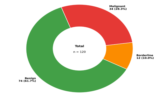
Figure 1: Distribution of benign, borderline and malignant epithelial ovarian tumors
Out of 120 tumors, 74 cases (61.7%) were benign, 12 cases (10.0%) were borderline and 34 cases (28.3%) were malignant. Serous tumors were the commonest histological group, accounting for 66 cases (55.0%), followed by mucinous tumors in 35 cases (29.2%). Benign serous cystadenoma was the most frequent individual diagnosis, seen in 38 cases (31.7%), followed by mucinous cystadenoma in 25 cases (20.8%). Among malignant tumors, high-grade serous carcinoma was the leading subtype, accounting for 18 cases (15.0% of all tumors and 52.9% of malignant tumors) (Table 2; Figure 2).
Table 2: Histopathological spectrum of epithelial ovarian tumors
|
Histopathological diagnosis |
Number of cases |
Percentage |
|
Serous cystadenoma |
38 |
31.7 |
|
Mucinous cystadenoma |
25 |
20.8 |
|
Benign Brenner tumor |
5 |
4.2 |
|
Seromucinous cystadenoma |
6 |
5.0 |
|
Serous borderline tumor |
7 |
5.8 |
|
Mucinous borderline tumor |
5 |
4.2 |
|
High-grade serous carcinoma |
18 |
15.0 |
|
Low-grade serous carcinoma |
3 |
2.5 |
|
Mucinous carcinoma |
5 |
4.2 |
|
Endometrioid carcinoma |
4 |
3.3 |
|
Clear cell carcinoma |
3 |
2.5 |
|
Malignant Brenner tumor |
1 |
0.8 |
|
Total |
120 |
100.0 |
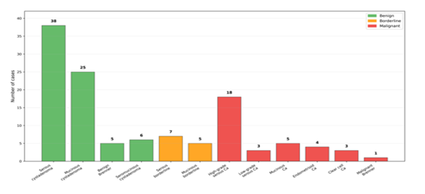
Figure 2: Histopathological spectrum of epithelial ovarian tumors
When tumors were grouped according to biological behavior, serous tumors formed the largest proportion of malignant cases. Of the 34 malignant tumors, 21 cases (61.8%) belonged to the serous group, including 18 high-grade serous carcinomas and 3 low-grade serous carcinomas. Mucinous tumors accounted for 5 malignant cases (14.7%). Endometrioid and clear cell carcinomas accounted for 4 cases (11.8%) and 3 cases (8.8%), respectively. The distribution of histological type differed significantly across benign, borderline and malignant categories (χ²=18.96, p=0.015) (Table 3).
Table 3: Distribution of major histological groups according to tumor behavior
|
Histological group |
Benign n=74 |
Borderline n=12 |
Malignant n=34 |
χ² value |
p-value |
|
Serous |
38 (51.4) |
7 (58.3) |
21 (61.8) |
18.96 |
0.015 |
|
Mucinous |
25 (33.8) |
5 (41.7) |
5 (14.7) |
||
|
Brenner |
5 (6.8) |
0 (0.0) |
1 (2.9) |
||
|
Seromucinous |
6 (8.1) |
0 (0.0) |
0 (0.0) |
||
|
Endometrioid |
0 (0.0) |
0 (0.0) |
4 (11.8) |
||
|
Clear cell |
0 (0.0) |
0 (0.0) |
3 (8.8) |
Data are expressed as n (%). Chi-square test was used. A p-value <0.05 was considered statistically significant
Serum tumor marker values showed a progressive rise from benign to borderline and malignant tumors. Median CA-125 level was 32 U/mL in benign tumors, 62 U/mL in borderline tumors and 248 U/mL in malignant tumors. Median HE4 level was 52 pmol/L in benign tumors, 73 pmol/L in borderline tumors and 168 pmol/L in malignant tumors. CA19-9 and CEA also showed higher median values in malignant tumors, although their rise was more prominent in mucinous tumors. The difference in marker levels across the three groups was statistically significant for CA-125, HE4, CA19-9 and CEA (p<0.001 for all markers) (Table 4; Figure 3).
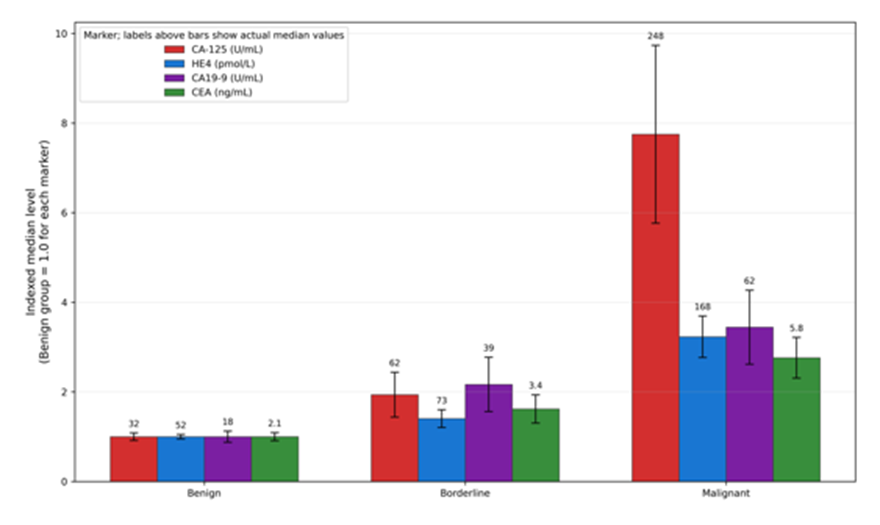
Figure 3: Tumor marker levels
Table 4: Serum tumor marker levels according to tumor category
|
Tumor marker |
Benign n=74 Median (IQR) |
Borderline n=12 Median (IQR) |
Malignant n=34 Median (IQR) |
Kruskal-Wallis H |
p-value |
|
CA-125, U/mL |
32 (18–49) |
62 (41–116) |
248 (112–612) |
58.74 |
<0.001 |
|
HE4, pmol/L |
52 (38–68) |
73 (56–104) |
168 (96–286) |
51.38 |
<0.001 |
|
CA19-9, U/mL |
18 (9–35) |
39 (21–72) |
62 (31–148) |
29.66 |
<0.001 |
|
CEA, ng/mL |
2.1 (1.2–3.4) |
3.4 (2.0–5.1) |
5.8 (3.1–10.6) |
26.84 |
<0.001 |
Marker values are expressed as median with interquartile range because values were not normally distributed. Kruskal-Wallis test was used. A p-value <0.05 was considered statistically significant.
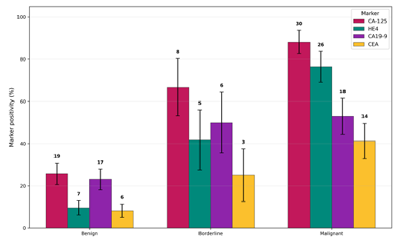
Figure 4: Tumor marker positivity across benign, borderline and malignant tumors
Marker positivity also increased significantly with tumor severity. CA-125 was elevated in 19 benign tumors (25.7%), 8 borderline tumors (66.7%) and 30 malignant tumors (88.2%). HE4 positivity was observed in 7 benign tumors (9.5%), 5 borderline tumors (41.7%) and 26 malignant tumors (76.5%). CA19-9 was positive in 17 benign tumors (23.0%), 6 borderline tumors (50.0%) and 18 malignant tumors (52.9%). CEA positivity was found in 6 benign tumors (8.1%), 3 borderline tumors (25.0%) and 14 malignant tumors (41.2%). All four markers showed statistically significant association with tumor category (Table 5; Figure 4).
Table 5: Tumor marker positivity in benign, borderline and malignant epithelial ovarian tumors
|
Marker positivity |
Benign n=74 |
Borderline n=12 |
Malignant n=34 |
χ² value |
p-value |
|
CA-125 positive, n (%) |
19 (25.7) |
8 (66.7) |
30 (88.2) |
39.42 |
<0.001 |
|
HE4 positive, n (%) |
7 (9.5) |
5 (41.7) |
26 (76.5) |
49.68 |
<0.001 |
|
CA19-9 positive, n (%) |
17 (23.0) |
6 (50.0) |
18 (52.9) |
10.91 |
0.004 |
|
CEA positive, n (%) |
6 (8.1) |
3 (25.0) |
14 (41.2) |
17.24 |
<0.001 |
Positivity was defined as CA-125 >35 U/mL, CA19-9 >37 U/mL, CEA >5 ng/mL and HE4 above menopausal status-specific cut-off. Chi-square test was used. A p-value <0.05 was considered statistically significant
Subtype-wise analysis showed that CA-125 and HE4 were more frequently elevated in serous carcinomas, particularly high-grade serous carcinoma. Among 18 cases of high-grade serous carcinoma, CA-125 was elevated in 17 cases (94.4%) and HE4 in 15 cases (83.3%). In mucinous tumors, CA19-9 and CEA showed more frequent elevation. CA19-9 was raised in 18 of 35 mucinous tumors (51.4%) compared with 23 of 85 non-mucinous tumors (27.1%). This association was statistically significant (χ²=6.67, p=0.010). CEA was elevated in 12 of 35 mucinous tumors (34.3%) compared with 11 of 85 non-mucinous tumors (12.9%) (χ²=7.46, p=0.006) (Table 6).
Table 6: Marker positivity according to major histological subtype
|
Histological subtype |
No. of cases |
CA-125 positive n (%) |
HE4 positive n (%) |
CA19-9 positive n (%) |
CEA positive n (%) |
|
Serous tumors |
66 |
39 (59.1) |
28 (42.4) |
16 (24.2) |
8 (12.1) |
|
Mucinous tumors |
35 |
12 (34.3) |
5 (14.3) |
18 (51.4) |
12 (34.3) |
|
Brenner tumors |
6 |
1 (16.7) |
1 (16.7) |
2 (33.3) |
1 (16.7) |
|
Seromucinous tumors |
6 |
1 (16.7) |
0 (0.0) |
1 (16.7) |
0 (0.0) |
|
Endometrioid carcinoma |
4 |
3 (75.0) |
2 (50.0) |
2 (50.0) |
1 (25.0) |
|
Clear cell carcinoma |
3 |
1 (33.3) |
2 (66.7) |
2 (66.7) |
1 (33.3) |
Data are expressed as n (%). Marker positivity was calculated within each histological subtype
Combined marker analysis demonstrated that dual positivity for CA-125 and HE4 was strongly associated with malignancy. Among malignant tumors, 25 of 34 cases (73.5%) showed combined CA-125 and HE4 positivity, compared with 5 of 74 benign tumors (6.8%) and 4 of 12 borderline tumors (33.3%). This association was statistically significant (χ²=54.73, p<0.001). CA-125 positivity alone was sensitive but less specific, whereas combined CA-125 and HE4 positivity better separated malignant tumors from benign tumors (Table 7; Figure 5).
Table 7: Combined CA-125 and HE4 expression pattern according to tumor category
|
CA-125/HE4 pattern |
Benign n=74 |
Borderline n=12 |
Malignant n=34 |
χ² value |
p-value |
|
Both negative |
53 (71.6) |
3 (25.0) |
3 (8.8) |
54.73 |
<0.001 |
|
CA-125 positive only |
14 (18.9) |
4 (33.3) |
5 (14.7) |
||
|
HE4 positive only |
2 (2.7) |
1 (8.3) |
1 (2.9) |
||
|
Both positive |
5 (6.8) |
4 (33.3) |
25 (73.5) |
Data are expressed as n (%). Chi-square test was used. A p-value <0.05 was considered statistically significant
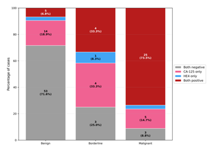
Figure 5: Combined CA-125 and HE4 pattern in epithelial ovarian tumors
The relationship between gross morphology and malignancy was also evident. Solid-cystic tumors, papillary excrescences, bilaterality, necrosis and surface involvement were more common among malignant tumors. Solid-cystic morphology was observed in 26 malignant tumors (76.5%) compared with 14 benign tumors (18.9%). Papillary excrescences were identified in 23 malignant tumors (67.6%) and 10 benign tumors (13.5%). These differences were statistically significant (Table 1). These findings supported the importance of careful gross examination and adequate sampling in ovarian epithelial tumors.
DISCUSSION:
The present study evaluated 120 epithelial ovarian tumors and correlated histopathological diagnosis with serum tumor marker levels. Benign tumors formed the largest group, accounting for 61.7% of cases, while borderline and malignant tumors accounted for 10.0% and 28.3%, respectively. This pattern is expected in a hospital-based surgical pathology setting, where many ovarian masses are removed because of symptoms, size or radiological suspicion, but not all represent malignant disease.
Serous tumors were the most common histological group in the present study. Serous cystadenoma was the leading benign tumor, while high-grade serous carcinoma was the most frequent malignant tumor. This observation is consistent with the known predominance of serous morphology among epithelial ovarian tumors. The biological importance of high-grade serous carcinoma lies in its aggressive behavior, frequent advanced-stage presentation and strong association with raised CA-125 levels.
Age showed a significant association with tumor behavior. Benign tumors were more common in younger and middle-aged women, while malignant tumors were more frequent in postmenopausal women. The mean age of patients with malignant tumors was significantly higher than that of patients with benign tumors. This finding reinforces the clinical need for careful evaluation of adnexal masses in postmenopausal women, especially when supported by suspicious imaging findings and elevated tumor markers.
Gross features such as bilaterality, solid-cystic morphology and papillary excrescences were significantly associated with malignant tumors. Although gross appearance alone cannot establish the final diagnosis, it guides tissue sampling and alerts the pathologist to areas that may show invasion. Papillary projections, solid areas, necrosis and capsular surface deposits require generous sampling because these areas are more likely to reveal borderline change or invasive carcinoma.
CA-125 showed a clear stepwise increase from benign to borderline and malignant tumors. It was elevated in 88.2% of malignant tumors, compared with 25.7% of benign tumors. The high positivity rate in malignant tumors supports the clinical utility of CA-125 in epithelial ovarian cancer, especially serous carcinoma. However, the marker was also elevated in nearly one-fourth of benign tumors, emphasizing its limited specificity. Benign serous tumors, endometriosis-associated lesions, inflammatory conditions and large cystic tumors may show CA-125 elevation because of peritoneal irritation or epithelial expression.
HE4 was less frequently positive in benign tumors and more frequently positive in malignant tumors. In the present study, HE4 positivity was observed in 76.5% of malignant tumors and only 9.5% of benign tumors. This pattern suggests that HE4 may be more useful than CA-125 in reducing false-positive interpretation, particularly when benign disease is part of the differential diagnosis. Combined CA-125 and HE4 positivity showed a strong association with malignancy, with dual positivity present in 73.5% of malignant tumors and only 6.8% of benign tumors.
CA19-9 and CEA showed a different pattern. Their positivity was more closely related to mucinous histology than to malignancy alone. CA19-9 was elevated in 51.4% of mucinous tumors, while CEA was elevated in 34.3% of mucinous tumors. This observation is clinically relevant because mucinous ovarian tumors may overlap with metastatic gastrointestinal tumors. In such cases, CA19-9 and CEA should be interpreted along with imaging, intraoperative findings, laterality, tumor size, histology and immunohistochemistry when required.\
The study highlights that no single tumor marker can reliably distinguish all benign, borderline and malignant ovarian epithelial tumors. CA-125 is sensitive but not completely specific. HE4 improves malignant discrimination in many cases. CA19-9 and CEA are useful adjuncts in mucinous tumors but are not ovarian-specific. Therefore, marker interpretation should always be integrated with clinical features, menopausal status, imaging findings and histopathological diagnosis.
The strength of the present study is that it evaluated multiple commonly used tumor markers and correlated them with histological tumor type and biological behavior. The inclusion of benign, borderline and malignant tumors allowed comparison across the full epithelial tumor spectrum. The study also emphasized subtype-specific marker behavior, which is useful for routine diagnostic practice.
The study had some limitations. It was a single-centre study with a moderate sample size. Follow-up data, stage-wise survival analysis and response to therapy were not included. Molecular testing and immunohistochemistry were not performed in all cases because the study focused mainly on routine histopathology and serum tumor markers. Future studies with larger sample size, stage-wise analysis, immunohistochemical markers and follow-up outcomes may provide stronger evidence regarding the prognostic role of tumor markers in ovarian epithelial tumors.
CONCLUSION:
The present study showed that ovarian epithelial tumors were predominantly benign, with serous tumors representing the most common histological group. High-grade serous carcinoma was the commonest malignant epithelial ovarian tumor. CA-125 and HE4 showed significant association with malignant tumors, while CA19-9 and CEA were more frequently elevated in mucinous tumors. Combined CA-125 and HE4 positivity was strongly associated with malignancy and may be useful in preoperative risk assessment. However, tumor markers should not be interpreted in isolation. Final diagnosis and classification depend on careful histopathological examination supported by clinical, radiological and laboratory correlation.
REFERENCES:
1. Sung H, Ferlay J, Siegel RL, Laversanne M, Soerjomataram I, Jemal A, et al. Global cancer statistics 2020: GLOBOCAN estimates of incidence and mortality worldwide for 36 cancers in 185 countries. CA Cancer J Clin. 2021;71(3):209-49.
2. Torre LA, Trabert B, DeSantis CE, Miller KD, Samimi G, Runowicz CD, et al. Ovarian cancer statistics, 2018. CA Cancer J Clin. 2018;68(4):284-96.
3. Hennessy BT, Coleman RL, Markman M. Ovarian cancer. Lancet. 2009;374(9698):1371-82.
4. Jayson GC, Kohn EC, Kitchener HC, Ledermann JA. Ovarian cancer. Lancet. 2014;384(9951):1376-88.
5. Prat J; FIGO Committee on Gynecologic Oncology. Staging classification for cancer of the ovary, fallopian tube and peritoneum. Int J Gynaecol Obstet. 2014;124(1):1-5.
6. Kurman RJ, Shih IM. The dualistic model of ovarian carcinogenesis: Revisited, revised and expanded. Am J Pathol. 2016;186(4):733-47.
7. Shih IM, Kurman RJ. Ovarian tumorigenesis: A proposed model based on morphological and molecular genetic analysis. Am J Pathol. 2004;164(5):1511-8.
8. De Leo A, Santini D, Ceccarelli C, Santandrea G, Palicelli A, Acquaviva G, et al. What is new on ovarian carcinoma: Integrated morphologic and molecular analysis following the new 2020 World Health Organization classification of female genital tumors. Diagnostics. 2021;11(4):697.
9. Köbel M, Kalloger SE, Boyd N, McKinney S, Mehl E, Palmer C, et al. Ovarian carcinoma subtypes are different diseases: Implications for biomarker studies. PLoS Med. 2008;5(12):e232.
10. Matsas A, Stefanoudakis D, Stefanoudakis G, Kokkali S, Papantoniou N, Boutas I. Tumor markers and their diagnostic significance in ovarian cancer. Life. 2023;13(8):1689.
11. Charkhchi P, Cybulski C, Gronwald J, Wong FO, Narod SA, Akbari MR. CA125 and ovarian cancer: A comprehensive review. Cancers. 2020;12(12):3730.
12. Jacobs I, Bast RC Jr. The CA 125 tumour-associated antigen: A review of the literature. Hum Reprod. 1989;4(1):1-12.
13. Moore RG, McMeekin DS, Brown AK, DiSilvestro P, Miller MC, Allard WJ, et al. A novel multiple marker bioassay utilizing HE4 and CA125 for the prediction of ovarian cancer in patients with a pelvic mass. Gynecol Oncol. 2009;112(1):40-6.
14. Hellström I, Raycraft J, Hayden-Ledbetter M, Ledbetter JA, Schummer M, McIntosh M, et al. The HE4 protein is a biomarker for ovarian carcinoma. Cancer Res. 2003;63(13):3695-700.
15. Dochez V, Caillon H, Vaucel E, Dimet J, Winer N, Ducarme G. Biomarkers and algorithms for diagnosis of ovarian cancer: CA125, HE4, RMI and ROMA, a review. J Ovarian Res. 2019;12(1):28.
16. Chen F, Shen J, Wang J, Cai P, Huang Y. Clinical analysis of four serum tumor markers in 458 patients with ovarian tumors: Diagnostic value of the combined use of HE4, CA125, CA19-9 and CEA in ovarian tumors. Cancer Manag Res. 2018;10:1313-8.
17. Lertkhachonsuk AA, Buranawongtrakoon S, Lekskul N, Rermluk N, Wee-Stekly WW, Charakorn C. Serum CA19-9, CA-125 and CEA as tumor markers for mucinous ovarian tumors. J Obstet Gynaecol Res. 2020;46(11):2287-91.
18. Zhang M, Cheng S, Jin Y, Zhao Y, Wang Y. Roles of CA125 in diagnosis, prediction and oncogenesis of ovarian cancer. Biochim Biophys Acta Rev Cancer. 2021;1875(2):188503.
19. Funston G, Hamilton W, Abel G, Crosbie EJ, Rous B, Walter FM. The diagnostic performance of CA125 for the detection of ovarian and non-ovarian cancer in primary care: A population-based cohort study. PLoS Med. 2020;17(10):e1003295.
20. Nolen BM, Lokshin AE. Biomarker testing for ovarian cancer: Clinical utility of multiplex assays. Mol Diagn Ther. 2013;17(3):139-46.