Correlation of Serum Malondialdehyde level with F wave parameters in tobacco users.
- Suman Sharma , PhD (Physiology), Designated Professor, Department of Physiology, ESIC Medical College and Hospital, Faridabad, Haryana, India.
- Mohan Babu , Assistant Professor, Department of Physiology, ESIC Medical College and Hospital, Faridabad, Haryana, India.
- Saroj Chaudhary , PhD (Physiology), Associate Professor, Department of Physiology, Pacific Medical College and Hospital, Udaipur, India.
- Manjinder Kaur , Professor and Head, Department of Physiology, Geetanjali Medical College & Hospital, Udaipur, India.
- Naren Kurmi , Professor, Department of Physiology, Geetanjali Medical College & Hospital, Udaipur, India.
Article Information:
Abstract:
Background: Tobacco is the most abused substance in India which not only exposes its users to nicotine but also harmful concoction of chemicals inducing the increased levels of circulating reactive oxygen species (ROS). Serum Malondialdehyde (S. MDA) being one of the markers for the same. The neurons, on the other hand are quite prone to the detrimental effects of ROS. Hence, the present study was aimed to find out the correlation of S.MDA levels and neural electrophysiological performance especially on the late responses (F-Wave). Methodology: The present study was conducted on 111 healthy volunteers, divided into two groups; nontobacco users (NTU; n=40) and tobacco users (TU; n=71). S.MDA levels were measured by Thiobarbituric acid assay method. All the parameters of F-Wave were recorded on Neuroperfect, EMG 2000, Medicaid, and Chandigarh. Data obtained was analyzed using SPSS version 23 for windows. Results: There was significant lengthening of all F Wave latencies such as Fmin (P=0.05), Fmax (p=0.013) & Fmean (0.004) and significant reduction (p=0.020) in the conduction velocity (p=0.003). The FM Ratio was significantly increased (p=0.047). F Wave index was also significantly prolonged (p=0.039) & significant reduction was observed in nerve conduction velocity. A significant (p=0.05) negative correlation was seen between oxidative stress marker S.MDA with persistence, F estimate & conduction velocity in tobacco consumers. However, the other parameters show a nonsignificant relationship with S.MDA. Conclusion: The tobacco smoke has a definitive effect on neural health, but the possible mechanism may not be through oxidative stress, but other toxic additives present in tobacco smoke, which are exhibiting direct neurotoxicity.
Keywords:
Article :
INTRODUCTION:
Tobacco is the most abused substance in India, enslaving all the sectors of society and affecting all the organs. Chemicals present in tobacco disturb the balance between the production of Reactive Oxygen Species (ROS) and antioxidant defense mechanism in our body resulting oxidative stress.1
Cigarette not only contains nicotine but is a toxic concoction of tar, carbon monoxide, toluene and other chemicals that affects the myelin sheath of peripheral nerves resulting demyelination, neural ischemia and decreased nerve conduction.2,3
To study the neural conduction and integrity, nerve conduction studies (NCS) have given the promising results owing to its sensitivity, reliability and noninvasiveness. It has been observed that long term exposure of tobacco leads to reduction of conduction velocity of sensory fibers of mixed nerves, whereas the motor fibers doesn’t show any electrophysiological change, especially in the distal portions of peripheral nerves.4
Although some studies have reported the effects of tobacco consumption on motor and sensory nerves, very little evidence is available regarding the impact of its toxic metabolites on late responses or proximal involvement of peripheral nerves. However, in our previous study, chronic tobacco consumption was associated with prolongation of all F-wave latencies, suggesting proximal demyelination and radiculopathy.5 we wanted to further explore the effect of oxidative stress, due to tobacco smoking, on proximal neural health, by exploring the late responses. Hence, the present study was conducted with an aim to find out the effect of tobacco related oxidative stress on the F-Wave parameters of median nerve, a prominent peripheral nerve.
MATERIALS AND METHODS:
Study design, setting and duration
This cross-sectional case-control study was conducted in the Clinical Neurophysiology Unit of the Department of Physiology at a tertiary care teaching hospital between 2013 and 2014. Written informed consent was obtained from all participants, and the study was initiated after obtaining approval from the Human Research Ethics Committee (Ref. No. GU/UCE/EC/2013/296 dated 15/05/2013).
Study population
The study population comprised apparently healthy adult volunteers working as contract labourers and housekeeping staff. Participants of both sexes aged more than 18 years were considered eligible for screening.
Sample size and participant selection
In our study, 111 eligible participants were enrolled in the study. The participants were categorized into two groups. Group NTU included 40 non-tobacco users, whereas Group TU included 71 tobacco users with a smoking exposure index above 400 and a history of cigarette/bidi smoking for more than five years. Participants with systemic disease, endocrine disorders, alcohol or substance abuse, severe anaemia, central nervous system disorders, or metabolic syndrome were excluded.
Data collection
Data were collected in predesigned case proforma. Information regarding age, socioeconomic status, family history, past and present medical history, smoking history, and substance abuse were collected. A basic clinical examination was performed in all participants before biochemical and electrophysiological evaluation.
Outcome variables
The primary outcome variables were serum malondialdehyde (S. MDA) level and F-wave parameters of the right median nerve. The electrophysiological parameters studied were F-min latency, F-max latency, and F-mean latency, motor latency, and amplitude, intensity of threshold stimulus, persistence, F-M ratio, F-wave index, chronodispersion, F-estimate, and nerve conduction velocity.
Biochemical assessment
For biochemical analysis, 5 mL of venous blood was collected from each participant in the morning after an overnight fast of 12 h. Serum was separated by centrifugation at 3000 rpm for 10 min and analysed for malondialdehyde using the thiobarbituric acid assay method.6
Electrophysiological assessment
Electrophysiological testing was performed in a noise-free room with ambient temperature maintained between 23°C and 26°C. Nerve conduction studies were carried out using a fully computerized electromyography and nerve conduction velocity system, Neuroperfect EMG 2000 (Medicaid, Chandigarh, India). Participants were positioned supine on the examination table with the arm extended alongside the body and the forearm supinated. After skin preparation with alcohol, surface electrodes were applied using conducting paste and secured with adhesive tape. The active recording electrode was placed over the belly of the abductor pollicis brevis muscle, the reference electrode over the distal phalanx of the thumb, and the ground electrode between the stimulating and recording electrodes.
In all participants, the right median nerve was selected for F-wave recording. The nerve was stimulated supramaximally at the wrist between the tendons of palmaris longus and flexor carpi radialis on the palmar aspect of the wrist near the distal wrist crease. Twenty consecutive stimuli were delivered, and artifact-free responses were recorded. Each response consisted of an M-wave followed by an F-wave. Upper limb length was measured from the point of stimulation to the C7 spinous process using a measuring tape for calculation of derived indices.
F-wave parameters studied
The F-wave parameters analysed included minimum latency, maximum latency, mean latency, amplitude, chronodispersion, F-M ratio, persistence, F-wave index, and F-estimate. Minimum latency represented conduction in the fastest fibres, whereas maximum latency represented conduction in the slowest fibres. Chronodispersion was defined as the difference between maximum and minimum latencies. Persistence was defined as the proportion of elicitable F- wave responses among the total number of stimuli delivered. F-wave index was calculated as:
F-wave index = (Persistence×arm length) / (Latency×chronodispersion) F-estimate was calculated using the formula:
F-estimate = (2×F distance / conduction velocity) + distal latency + 1 ms
Statistical analysis
Data were entered and analysed using Statistical Package for the Social Sciences for Windows, version 16.0 (SPSS Inc., Chicago, IL, USA) and Microsoft Office Excel 2007. Since the data were not normally distributed, non-parametric analysis was used and results were summarized using median values where appropriate. Intergroup comparisons were performed using the Mann-Whitney U test. Correlation between serum malondialdehyde level and F-wave parameters in tobacco users was assessed using Spearman correlation coefficient. A two-tailed P value of <0.05 was considered statistically significant.
RESULTS:
A total of 111 participants, including 71 tobacco users and 40 non-tobacco users, were studied. Tobacco users showed significantly prolonged F-min, F-max, and F-mean latencies, along with significantly higher F-estimate compared to non-tobacco users. Persistence and F-wave index were significantly reduced, whereas F-M ratio was significantly increased in tobacco users.
Nerve conduction velocity was significantly lower in tobacco users. No significant differences were observed in motor latency, amplitude, intensity of threshold stimulus, or chronodispersion between the groups (Table 1). Serum malondialdehyde level was significantly higher in tobacco users than in non-tobacco users (Table 2). In the tobacco user group, serum malondialdehyde level showed significant negative correlations with motor latency, intensity, F-estimate, and nerve conduction velocity, while no significant correlation was found with the remaining F-wave parameters, except persistence, which also showed statistical significance.
Table 1. Baseline demographic characteristics of study participants.
|
Variable |
Non-users (n = 40) |
Tobacco users (n = 71) |
p value |
|
Age (years), Mean ± SD |
32.92 ± 7.57 |
32.64 ± 6.28 |
0.845 |
|
Gender, n (%) |
0.001* |
||
|
Male |
28 (70.0%) |
67 (94.4%) |
|
|
Female |
12 (30.0%) |
4 (5.6%) |
Values are presented as mean±SD or n (%), as appropriate. Independent t test and Fisher exact test were used for tests of significance. * p value <0.05 was considered statistically significant.
Table 2. Comparison of F-wave parameters and serum MDA between non-tobacco users and tobacco users.
|
Variable |
Non-users (n = 40) |
Tobacco users (n = 71) |
p value |
|
Persistence |
80 (20) |
65 (25) |
0.020* |
|
M latency (ms) |
4.18 (1.94) |
3.88 (2.00) |
0.827 |
|
F-min latency (ms) |
27.94 (5.04) |
29.81 (4.25) |
0.005* |
|
F-max latency (ms) |
30.62 (4.32) |
32.38 (3.88) |
0.013* |
|
Mean latency (ms) |
29.12 ± 2.52 |
30.72 ± 2.95 |
0.003* |
|
Chronodispersion (ms) |
2.93 (2.22) |
2.87 (2.50) |
0.432 |
|
F-min–M (ms) |
23.63 (3.16) |
24.50 (4.62) |
0.047* |
|
Distance (mm) |
0.797 ± 0.06 |
0.792 ± 0.05 |
0.698 |
|
Amplitude (mV) |
15.63 ± 4.08 |
15.93 ± 4.80 |
0.725 |
|
Velocity (m/s) |
69.15 ± 7.20 |
64.53 ± 7.67 |
0.002* |
|
F-wave index |
0.74 (0.55) |
0.60 (0.49) |
0.039* |
|
Serum MDA (mmol/mL) |
1.52 (1.13) |
3.04 (1.82) |
<0.001* |
Data are presented as mean±SD or median (IQR), as appropriate. Mean latency, distance, amplitude and velocity were compared using Welch's test; remaining variables were compared using the Mann-Whitney U test. MDA, malondialdehyde. *p value <0.05 was considered statistically significant.
Table 3. Correlation of serum MDA with F-wave parameters among tobacco users (n=71).
|
Variable |
Spearman correlation coefficient (ρ) |
p value |
|
Persistence |
-0.097 |
0.423 |
|
M latency (ms) |
0.307 |
0.009* |
|
F-min latency (ms) |
0.087 |
0.468 |
|
F-max latency (ms) |
0.211 |
0.078 |
|
Mean latency (ms) |
0.218 |
0.068 |
|
Chronodispersion (ms) |
0.215 |
0.072 |
|
F-min–M (ms) |
-0.071 |
0.554 |
|
Distance (mm) |
-0.120 |
0.319 |
|
Velocity (m/s) |
-0.003 |
0.977 |
|
F-wave index |
0.037 |
0.760 |
Correlation was assessed using Spearman rank correlation because serum MDA was not normally distributed. MDA, malondialdehyde. *p value <0.05 was considered statistically significant.
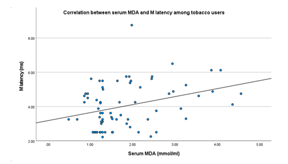
Figure 1. Scatter plot showing correlation between serum MDA and M latency among tobacco users.
DISCUSSION:
The present study showed significant F-wave abnormalities in smokers, including prolonged latencies, reduced persistence, increased F-min–M latency, reduced conduction velocity and altered F-wave index. These findings suggest subclinical motor nerve dysfunction, particularly involving proximal or late-response pathways. However, the lack of consistent correlation with serum MDA indicates that these changes may not be solely due to systemic lipid peroxidation. The significant prolongation of F-min, F-max and mean F-wave latencies among smokers indicates delayed conduction along the F-wave pathway. Since F-waves assess a long motor circuit involving distal motor fibres, proximal nerve segments, nerve roots, anterior horn cell activation and return conduction through the motor axon, latency prolongation may reflect early dysfunction beyond the distal nerve segment.
Panayiotopoulos and Chroni described F- waves as valuable late responses for assessing proximal conduction abnormalities, particularly when routine distal motor parameters may not adequately reflect nerve involvement.7 Cai et al. also emphasized that F-wave abnormalities provide additional information regarding motor neuron excitability and proximal motor conduction.8 In line with these observations, the prolonged F-wave latencies in the present study suggest early smoking-related impairment of proximal motor conduction.
Smokers also showed significantly reduced F-wave persistence. Persistence reflects the frequency of recordable late responses after repeated supramaximal stimulation and is influenced by motor neuron pool excitability and axonal integrity. Reduced persistence in this study may therefore indicate impaired motor neuron excitability, reduced consistency of motor unit recruitment or early axonal dysfunction. Sathya et al. considered persistence an important component of composite F-wave assessment in peripheral neuropathy.9 Thus, smoking appears to affect not only conduction time but also the reliability of late motor response generation. Conduction velocity was significantly reduced among smokers, suggesting early impairment of peripheral nerve conduction.
Similar findings were reported by Jeevalakshmi et al., who observed reduced nerve conduction velocity in smokers.3 Ahmad et al. also demonstrated poorer sensory nerve conduction among diabetic smokers compared with diabetic non- smokers.10 These findings support the view that tobacco exposure can adversely affect peripheral nerve function, and that smoking may aggravate nerve dysfunction in susceptible individuals.
The increase in F-min–M latency further supports proximal conduction slowing among smokers. As this parameter reduces the influence of distal motor latency, it provides a more focused estimate of proximal conduction time. Its increase, together with prolonged F-wave latencies, suggests that smoking-related nerve dysfunction may extend beyond distal motor segments. The significant alteration in F-wave index also supports this interpretation. Since F- wave index integrates persistence, latency, chronodispersion and limb length, it provides a broader assessment of motor nerve function than isolated latency measures. Sathya et al. reported the usefulness of F-wave index in detecting peripheral neuropathy and early neuropathic changes.11 In the present study, the altered F-wave index may reflect the combined effect of prolonged latency and reduced persistence among smokers.
Chronodispersion did not differ significantly between the two groups. As chronodispersion reflects variability between the fastest and slowest conducting motor fibres, the absence of a significant difference suggests that there was no marked conduction heterogeneity among motor axons. Panayiotopoulos and Chroni noted the role of chronodispersion in identifying demyelinating or dispersed conduction abnormalities.7 Therefore, the findings of the present study may represent early conduction slowing rather than established diffuse demyelinating neuropathy. M latency was comparable between smokers and non-smokers, suggesting relative preservation of distal motor conduction. However, several F-wave abnormalities were evident despite normal distal latency. This distinction is important, as it indicates that late responses may detect subtle or proximal motor nerve dysfunction earlier than conventional distal motor conduction measures. The findings therefore support the value of F-wave assessment in identifying early smoking-related motor nerve changes.
Serum MDA differed significantly between smokers and non-smokers, but its association with F-wave abnormalities was inconsistent. MDA is widely used as a marker of lipid peroxidation and oxidative stress. Isik et al. and Kashinakunti et al. reported higher MDA levels among smokers, supporting the role of tobacco exposure in oxidative injury.12,13 However, in the present study, serum MDA did not show a consistent relationship with most F-wave parameters. This suggests that although oxidative stress remains biologically relevant in smokers, serum MDA alone may not adequately reflect the mechanisms responsible for electrophysiological dysfunction. The lack of significant correlation between serum MDA and most F-wave parameters suggests that smoking-related neural dysfunction may be mediated through multiple pathways rather than systemic lipid peroxidation alone. Naik and Cucullo described tobacco-induced neural injury as a multifactorial process involving oxidative stress, inflammation and vascular dysfunction.14 Rodriguez-Fontan et al. further highlighted that tobacco exposure can influence peripheral nerve biology through cellular and immunomodulatory mechanisms affecting nerve repair.2 Thus, the weak MDA–F-wave relationship observed in the present study does not exclude smoking-related nerve injury, but indicates that toxic, vascular, inflammatory and metabolic mechanisms may act together. Overall, chronic smoking was associated with abnormal late motor responses, including prolonged F-wave latencies, reduced persistence, increased F-min–M latency, reduced conduction velocity and altered F-wave index. These findings suggest early subclinical motor nerve dysfunction with possible proximal involvement. The weak association with serum MDA indicates that lipid peroxidation alone may not explain these changes. Thus, F-wave analysis may help detect subtle smoking-related nerve dysfunction before routine distal parameters or clinical neuropathy become evident.
REFERENCES:
1. Kamceva G,Sarafinovska ZA, Ruskovska T, Zdravkovska M, Panova LK, Stikova E. Cigarette Smoking and Oxidative Stress in Patients with Coronary Artery Disease.Open Access Maced J Med Sci.2016;4(4):636–40.
2. Jeevalakshmi R., Shanmughavadivu R. and Kavitha S. Effect of Cigarette Smoking on Nerve Conduction Velocity. Egypt. Acad. J. Biolog. Sci., 2023;15(1):439-446
3. Sharma S, Shrivastav C, Suhalka ML, Kaur M. Influence of tobacco on median and ulnar nerve in the population of South Rajasthan. Int J Res Med Sci 2016;4:74-7.
4. Sharma S, Kurmi N, Kaur M. F-Wave latencies of median nerve in smokers from southern Rajasthan: A case-control study. Natl J Pharm Pharmacol2019;(10):983-86.
5. Buege and Aust. Method for estimation of serum Malondialdehyde levels. Methods in Enzymology.1978;105:302-10.
6. Panayiotopoulos CP, Chroni E. F-waves in clinical neurophysiology: a review, methodological issues and overall value in peripheral neuropathies. Electroencephalogr Clin Neurophysiol. 1996;101(5):365-74.
7. Cai Q, Niu J, Zhang Y, Wang Q, Liu H. Clinical and physiological significance of F-wave in neurological diseases. Front Neurol.2020;11:571341.
8. Sathya GR, Krishnamurthy N, Veliath S, Arulneyam J, Venkatachalam J. F-wave index: a diagnostic tool for peripheral neuropathy. Indian J Med Res. 2017;145(3):353-7.
9. Rodriguez-Fontan F, Tuaño BRK, Colakoglu S, Agostino LD, Banegas R. Tobacco use and neurogenesis: A theoreticalreview of pathophysiological mechanism affecting the outcome of peripheral nerve regeneration. Journal of Orthopaedics.2020;22:59–63.