ASSESSMENT OF THYROID FUNCTION STATUS IN MULTI-TRANSFUSED THALASSEMIA PATIENTS.
- Sourav Pati , Senior Resident, MD, DNB (Paediatrics), Department of Paediatrics, Bankura Sammilani Medical College, Gobindanagar, Bankura, Pin-722102, West Bengal, India.
- Toshibananda Bag , Associate Professor, MD (Paediatrics), Department of Paediatrics, Bankura Sammilani Medical College, Gobindanagar, Bankura, Pin-722102, West Bengal, India.
Article Information:
Abstract:
Introduction: Thalassemia major is a hereditary Hemoglobinopathy characterized by chronic anemia requiring regular blood transfusions. Repeated transfusions lead to iron overload, which can deposit in endocrine glands, including the thyroid, resulting in thyroid dysfunction. Early detection of thyroid abnormalities is essential to prevent growth retardation, metabolic complications, and impaired quality of life. Aims: To assess thyroid function status in multi-transfused thalassemia patients and determine the prevalence of thyroid dysfunction in this population. Materials and Methods: This cross-sectional observational study was conducted over a period of 18 months in the Department of Pediatric Medicine, Bankura Sammilani Medical College and Hospital, a rural-based tertiary care center. The study population comprised thalassemia patients aged 3–12 years who had been receiving regular blood transfusions for at least three years. A total of 113 multi-transfused thalassemia patients were included in the study. Results: In this study of 120 multi-transfused thalassemia patients, the majority (79.2%) were euthyroid, while 17.5% had subclinical hypothyroidism and 3.3% had overt hypothyroidism. The mean age of euthyroid and hypothyroid patients was comparable (8.4 ± 2.9 vs. 9.2 ± 2.5 years; P = 0.18). Hypothyroid patients had received significantly more transfusions than euthyroid patients (232 ± 146 vs. 148 ± 97; P = 0.01). Among the participants, 63% were on chelation therapy. Analysis by sex showed no significant difference in mean TSH levels between males and females (5.6 ± 15.6 vs. 3.9 ± 2.5 μIU/ml; P = 0.9). Conclusion: Multi-transfused thalassemia patients are at significant risk of developing thyroid dysfunction, primarily hypothyroidism. Regular screening of thyroid function should be incorporated into routine management of thalassemia to allow early detection and timely intervention.
Keywords:
Article :
INTRODUCTION:
Thalassemia major is a hereditary hemoglobinopathy characterized by defective synthesis of globin chains, leading to chronic hemolytic anemia that necessitates regular lifelong blood transfusions for survival [1]. Repeated transfusions, while life-saving, cause progressive iron overload, as the human body lacks a physiological mechanism for iron excretion [2]. Excess iron accumulates in vital organs including the liver, heart, and endocrine glands, leading to significant morbidity and mortality in these patients [3]. Among the endocrine complications, thyroid dysfunction is a well-documented consequence of iron overload in multi-transfused thalassemia patients [4]. Iron deposition in the thyroid gland, coupled with oxidative stress and free radical formation, disrupts normal thyroid hormone synthesis and regulation [5].
The spectrum of thyroid abnormalities ranges from subclinical hypothyroidism to overt hypothyroidism, while hyperthyroidism is less frequently encountered [6]. The prevalence of thyroid dysfunction in thalassemia major varies widely across different studies, with reports ranging from 6% to 30%, depending on patient age, transfusion burden, chelation therapy compliance, and regional factors [7,8]. Subclinical hypothyroidism is the most common abnormality observed, particularly in adolescents and young adults, and if untreated, it may contribute to growth retardation, delayed puberty, metabolic disturbances, and impaired quality of life [9].
Early recognition and monitoring of thyroid dysfunction are therefore critical in the comprehensive care of thalassemia patients. Routine assessment of thyroid function tests, including serum TSH and free thyroid hormones, should be incorporated into long-term follow-up protocols for these patients [10]. The consequences of thalassemia extend to multiple organ systems, with thyroid dysfunction being a common endocrine complication among multi-transfused children. Since the clinical manifestations of thyroid dysfunction in these patients are often subtle and non-specific, early detection through laboratory evaluation is essential to prevent complications and enhance quality of life. The present study was undertaken with the general objective of assessing serum thyroid hormone status in multi-transfused thalassemia patients. Specifically, it aimed to determine serum T3, T4, and TSH levels in children receiving blood transfusions for at least three years, and to analyze the association of hypothyroidism with serum ferritin levels, frequency of blood transfusions, age, and sex.
MATERIALS AND METHODS:
Study Design: Institution-based cross-sectional observational study.
Study Setting and Timeline: The study was conducted at Bankura Sammilani Medical College and Hospital, a rural-based tertiary care center, over a period of approximately 18 months from the acceptance of the synopsis.
Place of Study: Thalassemia Clinic, Pediatric Medicine, Bankura Sammilani Medical College & Hospital, Bankura.
Period of Study: The study was conducted over a period of 18 months following university approval of the synopsis, spanning from July 2020 to June 2021.
Study Population:
• Thalassemia patients aged 3–12 years.
• Receiving blood transfusions for a minimum of three years.
Sample Size:
• Calculated sample size: 103.
• Considering 10% non-response, the estimated sample size: 113.
Inclusion Criteria:
• Thalassemic patients aged >3 years and <12 years.
• Receiving blood transfusions for at least three years at a frequency of every 2–4 weeks.
• Willing to participate in the study.
Exclusion Criteria:
• Patients aged <3 years or >12 years.
• Known patients with congenital hypothyroidism.
• Patients receiving blood transfusions for less than three years.
• Patients with co-existing chronic diseases.
• Patients unwilling to participate in the study.
Study Variables:
• Clinical Variables:
· Age
· Sex
· Weight
· Height
· Age at first blood transfusion
· Frequency of blood transfusions
· Total number of blood transfusions
Signs and symptoms of hypothyroidism
• Laboratory Variables:
· Serum T3
· Serum T4
· Serum TSH
· Serum Ferritin
Data Collection and Interpretations
The study was conducted after obtaining approval from the Institutional Ethics Committee and The West Bengal University of Health Sciences. Thalassemia patients aged 3–12 years, receiving blood transfusions for at least three years, were included based on predefined inclusion and exclusion criteria. Written informed consent was obtained from the parents after explaining the study procedure in their native language. Data collection was carried out over approximately nine months (37 weeks) on two randomly selected days per week using a lottery-based Simple Random Sampling method, enrolling one or two eligible patients per day to achieve the target sample size of 113. Parents were interviewed using a pre-designed proforma to record history of acute illness, clinical features, and family history of hypothyroidism, followed by detailed physical examination. After 12-hour overnight fasting, 5 ml of pre-transfusion venous blood was collected from each patient, transported to the Biochemistry Department, and centrifuged. Serum was analyzed for T3, T4, TSH, and ferritin using Chemiluminescence Immunoassay (CLIA) on the Advia Centaur CP (Siemens). Thyroid status was classified as euthyroid (normal T3, T4, TSH), subclinical hypothyroidism (normal T3, T4, elevated TSH), or overt hypothyroidism (low T3, T4, elevated TSH), with age-specific reference ranges. Serum ferritin values up to 1650 ng/ml were recorded as measured, while values above 1650 ng/ml were categorized as >1650 ng/ml for statistical analysis.
Statistical Analysis:
Data were analysed using SPSS software. Continuous variables, such as age and liver enzyme levels, were expressed as mean ± standard deviation (SD) and compared using Student’s t-test or Mann–Whitney U test, as appropriate. Categorical variables, including gender, presence of CBD stones, and complications, were presented as frequencies and percentages and compared using Chi-square or Fisher’s exact tests. Diagnostic performance of MRCP-first and EUS-first strategies was evaluated in terms of sensitivity, specificity, positive predictive value (PPV), negative predictive value (NPV), and overall accuracy, using ERCP or intraoperative findings as the reference standard. Time-to-intervention comparisons were analysed using Kaplan–Meier survival analysis. A p-value < 0.05 was considered statistically significant.
RESULTS:
Table 1: Classification of hypothyroidism and their frequency in study population (n=120).
|
Classification |
Number of cases |
Percentage (%) |
|
Euthyroid |
95 |
79.20% |
|
Subclinical Hypothyroid |
21 |
17.50% |
|
Overt Hypothyroid |
4 |
3.30% |
|
Total |
120 |
100% |
Table 2: Distribution of mean age of Thalassemia children in study population according to Thyroid function status.
|
Thyroid Profile |
Number of Cases |
Mean Age ± SD (years) |
P value |
|
Euthyroid |
95 |
8.4 ± -2.9 |
0.18 |
|
Hypothyroid |
25 |
9.2 ± -2.5 |
Table 3: Mean number of transfusions of hypothyroid and euthyroid thalassemia patients.
|
Thyroid Profile |
Number of Patients |
Mean Number of Transfusions |
Std. Deviation |
Minimum Number of Transfusions |
Maximum Number of Transfusions |
P value |
|
Euthyroid |
95 |
148 |
97 |
36 |
492 |
0.01 |
|
Hypothyroid |
25 |
232 |
146 |
41 |
528 |
— |
|
Total |
120 |
165 |
114 |
— |
— |
— |
Table 4: Distribution of study population according to chelation therapy.
|
Chelation |
Number of Patients |
Percentage (%) |
|
Yes |
76 |
63 |
|
No |
44 |
37 |
|
Total |
120 |
100 |
Table 5: Mean TSH among the thalassemia patients of different sex.
|
Sex |
Mean TSH (μIU/ml) |
SD (μIU/ml) |
Minimum (μIU/ml) |
Maximum (μIU/ml) |
P value |
|
Male |
5.6 |
15.6 |
0.6 |
124.2 |
0.9 |
|
Female |
3.9 |
2.5 |
0.2 |
11.8 |
— |
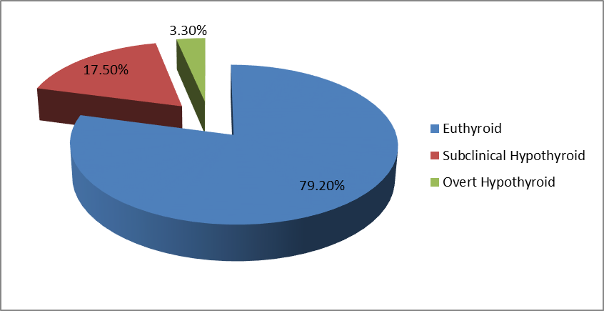
Figure: 1 Distribution of Thyroid Function Status in the Population.
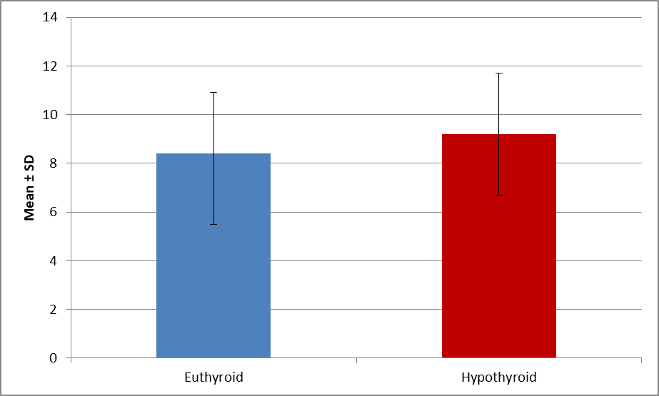
Figure: 2 Comparison between Euthyroid and Hypothyroid Groups
In our study, Out of a total of 120 patients, the majority, 95 patients (79.2%), were found to be euthyroid. Subclinical hypothyroidism was observed in 21 patients (17.5%), while overt hypothyroidism was present in 4 patients (3.3%).
In our study, the mean age of euthyroid patients was 8.4 ± 2.9 years, while hypothyroid patients had a mean age of 9.2 ± 2.5 years. The difference was not statistically significant (P = 0.18).
In our study, the analysis of transfusion burden among multi-transfused thalassemia patients revealed that euthyroid patients (n = 95) had received a mean of 148 ± 97 transfusions, whereas hypothyroid patients (n = 25) had a higher mean number of transfusions at 232 ± 146. Overall, the total study population (n = 120) had a mean of 165 ± 114 transfusions. The difference was statistically significant (P = 0.01)
In our study, Among the 120 multi-transfused thalassemia patients, 76 patients (63%) were receiving chelation therapy, while 44 patients (37%) were not undergoing chelation.
In our study, the analysis of thyroid-stimulating hormone (TSH) levels by sex showed that male patients had a mean TSH of 5.6 ± 15.6 μIU/ml, while female patients had a mean TSH of 3.9 ± 2.5 μIU/ml. The difference was not statistically significant (P = 0.9).
DISCUSSION:
In this study of 120 multi-transfused thalassemia patients, 95 patients (79.2%) were euthyroid, 21 patients (17.5%) had subclinical hypothyroidism, and 4 patients (3.3%) had overt hypothyroidism, which is consistent with previous reports showing hypothyroidism prevalence ranging from 6% to 30% in β-thalassemia major patients [11]. The mean age of euthyroid patients was 8.4 ± 2.9 years, while hypothyroid patients had a mean age of 9.2 ± 2.5 years, with no statistically significant difference (P = 0.18), supporting the observation that thyroid dysfunction can occur at various ages in thalassemia patients [12,13].Hypothyroid patients had received a significantly higher number of transfusions (232 ± 146) compared to euthyroid patients (148 ± 97; P = 0.01), highlighting the role of iron overload from repeated transfusions in thyroid impairment [14,15].
Regarding chelation therapy, 63% of patients were receiving treatment; while chelation reduces iron burden, inadequate chelation has been linked to higher risk of endocrine dysfunction, including hypothyroidism [16].Analysis of thyroid-stimulating hormone (TSH) levels by sex showed no significant difference between males (5.6 ± 15.6 μIU/ml) and females (3.9 ± 2.5 μIU/ml; P = 0.9), aligning with other studies that found no gender-based disparity in thyroid dysfunction among thalassemia patients [17]. Collectively, these findings emphasize the need for routine thyroid function monitoring in multi-transfused thalassemia patients, especially those with higher transfusion frequency and suboptimal chelation [18 -20].
CONCLUSION:
Thyroid dysfunction is a common endocrine complication in multi-transfused thalassemia patients, with subclinical hypothyroidism being more frequent than overt hypothyroidism. Age and sex were not significantly associated with thyroid abnormalities, while a higher transfusion burden was linked to an increased risk of thyroid dysfunction. Regular monitoring of thyroid function, along with appropriate chelation therapy, is essential for early detection and management to improve the overall health and quality of life in these patients.
REFERENCES:
1. Weatherall DJ, Clegg JB. The thalassaemia syndromes. 4th ed. Oxford: Blackwell Science; 2001.
2. Cappellini MD, Cohen A, Porter J, Taher A, Viprakasit V. Guidelines for the management of transfusion dependent thalassaemia (TDT). 3rd ed. Nicosia: Thalassaemia International Federation; 2014.
3. Borgna-Pignatti C, Galanello R. Thalassemias and related disorders: Quantitative disorders of hemoglobin synthesis. In: Orkin SH, Nathan DG, Ginsburg D, Look AT, Fisher DE, Lux SE, editors. Nathan and Oski’s Hematology of Infancy and Childhood. 7th ed. Philadelphia: Elsevier; 2009. p. 1011–66.
4. De Sanctis V, Soliman AT, Elsedfy H, Skordis N, Kattamis C, Angastiniotis M, et al. Growth and endocrine disorders in thalassemia: The international network on endocrine complications in thalassemia (I-CET) position statement and guidelines. Indian J Endocrinol Metab. 2013;17(1):8–18.
5. Hershko C. Pathogenesis and management of iron toxicity in thalassemia. Ann N Y Acad Sci. 2010;1202:1–9.
6. Farmaki K, Tzoumari I, Pappa C, Chouliaras G, Berdoukas V. Normalisation of total body iron load with very intensive combined chelation reverses cardiac and endocrine complications of thalassaemia major. Br J Haematol. 2010;148(3):466–75.
7. Soliman AT, Eldabbagh M, Al Yafei F, Al-Naimi L, Aziz Bedair EM, Elsiddig S, et al. Thyroid hormone changes in thalassemia major: Thyroid function, ferritin level, and pattern of pituitary-thyroid axis. Pediatr Endocrinol Rev. 2013;11(1):88–95.
8. Shamshirsaz AA, Bekheirnia MR, Kamgar M, Pourzahedgilani N, Bouzari N, Habibzadeh M, et al. Metabolic and endocrinologic complications in beta-thalassemia major: A multicenter study in Tehran. BMC Endocr Disord. 2003;3:4.
9. De Sanctis V, Vullo C, Katz M, Wonke B. Endocrine aspects of thalassaemia. In: Cappellini MD, Cohen A, Eleftheriou A, Piga A, Porter J, Taher A, editors. Guidelines for the clinical management of thalassaemia. 2nd ed. Nicosia: Thalassaemia International Federation; 2008. p. 46–54.
10. Chern JP, Lin KH, Tsai WY, Wang SC, Lu MY, Lin DT, et al. Hypothyroidism and glucose intolerance in beta-thalassemic patients with iron overload. Arch Dis Child. 2001;85(5):397–400.
11. Upadya SH, Rukmini MS, Sundararajan S, Baliga BS, Kamath N. Thyroid function in chronically transfused children with beta thalassemia major: a cross-sectional hospital-based study. Int J Pediatr. 2018;2018:9071213.
12. Soliman AT, Al-Saleh S, Al-Sayed M, et al. Longitudinal study on thyroid function in patients with thalassemia major. Indian J Endocrinol Metab. 2013;17(2):300-305.
13. De Sanctis V, Soliman AT, Elsedfy H, et al. Growth and endocrinopathies among children with β-thalassemia major. Acta Biomed. 2024;95(1):e2024004.
14. Eshragi P, Movahedi M, Movahedi M, et al. Thyroid function in major thalassemia patients: Is it related to iron overload? Caspian J Intern Med. 2011;2(4):168-172.
15. Zervas A, Katopodi A, Protonotariou A, et al. Assessment of thyroid function in two hundred patients with beta-thalassemia major. Thyroid. 2002;12(2):151-154.
16. Soliman AT, Elsedfy H, Al-Sayed M, et al. Study on linear growth and thyroid function for 12 years in patients with thalassemia major. Endocrine Abstracts. 2013;32:P992.
17. Ansaf AI, Al-Jubouri S, Al-Mashhadani SA. Evaluation of the thyroid status and types of thyroid dysfunction in thalassemia major patients. Open Access Maced J Med Sci. 2021;9(1):67-72.
18. Fessas P, Loukopoulos D, Kaltsoya A. Peptide analysis of the inclusions of erythroid cells in B–thalassemia. Biochim Biophys Acta. 1966;124(2):430-432.
19. Delaporta P, Kattamis C, Papassotiriou I, et al. Thyroid function in Greek patients with thalassemia major. Eur J Haematol. 2012;88(5):429-433.
20. Rindang C, Suryana K, Suryadi S. Some aspects of thyroid dysfunction in thalassemia major patients. Paediatrica Indonesiana. 2011;51(5-6):157-161.