CLINICOPATHOLOGICAL AND MICROBIOLOGICAL CORRELATION OF INFLAMMATORY BIOMARKERS IN PATIENTS WITH PULMONARY TUBERCULOSIS: A PROSPECTIVE OBSERVATIONAL STUDY.
- Vivekanand , Associate Professor, Department of General Medicine, Chhattisgarh Institute Of Medical Sciences, (CIMS), Bilaspur, Chhattisgarh, India.
- Shubham Dixit , Assistant Professor, Department of Respiratory Medicine, Ram Krishna Medical College, Bhopal, Madhya Pradesh, India.
- Shashank Tyagi , Professor and Head, Department of Biochemistry, SRVS Government Medical College, Shivpuri, Madhya Pradesh, India.
Article Information:
Abstract:
Background: Inflammatory biomarkers may provide valuable information regarding disease activity in pulmonary tuberculosis (PTB) and complement conventional microbiological investigations. This study evaluated the clinicopathological and microbiological correlation of routinely available inflammatory biomarkers in patients with PTB. Methods: A prospective observational study was conducted among 300 newly diagnosed adult patients with pulmonary tuberculosis at a tertiary care hospital over 12 months. Clinical characteristics, radiological findings, sputum smear microscopy, and Cartridge-Based Nucleic Acid Amplification Test (CBNAAT) results were recorded. Baseline inflammatory biomarkers, including erythrocyte sedimentation rate (ESR), C-reactive protein (CRP), neutrophil-to-lymphocyte ratio (NLR), platelet-to-lymphocyte ratio (PLR), lymphocyte-to-monocyte ratio (LMR), hemoglobin, and total leukocyte count, were assessed before initiation of anti-tubercular therapy. Associations between biomarkers and microbiological findings were analyzed using appropriate statistical tests. Results: The mean age of participants was 44.9 ± 15.2 years, and 65.3% were male. Smear microscopy was positive in 74.0% of patients, while all cases were confirmed by CBNAAT; rifampicin resistance was detected in 5.3%. Mean ESR (67.5 ± 24.6 mm/h), CRP (39.8 ± 18.5 mg/L), NLR (5.12 ± 2.24), and PLR (228 ± 82) were elevated, whereas mean LMR was 2.18 ± 0.91. Smear-positive patients demonstrated significantly higher ESR, CRP, NLR, and PLR and significantly lower LMR than smear-negative patients (all p < 0.001). ESR (r=0.54), CRP (r=0.59), NLR (r=0.48), and PLR (r=0.44) showed significant positive correlations with bacillary burden, whereas LMR demonstrated a significant inverse correlation (r=−0.41; all p < 0.001). Conclusions: Routinely available inflammatory biomarkers, particularly ESR, CRP, NLR, PLR, and LMR, correlate significantly with microbiological positivity and bacillary burden in pulmonary tuberculosis. These inexpensive and widely accessible markers may serve as useful adjuncts for assessing disease severity and supporting clinical decision-making alongside microbiological investigations, especially in resource-limited settings.
Keywords:
Article :
INTRODUCTION:
Tuberculosis (TB) remains a major global public health problem despite substantial advances in diagnosis and treatment. In 2023, an estimated 10.8 million people developed TB and 1.25 million died from the disease, with India contributing the highest number of cases worldwide [1]. Early diagnosis and accurate assessment of disease severity remain essential for reducing transmission, improving treatment outcomes, and achieving the goals of TB elimination.
Pulmonary tuberculosis (PTB), the commonest form of TB, results from a complex interaction between Mycobacterium tuberculosis and the host immune system. The inflammatory response generated during infection plays a central role in bacterial containment but may also contribute to pulmonary tissue injury and disease progression. Consequently, the intensity of systemic inflammation often reflects the underlying disease activity and clinical severity, making inflammatory biomarkers potential adjuncts in patient evaluation [2, 3].
Microbiological techniques, including sputum smear microscopy, culture, and molecular assays such as Xpert MTB/RIF, remain the cornerstone of PTB diagnosis. Although these methods reliably confirm infection, they provide limited information regarding the host inflammatory response or disease severity. Furthermore, culture requires prolonged incubation, while smear microscopy has limited sensitivity in patients with low bacillary burden. These limitations have prompted interest in readily available laboratory biomarkers that may complement conventional diagnostic methods [4].
Inflammatory biomarkers such as erythrocyte sedimentation rate (ESR), C-reactive protein (CRP), total leukocyte count, neutrophil-to-lymphocyte ratio (NLR), platelet-to-lymphocyte ratio (PLR), and lymphocyte-to-monocyte ratio (LMR) are inexpensive, widely available, and routinely measured in clinical practice. Growing evidence suggests that these markers correlate with bacterial burden, radiological extent of disease, treatment response, and clinical outcomes. However, published findings remain inconsistent because of differences in study populations, biomarker thresholds, and study designs, limiting their routine clinical application.
Prospective data examining the relationship between inflammatory biomarkers and clinicopathological as well as microbiological characteristics of PTB are still limited, particularly in high-burden countries such as India. Establishing these correlations may improve disease assessment and help identify simple biomarkers that can support clinical decision-making alongside microbiological investigations [5, 6].
Therefore, the present prospective observational study was conducted to evaluate the clinicopathological and microbiological correlation of inflammatory biomarkers in patients with pulmonary tuberculosis. The study aimed to determine the association of routinely measured inflammatory biomarkers with clinical presentation, radiological findings, microbiological status, and disease severity, thereby assessing their potential role as adjunctive markers in the evaluation of pulmonary tuberculosis.
MATERIALS AND METHODS:
Study Design and Setting: This prospective observational study was conducted in the Department of Pulmonary Medicine in collaboration with the Departments of General Medicine in a tertiary care Indian hospital, over a period of 12 months.
Study Population: 300 Adult patients (≥18 years) with newly diagnosed pulmonary tuberculosis attending the outpatient or inpatient services during the study period were consecutively recruited. Pulmonary tuberculosis was diagnosed based on clinical features, radiological findings, and microbiological confirmation by sputum smear microscopy and/or Cartridge-Based Nucleic Acid Amplification Test (CBNAAT/Xpert MTB/RIF). Patients already receiving anti-tubercular therapy for more than seven days, those with extrapulmonary tuberculosis alone, pregnancy, active malignancy, autoimmune disorders, chronic inflammatory diseases, or concurrent acute bacterial or viral infections were excluded to minimize confounding of inflammatory biomarkers.
Data Collection: Demographic characteristics, smoking status, alcohol consumption, body mass index, comorbidities (including diabetes mellitus and HIV infection), presenting symptoms, duration of illness, and physical examination findings were recorded using a standardized case record form. Chest radiographs were evaluated for the extent of pulmonary involvement, cavitary lesions, and laterality by experienced physicians blinded to laboratory results.
Laboratory Investigations: Before initiation of anti-tubercular treatment, venous blood samples were collected under aseptic precautions. Complete blood counts were analyzed using an automated hematology analyzer. Erythrocyte sedimentation rate (ESR) was measured using the Westergren’s method, while serum C-reactive protein (CRP) was estimated by an immunoturbidimetric assay. Neutrophil-to-lymphocyte ratio (NLR), platelet-to-lymphocyte ratio (PLR), and lymphocyte-to-monocyte ratio (LMR) were calculated from absolute blood cell counts. Microbiological diagnosis was established using Ziehl–Neelsen smear microscopy and CBNAAT (Xpert MTB/RIF) according to the National Tuberculosis Elimination Programme (NTEP) guidelines.
Outcome Measures: The primary outcome was the correlation of inflammatory biomarkers with microbiological positivity. Secondary outcomes included associations between inflammatory biomarkers and clinicopathological variables, including symptom duration, radiological severity, cavitary disease, bacillary burden, and relevant comorbidities.
Statistical Analysis: Data were analyzed using IBM SPSS Statistics version 26.0 (IBM Corp., Armonk, NY, USA). Continuous variables were expressed as mean ± standard deviation whereas Categorical variables were summarized as frequencies and percentages. Comparisons between groups were performed using the independent-samples t-test or Mann–Whitney U test for continuous variables and the Chi-square or Fisher's exact test for categorical variables. Correlations between inflammatory biomarkers and clinical or microbiological parameters were evaluated using Pearson's or Spearman's correlation coefficients. A p value <0.05 was considered statistically significant.
RESULTS:
A total of 300 patients with pulmonary tuberculosis were included. The mean age was 44.9 ± 15.2 years, with the highest proportion belonging to the 31–45-year age group (34.7%). Males predominated (65.3%), and most patients were from rural areas (62.0%). Nearly half (49.3%) had a body mass index (BMI) below 18.5 kg/m². Current smoking (37.3%) and alcohol consumption (31.7%) were common risk factors, while diabetes mellitus and HIV infection were present in 20.7% and 6.0% of patients, respectively.
Table 1: Baseline clinico-demographic characteristics of study participants (N=300)
|
Variable |
Frequency (%) |
|
|
Age (years) |
18–30 |
52 (17.3) |
|
31–45 |
104 (34.7) |
|
|
46–60 |
92 (30.7) |
|
|
>60 |
52 (17.3) |
|
|
Mean age (years) |
44.9 ± 15.2 |
|
|
Gender |
Male |
196 (65.3) |
|
Female |
104 (34.7) |
|
|
Residential status |
Rural residence |
186 (62.0) |
|
Urban residence |
114 (38.0) |
|
|
Risk Factors |
Current smoker |
112 (37.3) |
|
Alcohol use |
95 (31.7) |
|
|
Diabetes mellitus |
62 (20.7) |
|
|
HIV positive |
18 (6.0) |
|
|
BMI <18.5 kg/m² |
148 (49.3) |
|
Cough was the most common presenting symptom (95.3%), followed by fever (84.7%), weight loss (76.0%), and loss of appetite (70.7%), indicating that constitutional and respiratory symptoms predominated in patients with pulmonary tuberculosis.
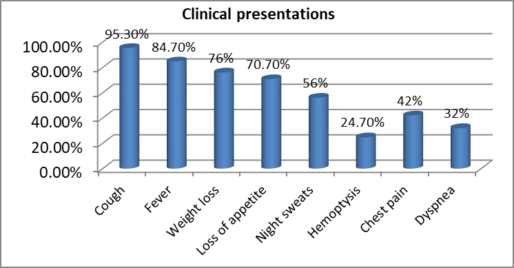
Graph 1: Clinical presentation of the study patients
Sputum smear microscopy was positive in 222 patients (74.0%), all patients were confirmed positive by CBNAAT. Based on bacillary load, 45.3% had a medium bacillary burden, and Rifampicin resistance was detected in 16 patients (5.3%).
Table 2; Microbiological findings among study patients
|
Variable |
N (%) |
|
Smear Positive |
222 (74.0) |
|
Smear Negative |
78 (26.0) |
|
CBNAAT Positive |
300 (100) |
|
Low bacillary burden |
62 (20.7) |
|
Medium bacillary burden |
136 (45.3) |
|
High bacillary burden |
102 (34.0) |
|
Rifampicin Resistant |
16 (5.3) |
Patients demonstrated elevated inflammatory markers at baseline. The mean ESR was 67.5 ± 24.6 mm/hour, CRP was 39.8 ± 18.5 mg/L, NLR was 5.12 ± 2.24, and PLR was 228 ± 82, whereas the mean LMR was 2.18 ± 0.91. The mean hemoglobin concentration was 10.9 ± 1.8 g/dL, indicating frequent anemia, and the total leukocyte count averaged 10.8 ± 3.4 ×10⁹/L, consistent with systemic inflammation.
Table 3; inflammatory biomarker profile
|
Biomarker |
Mean ± SD |
|
Hemoglobin (g/dL) |
10.9 ±1.8 |
|
Total leukocyte count (×10⁹/L) |
10.8 ±3.4 |
|
ESR (mm/hr) |
67.5 ±24.6 |
|
CRP (mg/L) |
39.8 ±18.5 |
|
NLR |
5.12 ±2.24 |
|
PLR |
228 ±82 |
|
LMR |
2.18 ±0.91 |
Patients with smear-positive tuberculosis exhibited significantly higher inflammatory activity than smear-negative patients. ESR, CRP, NLR, and PLR were all significantly elevated in the smear-positive group, whereas LMR was significantly lower (all p < 0.001). These findings indicate a strong association between increased inflammatory response and microbiological positivity.
Table 4: Comparison of inflammatory biomarkers according to smear microscopy
|
Biomarker |
Smear Positive (n=222) |
Smear Negative (n=78) |
p value |
|
ESR |
72.6±22.1 |
53.2±19.8 |
<0.001 |
|
CRP |
44.7±17.4 |
26.5±13.9 |
<0.001 |
|
NLR |
5.78±2.13 |
3.71±1.64 |
<0.001 |
|
PLR |
244±79 |
183±61 |
<0.001 |
|
LMR |
1.94±0.71 |
2.88±0.82 |
<0.001 |
ESR, CRP, NLR, and PLR demonstrated significant positive correlations with bacillary burden (all p < 0.001). In contrast, LMR showed a significant negative correlation (r = −0.41, p < 0.001). These results suggest that increasing inflammatory biomarker levels are associated with greater mycobacterial burden, while LMR decreases with increasing disease severity.
Table 5: Correlation of inflammatory biomarkers with bacillary burden
|
Biomarker |
Correlation coefficient (r) |
p value |
|
ESR |
0.54 |
<0.001 |
|
CRP |
0.59 |
<0.001 |
|
NLR |
0.48 |
<0.001 |
|
PLR |
0.44 |
<0.001 |
|
LMR |
-0.41 |
<0.001 |
DISCUSSION:
The current study findings demonstrated that elevated inflammatory biomarkers, particularly ESR, CRP, NLR, and PLR, were significantly associated with smear positivity and increasing bacillary burden, whereas LMR showed an inverse relationship, supporting their utility as adjunctive indicators of disease severity.
The majority of patients were middle-aged males from rural backgrounds, with a high prevalence of undernutrition and behavioral risk factors such as smoking and alcohol use. These demographic characteristics are consistent with reports from India and other high-burden countries, where tuberculosis disproportionately affects economically productive age groups and populations with nutritional deficiencies and adverse socioeconomic conditions. Similar observations have been reported by Bhargava et al. and Sarkar et al., who identified male sex, rural residence, and low BMI as important determinants of active pulmonary tuberculosis and poor clinical outcomes [7, 8].
Cough, fever, weight loss, and loss of appetite were the predominant presenting symptoms in the present study, reflecting the classical clinical manifestations of active pulmonary tuberculosis. Comparable symptom profiles have been described by Migliori et al., who emphasized that constitutional symptoms often correlate with increasing inflammatory activity and disease progression rather than microbiological status alone [9].
Microbiological evaluation showed smear positivity in nearly three-fourths of patients, while all cases were confirmed by CBNAAT. Rifampicin resistance was detected in a small proportion of patients, consistent with current epidemiological data from India. Molecular diagnostic techniques provide rapid confirmation of tuberculosis and drug resistance; however, they offer limited information regarding the host inflammatory response. Consequently, host-derived inflammatory biomarkers may provide complementary information regarding disease activity and treatment monitoring [10].
The present study demonstrated markedly elevated ESR and CRP concentrations among patients with pulmonary tuberculosis, with significantly higher values in smear-positive disease. Moreover, both biomarkers showed moderate positive correlations with bacillary burden. These findings agree with recent evidence from Yoon et al., who reported that CRP and ESR increase proportionally with mycobacterial load and radiological severity because of enhanced cytokine-mediated acute-phase responses. Persistent elevation of these markers reflects systemic inflammation induced by interleukin-6 and other pro-inflammatory mediators released during active infection [11].
Among the hematological indices, NLR and PLR were significantly increased in smear-positive patients and correlated positively with bacillary burden, whereas LMR demonstrated a significant inverse correlation. These observations support the concept that tuberculosis induces neutrophilia and relative lymphopenia through activation of innate immune responses and redistribution of lymphocytes to infected tissues. Monocyte expansion accompanying mycobacterial infection further contributes to reduce LMR. A recent systematic review and meta-analysis by Fritschi et al. concluded that NLR and LMR are reliable, inexpensive biomarkers for assessing disease severity and treatment response in tuberculosis, findings that closely parallel the present results [12]. Similar conclusions were reported by Wang et al., who observed that elevated NLR independently predicted extensive pulmonary involvement and delayed sputum conversion during anti-tubercular therapy [13].
The significant correlations observed between inflammatory biomarkers and bacillary burden suggest that these parameters reflect the intensity of host immune activation. Higher bacillary loads stimulate macrophage activation, cytokine production, and acute-phase protein synthesis, resulting in elevated CRP, ESR, and alterations in circulating leukocyte subsets. Conversely, declining LMR may indicate greater monocyte recruitment to sites of infection and impaired adaptive immune responses. These biological mechanisms provide a plausible explanation for the observed associations and support the potential role of inflammatory biomarkers as surrogate indicators of disease severity [14].
From a clinical perspective, inflammatory biomarkers possess several advantages, including low cost, widespread availability, and rapid turnaround time. Although they cannot replace microbiological confirmation, they may facilitate early risk stratification, identification of patients with higher bacillary burden, and monitoring of treatment response, particularly in resource-limited settings where advanced diagnostic facilities may not always be available. Combining inflammatory biomarkers with microbiological investigations could therefore improve comprehensive patient assessment and optimize clinical decision-making [15].
CONCLUSION:
Routinely available inflammatory biomarkers, particularly ESR, CRP, NLR, and PLR, showed significant positive associations with smear positivity and bacillary burden, while LMR demonstrated a significant inverse correlation in patients with pulmonary tuberculosis. These findings indicate that inflammatory biomarkers reflect disease activity and microbiological severity and may serve as inexpensive, readily accessible adjuncts to conventional microbiological investigations. Their integration into routine clinical assessment could facilitate early risk stratification and disease severity evaluation, especially in resource-limited settings. Further multicenter longitudinal studies are warranted to validate their prognostic value and establish standardized clinical cut-off values.
REFERENCES:
1. World Health Organization. Global tuberculosis report 2024. Geneva: WHO; 2024.
2. Leo S, Narasimhan M, Rathinam S. Biomarkers in diagnosing and therapeutic monitoring of tuberculosis: a review. J Intern Med Res. 2024.
3. Gaeddert M, Glaser K, Chendi BH, et al. Host blood protein biomarkers to screen for tuberculosis disease: a systematic review and meta-analysis. J Clin Microbiol. 2024.
4. Schildkraut JA, Köhler N, Lange C, et al. Advances in tuberculosis biomarkers: unravelling risk factors, active disease and treatment success. Breathe (Sheff). 2024.
5. Li Z, Hu Y, Wang W, et al. Integrating pathogen- and host-derived blood biomarkers for enhanced tuberculosis diagnosis: a comprehensive review. Front Immunol. 2024.
6. Fritschi N, Vaezipour N, Buettcher M, et al. Ratios from full blood count as markers for TB diagnosis, treatment and prognosis: a systematic review. Int J Tuberc Lung Dis. 2023.
7. Bhargava A, Benedetti A, Oxlade O, et al. Undernutrition and the incidence of tuberculosis in India: national and subnational estimates. Lancet Glob Health. 2023;11:e1023–e1032.
8. Sarkar S, Sinha P, Gupta A, et al. Clinical profile and determinants of pulmonary tuberculosis severity in India. Indian J Tuberc. 2023; 70:356–363.
9. Migliori GB, Tiberi S, Zumla A, et al. Clinical management of pulmonary tuberculosis in adults. Int J Infect Dis. 2023; 130:S1–S12.
10. Dheda K, Gumbo T, Maartens G, et al. The epidemiology, pathogenesis, transmission, diagnosis, and management of multidrug-resistant tuberculosis. Lancet Respir Med. 2024; 12:196–218.
11. Yoon C, Chaisson LH, Patel SM, et al. Host inflammatory biomarkers for tuberculosis disease severity and treatment monitoring. Clin Infect Dis. 2023;77:1198–1207.
12. Fritschi N, Vaezipour N, Buettcher M, et al. Ratios from full blood count as markers for TB diagnosis, treatment and prognosis: a systematic review. Int J Tuberc Lung Dis. 2023; 27:785–795.
13. Wang Y, Zhang H, Liu X, et al. Neutrophil-to-lymphocyte ratio predicts disease severity and sputum conversion in pulmonary tuberculosis. BMC Infect Dis. 2022; 22:826.
14. Kumar NP, Moideen K, Banurekha VV, et al. Host immune biomarkers and inflammatory pathways associated with tuberculosis disease severity. Front Immunol. 2023; 14:1215634.
15. Wallis RS, Peppard T, Hermann D. Biomarkers for tuberculosis disease activity, treatment response and cure. Clin Chest Med. 2023; 44:467–481.