Cytogenetic Complexity in Translocation Down Syndrome: Apparently De Novo der(13;21)(q10;q10),+21 with Additional t(6;7)(q13;p15).
- Ajay Suryakant Wani , Sr. Consultant & In-charge Clinical Cytogenomics, Dr Lal PathLabs Ltd., Kolkata Reference Lab (KRL), Kolkata, India.
- Ms Taniya Dasgupta , Research Assistant-1, Dr Lalpathlabs ltd, Kolkata Reference lab [KRL], Kolkata, India.
- Sumedha Dey , Associate Head of Lab Ops, Dr Lal PathLabs Ltd., Kolkata Reference Lab (KRL), Kolkata, India.
- Mallika Ghosh , Lab Operations – Zonal Head – West Bengal & North East, Dr Lal PathLabs Ltd., Kolkata Reference Lab (KRL), Kolkata, India.
- Reena Nakra , Principal Director – Lab Management & Technical Excellence, Dr Lal PathLabs Ltd., National Reference Lab (NRL), New Delhi, India.
- Vandana Lal , Executive Director, Dr Lal PathLabs Ltd., National Reference Lab (NRL), New Delhi, India.
Article Information:
Abstract:
Background: Translocation Down syndrome requires complete cytogenetic definition because recurrence-risk counselling depends on whether the chromosome 21 dosage imbalance results from free trisomy 21, mosaicism, a Robertsonian translocation or another structural rearrangement. A second visible autosomal translocation further increases the interpretative and counselling complexity. Case presentation: A two-year old female with clinical features suggestive of Down syndrome, including dysmorphic facies, low birth weight, ventricular septal defect, impaired auditory response and developmental delay, underwent peripheral blood GTG-banded chromosome analysis. Thirty metaphases were counted in the proband. The final band-level karyotype was interpreted as 46,XX,t(6;7)(q13;p15),der(13;21)(q10;q10),+21[30]. The der(13;21)(q10;q10), together with an additional chromosome 21, resulted in chromosome 21 dosage imbalance and explained the Down syndrome phenotype. The t(6;7)(q13;p15) appeared balanced at GTG-band resolution. Parental karyotypes were normal. Discussion: The patient showed apparently de novo Robertsonian-translocation Down syndrome. The proband is the only affected individual and both parental karyotypes are normal. De novo Robertsonian formation is a recognized mechanism within translocation Down syndrome. Peripheral blood karyotyping cannot exclude low-level parental gonadal mosaicism. If either parent had been a balanced der(13;21) carrier, the recurrence-risk discussion would be sex-dependent and substantially different; if a parent had carried the reciprocal t(6;7), counselling would additionally address quadrivalent segregation and the possibility of miscarriage or unbalanced offspring. Therefore, this case is an apparently de novo dual structural rearrangement with low but non-zero recurrence risk and a need for diagnostic prenatal testing in future pregnancies. Conclusion: This case highlights the continued value of conventional cytogenetics for structurally complex constitutional abnormalities. Accurate ISCN, parental studies, recurrence-risk counselling and cytogenomic follow-up where indicated are essential for interpretation.
Keywords:
Article :
INTRODUCTION:
Down syndrome is the most common viable autosomal aneuploidy and results from increased dosage of chromosome 21 material. The phenotype reflects chromosome 21 gene dosage across neurodevelopmental, craniofacial, cardiac, immune, hematopoietic and ageing-related pathways rather than a single-gene mechanism.1,2
Cytogenetically, Down syndrome may result from free trisomy 21, mosaic trisomy 21 or a structural rearrangement that produces extra chromosome 21 material. Approximately 3%-4% of individuals with the Down syndrome phenotype have additional chromosome 21 material due to an unbalanced translocation, most often involving chromosome 21 and another acrocentric chromosome.2 A complete karyotype is therefore important because rapid aneuploidy assays or FISH may confirm extra chromosome 21 dosage but do not reliably define the translocation mechanism required for recurrence-risk counselling.2
Robertsonian translocations are among the most frequent constitutional structural rearrangements and arise through centromeric or whole-arm fusion of acrocentric chromosomes. Their population frequency has been estimated at approximately 1 in 1000 newborns.17 In contrast, autosomal reciprocal translocations are also important in reproductive genetics; large PGT datasets cite a prevalence of approximately 0.14% among newborns and increased representation in infertility and recurrent pregnancy-loss cohorts.19
The present case combines translocation Down syndrome caused by der(13;21)(q10;q10),+21 with an additional apparently balanced reciprocal t(6;7)(q13;p15) where a segment from the long arm of Chromosome 6 at band (q13) has swapped places with a segment on the short arm of Chromosome 7 at band (p15) . This dual-rearrangement setting requires careful separation of the mechanism causing Down syndrome from the second rearrangement.
CASE PRESENTATION:
A two-year-old female was referred for constitutional cytogenetic evaluation because of clinical features suggestive of Down syndrome. The history included Brachycephaly, dysmorphic facies, macroglossia, low birth weight, ventricular septal defect, impaired auditory response to noise and developmental delay.
A three-generation pedigree was constructed from the available family information. The proband's parents were clinically unaffected and peripheral blood karyotypes were normal. The proband has one clinically unaffected brother. On the maternal side, the maternal grandfather was deceased due to cardiac arrest, the maternal grandmother was alive and unaffected, and the mother had two unaffected sisters; each maternal aunt had one unaffected son and one unaffected daughter. On the paternal side, the paternal grandfather was deceased due to cardiac arrest, the paternal grandmother was unaffected, and the father had one unaffected sister with two unaffected sons. No other family member with Down syndrome, congenital anomaly or a known chromosomal rearrangement was reported. This pedigree pattern is compatible with an apparently de novo Robertsonian translocation event in the proband and does not support a proven familial Robertsonian carrier state.3,4
Cytogenetic methods
Conventional cytogenetic analysis was performed on peripheral blood lymphocytes at 450–550 band resolution using GTG banding according to standard protocols23. Peripheral blood lymphocytes were cultured in RPMI-1640 medium supplemented with 20% fetal calf serum, L-glutamine, antibiotics (penicillin and streptomycin), and phytohemagglutinin. Cultures were incubated for 72 hours at 37°C in a humidified atmosphere with 5% CO₂. Colcemid was added for 45 minutes to achieve metaphase arrest prior to chromosome harvesting. Cells were exposed to hypotonic solution (0.075 mol/L KCl) and fixed with methanol: acetic acid (3:1). Slides were prepared and stained using G-banding. A minimum of 30 metaphases were analyzed for each individual using an Olympus BX-63 microscope (ASI software, version 8.3.2). Karyotypes were assigned according to ISCN 2024 recommendations12.
Cytogenetic findings
GTG-banded chromosome analysis of the proband showed a female chromosome complement with 46 chromosomes and two visible structural chromosomal abnormalities. The final band-level karyotype was interpreted as:
46,XX,t(6;7)(q13;p15),der(13;21)(q10;q10),+21[30]. Clinical history provided as parental karyotypes were normal.
Reciprocal translocation t(6;7)(q13;p15) where a segment from the long arm of Chromosome 6 at band (q13) has swapped places with a segment on the short arm of Chromosome 7 at band (p15).
The 13;21 rearrangement was described as a Robertsonian translocation because the abnormal chromosome was interpreted as a centromeric or whole-arm acrocentric fusion. The q10;q10 notation is used to denote fusion at the centromeric/whole-arm level. 12.
The additional chromosome 21 material explains the Down syndrome phenotype through chromosome 21 dosage imbalance. The reciprocal translocation between chromosomes 6 and 7 appeared balanced at GTG-band resolution. Because both parental karyotypes were normal, the dual rearrangement is best interpreted as apparently de novo; however, very low-level gonadal mosaicism cannot be excluded by routine peripheral blood karyotyping.3,4.
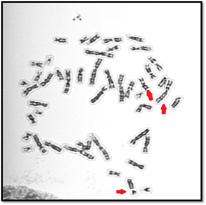
Figure 1. Representative GTG-banded metaphase spread of the proband. Red arrows highlight the chromosomes involved in the visible structural rearrangements. The final band-level karyotype was 46,XX,t(6;7)(q13;p15),der(13;21)(q10;q10),+21[30].
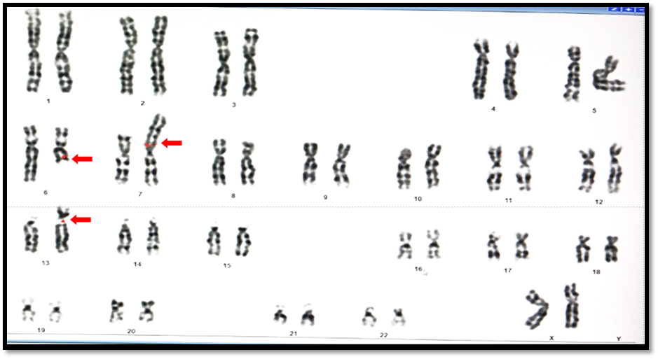
Figure 2. GTG-banded karyogram of the proband showing a female complement with reciprocal t(6;7)(q13;p15), der(13;21)(q10;q10), and additional chromosome 21 material.
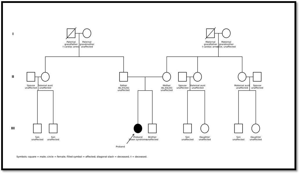
Figure 3. Three-generation pedigree of the proband's family. The proband is indicated by a filled circle and arrow. Both parents are clinically unaffected and have normal peripheral blood karyotypes; The proband has one unaffected brother. The maternal grandmother and paternal grandmother are unaffected, while both grandfathers are deceased due to cardiac arrest as per family history. The mother has two unaffected sisters, each with one unaffected son and one unaffected daughter; the father has one unaffected sister with two unaffected sons. No other family member with Down syndrome, congenital anomaly or known chromosomal rearrangement was reported. The pedigree supports an apparently de novo event in the proband, although very low-level gonadal mosaicism cannot be excluded by peripheral blood karyotyping.
DISCUSSION:
Translocation Down syndrome is uncommon relative to free trisomy 21 but has disproportionate counselling importance. Approximately 3%-4% of individuals with the Down syndrome phenotype have additional chromosome 21 material due to an unbalanced translocation, most often involving chromosome 21 and another acrocentric chromosome.2 A large cytogenetic registry from England and Wales including 29,256 Down syndrome cases reported that nearly 97% were free trisomy 21, while contributory trisomy 21 accounted for 2.9%; the majority of contributory translocations were Robertsonian or rearrangements involving chromosome 21.22 Robertsonian translocations as a class occur in approximately 1 in 1000 newborns, whereas reciprocal translocations have been reported in approximately 0.14% of newborns and are enriched in infertility or recurrent pregnancy-loss settings.17,19
The de novo possibility in the present family is therefore scientifically plausible. In the same national registry, among Robertsonian der(14;21) translocations with known parental origin, 54% were de novo, 41% were maternally inherited and 5% were paternally inherited.22 These proportions should not be applied numerically to every Robertsonian subtype, and they are best established for der(14;21), not specifically for der(13;21). Nevertheless, they support the broader counselling principle that de novo Robertsonian formation is well recognized among translocation Down syndrome cases. In this case, the normal maternal & paternal karyotype support an apparently de novo origin of the proband's der(13;21)(q10;q10),+21 and additional t(6;7)(q13;p15) where a segment from the long arm of Chromosome 6 at band (q13) has swapped places with a segment on the short arm of Chromosome 7 at band (p15) . This combination is rare because it places two different structural-rearrangement mechanisms in the same constitutional karyotype.
Role of karyotyping, FISH, chromosomal microarray and cytogenomics
This case illustrates why conventional cytogenetics remains clinically useful in the genomic era. Karyotyping can detect balanced and unbalanced visible rearrangements, define overall chromosomal architecture and guide parental studies. Rapid interphase FISH or QF-PCR may confirm increased chromosome 21 dosage, but they may not reconstruct the full rearrangement. CMA can detect copy-number imbalance at higher resolution, but it generally will not define a truly balanced reciprocal translocation.6,7
For structurally complex constitutional cases, the most robust interpretation combines conventional karyotyping, parental studies, phenotype correlation and targeted cytogenomic testing. FISH could verify chromosome 21 material on the Robertsonian chromosome and clarify derivative architecture. CMA can assess for cryptic copy-number imbalance, and optical genome mapping or genome sequencing may define breakpoint architecture beyond routine karyotype resolution.6-8,14-16
Clinical management should follow standard Down syndrome health supervision, including cardiac, hearing, developmental, thyroid, growth and other surveillance according to paediatric guidance.2 The cytogenetic complexity should be handled through a genetics service so that medical management and recurrence-risk counselling are not confused.
Genetic counselling implications:
The family should be counselled that the child has extra genetic material from chromosome 21, which explains the Down syndrome phenotype. The extra chromosome 21 material is present because of a structural chromosome rearrangement involving chromosomes 13 and 21. A second separate translocation involving chromosomes 6 and 7 is also present. The result is chromosomal in origin and should not be attributed to parental behaviour, diet, stress, infection, routine medication exposure or any action by either parent.
The pedigree shows unaffected parents, one unaffected brother, unaffected maternal and paternal aunts and unaffected available cousins. This pattern is scientifically compatible with a de novo Robertsonian translocation Down syndrome case; it is not necessary for other relatives to be affected when the rearrangement has arisen in the proband. Therefore, the most appropriate inheritance interpretation is apparently de novo dual structural rearrangement with a low but non-zero recurrence risk due to the residual possibility of parental gonadal mosaicism.3,4
If Robertsonian translocation der(13;21) had been inherited from a parent
If either parent had carried a balanced der(13;21)(q10;q10), the parent would usually have had 45 chromosomes and could be phenotypically normal. During meiosis, the Robertsonian chromosome and the two homologous acrocentric chromosomes form a trivalent. Alternate segregation can produce normal or balanced gametes, whereas adjacent or 2:1 segregation can produce gametes with monosomy or trisomy for the involved acrocentric long arms. A gamete carrying the der(13;21) chromosome together with a normal chromosome 21 can lead to translocation trisomy 21 after fertilization.3,4,17,18
If the balanced carrier were the mother, recurrence risk for a liveborn child with translocation Down syndrome would generally be higher than for a paternal carrier, although the exact empirical risk depends on the chromosomes involved and the specific rearrangement. If the balanced carrier were the father, the liveborn recurrence risk is generally lower, but miscarriage, infertility and unbalanced conceptions remain possible. If the rearrangement were a homologous 21;21 Robertsonian or isochromosome 21q, viable offspring would be expected to have either trisomy 21 or, alternatively, conceptions with monosomy 21 would be non-viable; therefore recurrence risk is far higher than for most non-homologous Robertsonian carriers.3,4
If t(6;7)(q13;p15) had been inherited from a parent
If one parent carried a balanced reciprocal t(6;7)(q13;p15), that rearrangement would not explain the Down syndrome phenotype by itself. However, it would be highly relevant for reproductive risk. In reciprocal translocation carriers, the two derivative chromosomes and the two normal homologues form a quadrivalent during meiosis. The quadrivalent may segregate through alternate, adjacent-1, adjacent-2, 3:1 or rarely 4:0 patterns. Alternate segregation gives either a normal or balanced gamete. Adjacent-1 and adjacent-2 segregation usually create partial monosomy for one segment and partial trisomy for the other. A 3:1 pattern can create a 47- or 45-chromosome conceptus, and 4:0 segregation can create double trisomic or double monosomic products.19,20
Because the parental karyotypes are normal in this case, t(6;7) is not currently shown to be familial. Nevertheless, its presence in the proband is important for future counselling because balanced reciprocal translocation carriers may have risks of infertility, recurrent pregnancy loss or chromosomally unbalanced offspring.5,19,20
Future pregnancy planning for the parents
For any future pregnancy, the couple should be offered preconception or early antenatal genetic counselling. The recurrence risk is expected to be low because both parental karyotypes are normal, but it should not be described as zero. Screening tests, including cell-free DNA screening, assess risk for common aneuploidies but do not define the precise fetal structural chromosomal architecture in a family with a previously affected child and a known structural rearrangement.9,10
Chorionic villus sampling or amniocentesis with fetal karyotyping should be discussed as the key diagnostic approach for determining whether the fetus has a normal chromosome complement, a balanced rearrangement or an unbalanced rearrangement involving chromosome 21. Rapid QF-PCR or interphase FISH may provide early information about chromosomes 13, 18, 21 and sex chromosomes, but it should be considered an adjunct rather than a replacement for full fetal karyotyping when the family issue is structural.2,9,10
Future counselling for the proband
The proband is currently a child, so counselling should be revisited over time. In childhood, the focus should be health supervision, early developmental support, hearing and cardiac follow-up, and family support.2 In adolescence and adulthood, specialist genetic counselling will be required if reproductive planning becomes relevant. The proband carries translocation trisomy 21 and an additional reciprocal translocation; theoretically, meiosis would involve a trivalent related to der(13;21) and a quadrivalent related to t(6;7), producing risks that cannot be summarized by a simple single-translocation recurrence estimate.3,5,19,21
Table 1. Genetic counselling summary
|
Person/group |
Main issue |
Recommended counselling |
|
Parents |
Apparently de novo translocation Down syndrome with der(13;21)(q10;q10),+21 and a second apparently balanced reciprocal t(6;7). Parental karyotypes are documented normal. |
Explaining chromosome 21 dosage imbalance; low but not zero recurrence risk because gonadal mosaicism is not excluded; recommend early genetic counselling and diagnostic prenatal testing in future pregnancies. |
|
Future pregnancies |
Screening alone cannot define fetal structural chromosomal architecture. |
Discussing CVS or amniocentesis with fetal karyotype. Using QF-PCR/FISH only as rapid adjuncts. Adding CMA if ultrasound anomalies or cryptic imbalance concerns exist. |
|
Siblings |
Not expected to be obligate carriers when both parents have documented normal blood karyotypes. |
No mandatory childhood testing solely due to this case. Offering karyotyping later for reproductive planning or if clinical indications arise. |
|
Proband |
Long-term reproductive risk may be complex because of translocation trisomy 21 plus t(6;7)(q13;p15). |
Revisiting counselling in adolescence/adulthood. Future pregnancy would require specialist genetic counselling and diagnostic prenatal testing. |
|
Clinical team |
Separating Down syndrome health supervision from cytogenetic recurrence-risk counselling. |
Continuing Down syndrome health supervision, cardiac and hearing follow-up, developmental support and genetics follow-up as appropriate. |
CONCLUSION:
The der(13;21)(q10;q10),+21 explains the Down syndrome phenotype through chromosome 21 dosage imbalance, while the additional apparently balanced t(6;7)(q13;p15) adds cytogenetic and future reproductive counselling complexity. The case emphasizes the continued importance of conventional karyotyping, parental studies, pedigree assessment and individualized genetic counselling. Although the rearrangement is apparently de novo, recurrence risk should be considered low but not zero because parental gonadal mosaicism cannot be completely excluded.
REFERENCES:
1. Antonarakis SE, Skotko BG, Rafii MS, Strydom A, Pape SE, Bianchi DW, Sherman SL, Reeves RH. Down syndrome. Nat Rev Dis Primers. 2020;6(1):9. doi:10.1038/s41572-019-0143-7.
2. Bull MJ, Trotter T, Santoro SL, Christensen C, Grout RW; Council on Genetics. Health supervision for children and adolescents with Down syndrome. Pediatrics. 2022;149(5):e2022057010. doi:10.1542/peds.2022-057010.
3. Gardner RJM, Amor DJ. Gardner and Sutherland's Chromosome Abnormalities and Genetic Counseling. 5th ed. Oxford: Oxford University Press; 2018. doi:10.1093/med/9780199329007.001.0001.
4. Scriven PN, Flinter FA, Braude PR, Ogilvie CM. Robertsonian translocations: reproductive risks and indications for preimplantation genetic diagnosis. Hum Reprod. 2001;16(11):2267-2273. doi:10.1093/humrep/16.11.2267.
5. Madan K. Balanced complex chromosome rearrangements: reproductive aspects. A review. Am J Med Genet A. 2012;158A(4):947-963. doi:10.1002/ajmg.a.35220.
6. Feenstra I, Hanemaaijer N, Sikkema-Raddatz B, Yntema HG, Dijkhuizen T, Lugtenberg D, et al. Balanced into array: genome-wide array analysis in 54 patients with an apparently balanced de novo chromosome rearrangement and a meta-analysis. Eur J Hum Genet. 2011;19(11):1152-1160. doi:10.1038/ejhg.2011.120.
7. Miller DT, Adam MP, Aradhya S, Biesecker LG, Brothman AR, Carter NP, et al. Consensus statement: chromosomal microarray is a first-tier clinical diagnostic test for individuals with developmental disabilities or congenital anomalies. Am J Hum Genet. 2010;86(5):749-764. doi:10.1016/j.ajhg.2010.04.006.
8. Sismani C, Kitsiou-Tzeli S, Ioannides M, Christodoulou C, Anastasiadou V, Stylianidou G, Papadopoulou E, Kanavakis E, Kosmaidou-Aravidou Z, Patsalis PC. Cryptic genomic imbalances in patients with de novo or familial apparently balanced translocations and abnormal phenotype. Mol Cytogenet. 2008;1:15. doi:10.1186/1755-8166-1-15.
9. American College of Obstetricians and Gynecologists. Screening for fetal chromosomal abnormalities: Practice Advisory. Published January 7, 2026. Available at: https://www.acog.org/clinical/clinical-guidance/practice-advisory/articles/2026/01/screening-for-fetal-chromosomal-abnormalities. Accessed July 6, 2026.
10. Rink BD, Dugoff L, Kuller JA; Society for Maternal-Fetal Medicine Publications Committee. Society for Maternal-Fetal Medicine Consult Series #74: Cell-free DNA screening for aneuploidies: updated guidance. Pregnancy. 2025;1:e70139. doi:10.1002/pmf2.70139.
11. American College of Obstetricians and Gynecologists. Preimplantation genetic testing: ACOG Committee Opinion, Number 799. Obstet Gynecol. 2020;135(3):e133-e137. doi:10.1097/AOG.0000000000003714.
12. ISCN 2024 - An International System for Human Cytogenomic Nomenclature (2024). Cytogenet Genome Res. 2024;164(Suppl 1):1-224. doi:10.1159/000538512.
13. Gagnier JJ, Kienle G, Altman DG, Moher D, Sox H, Riley DS; CARE Group. The CARE guidelines: consensus-based clinical case reporting guideline development. J Clin Epidemiol. 2014;67(1):46-51. doi:10.1016/j.jclinepi.2013.08.003.
14. Schluth-Bolard C, Diguet F, Chatron N, Rollat-Farnier PA, Bardel C, Afenjar A, et al. Whole genome paired-end sequencing elucidates functional and phenotypic consequences of balanced chromosomal rearrangement in patients with developmental disorders. J Med Genet. 2019;56(8):526-535. doi:10.1136/jmedgenet-2018-105778.
15. Mantere T, Neveling K, Pebrel-Richard C, Benoist M, van der Zande G, Kater-Baats E, et al. Optical genome mapping enables constitutional chromosomal aberration detection. Am J Hum Genet. 2021;108(8):1409-1422. doi:10.1016/j.ajhg.2021.05.012.
16. Zhang S, Pei Z, Lei C, Zhu S, Deng K, Zhou J, et al. Detection of cryptic balanced chromosomal rearrangements using high-resolution optical genome mapping. J Med Genet. 2023;60(3):274-284. doi:10.1136/jmedgenet-2022-108553.
17. Wang B, Nie B, Tang D, Li R, Liu X, Song J, et al. Analysis of meiotic segregation patterns and interchromosomal effects in sperm from 13 Robertsonian translocations. Balkan J Med Genet. 2017;20(1):43-50. doi:10.1515/bjmg-2017-0003.
18. Zhang S, Lei C, Wu J, Zhou J, Xiao M, Zhu S, et al. Meiotic heterogeneity of trivalent structure and interchromosomal effect in blastocysts with Robertsonian translocations. Front Genet. 2021;12:609563. doi:10.3389/fgene.2021.609563.
19. Xie P, Hu L, Peng Y, Tan YQ, Luo K, Gong F, et al. Risk factors affecting alternate segregation in blastocysts from preimplantation genetic testing cycles of autosomal reciprocal translocations. Front Genet. 2022;13:880208. doi:10.3389/fgene.2022.880208.
20. Verdoni A, Hu J, Surti U, Babcock M, Sheehan E, Clemens M, et al. Reproductive outcomes in individuals with chromosomal reciprocal translocations. Genet Med. 2021;23(9):1753-1760. doi:10.1038/s41436-021-01195-w.
21. Pellestor F, Anahory T, Lefort G, Puechberty J, Liehr T, Hédon B, Sarda P. Complex chromosomal rearrangements: origin and meiotic behavior. Hum Reprod Update. 2011;17(4):476-494. doi:10.1093/humupd/dmr010.
22. Morris JK, Alberman E, Mutton D, Jacobs P. Cytogenetic and epidemiological findings in Down syndrome: England and Wales 1989-2009. Am J Med Genet A. 2012;158A(5):1151-1157. doi:10.1002/ajmg.a.35248.
23. Rawal, L., Kumar, S., Mishra, S. R., Lal, V., & Bhattacharya, S. K. (2020).
Clinical manifestations of chromosomal anomalies and polymorphic variations in patients suffering from reproductive failure. Journal of Human Reproductive Sciences, 13(3), 209–215.
https://doi.org/10.4103/jhrs.JHRS_89_20.