A Study of Association of ABO Blood Group Types and Secretor Status in Patients with Oral Potentially Malignant Disorders (OPMDs) in Government General Hospital, Kadapa.
- Parveen Shaik , Associate Professor, Department of Physiology, Government Medical College, Kadapa, Andhra Pradesh, India.
- P. Nagajyothi , Assistant Professor, Department of Dentistry, Government Medical College, Kadapa, Andhra Pradesh, India.
- Nagamani Mekala , Assistant Professor, Department of Dentistry, Government Medical College, Pulivendula, Y.S.R. District, Andhra Pradesh, India.
Article Information:
Abstract:
Background: Oral potentially malignant disorders (OPMDs) are precursor lesions with a risk of progression to oral cancer. ABO blood group antigens and salivary secretor status have been proposed as host-related factors influencing susceptibility to these disorders. This study evaluated the association of ABO blood groups and secretor status with OPMDs. Aim: To assess the association between ABO blood group types, salivary secretor status, and oral potentially malignant disorders in patients attending Government General Hospital, Kadapa. Materials and Methods: A hospital-based case–control study was conducted from June 2023 to April 2025 among 90 participants, comprised clinically diagnosed OPMD cases (n=45) and age and sex-matched healthy controls (n=45). ABO blood grouping was performed using the slide agglutination method. Salivary secretor status was determined by the hemagglutination inhibition technique. Statistical analysis was performed using the Chi-square test and odds ratio, with a p-value <0.05 considered statistically significant. Results: The majority of OPMD patients were aged 36–45 years (28.9%), and tobacco chewing was the predominant habit (68.9%). Blood group A was the most common blood group among OPMD patients (40.0%), followed by O (26.7%), B (22.2%), and AB (11.1%); however, no significant association was observed between ABO blood groups and OPMDs (p>0.05). In contrast, salivary non-secretor status was significantly associated with OPMDs. Among the OPMD group, 39 (86.7%) were non-secretors compared with 8 (17.8%) among controls (p<0.001). Although all patients with multiple-site lesions were non-secretors, the association between secretor status and lesion multiplicity was not statistically significant (p=0.098). Conclusion: Salivary non-secretor status is strongly associated with oral potentially malignant disorders and may serve as a useful host-related risk marker for identifying individuals at increased risk. Although blood group A was more frequent among patients with OPMDs, ABO blood group was not significantly associated with disease occurrence. Larger multicentre studies are needed to validate these findings.
Keywords:
Article :
INTRODUCTION:
Oral cancer is one of the most common malignancies worldwide and a major public health concern, particularly in developing countries. India accounts for a significant proportion of the global burden of oral cancer, with most cases arising from oral potentially malignant disorders (OPMDs) such as leukoplakia, oral submucous fibrosis (OSF), oral lichen planus, and erythroplakia. Early identification of individuals at increased risk of malignant transformation is essential for improving prognosis and reducing mortality.1,2,3
The development of OPMDs is multifactorial. Although tobacco use, areca nut chewing, alcohol consumption, poor oral hygiene, and nutritional deficiencies are well-established risk factors, genetic susceptibility also contributes to disease development.⁴ The ABO blood group system, one of the most extensively studied genetic polymorphisms, has been implicated in susceptibility to several malignancies, including oral cancer. While some studies have reported a higher prevalence of blood group A among patients with oral cancer and oral lichen planus, others have found no significant association.5-8
Another genetically determined host factor is salivary secretor status, which is controlled by the FUT2 (Se) gene. Secretors express ABO(H) blood group antigens in saliva and other body fluids, whereas non-secretors lack these antigens. These salivary antigens contribute to mucosal integrity, microbial homeostasis, and local immune defence. Several studies have reported a significantly higher prevalence of non-secretor status among patients with OPMDs, suggesting that the absence of salivary ABO antigens may increase susceptibility to oral premalignant lesions.9-12 However, conflicting findings have also been reported, warranting further investigation.13,14
Evidence regarding the association between ABO blood groups, secretor status, and OPMDs remains limited, particularly in the Indian population. Therefore, the present study was undertaken to evaluate the association between ABO blood group types, salivary secretor status, and oral potentially malignant disorders among patients attending Government General Hospital, Kadapa.
Study Aim:
To evaluate the association between ABO blood group types and secretor status among patients with oral potentially malignant disorders (OPMDs) attending Government General Hospital, Kadapa, and to compare these findings with those of healthy controls.
MATERIALS AND METHODS:
Study Design and Setting
A hospital-based case–control observational study was conducted from June 2023 to April 2025 in the Departments of Physiology and Dentistry at Government General Hospital, Kadapa, Andhra Pradesh. The study was approved by the Institutional Ethics Committee (IEC No.: ACAD/E3B/2022-2023), Government Medical College, Kadapa and written informed consent was obtained from all participants.
Study Population
A total of 90 participants were enrolled, comprising 45 patients with oral potentially malignant disorders (OPMDs) (cases) and 45 age- and sex-matched healthy individuals (controls). Participants were selected by simple random sampling.
Inclusion Criteria
Patients with clinically diagnosed OPMDs, including oral submucous fibrosis (OSMF), oral lichen planus (OLP), and leukoplakia, were included. Clinically suspicious lesions were confirmed by histopathological examination.
Exclusion Criteria
Patients with previously diagnosed oral premalignant or malignant lesions receiving treatment and those unwilling to participate were excluded.
ABO Blood Group Determination
ABO blood grouping was performed using the slide agglutination method. Capillary blood obtained by finger prick was mixed separately with anti-A and anti-B antisera on a glass slide. Blood groups were identified based on the agglutination pattern: agglutination with anti-A indicated blood group A, with anti-B indicated blood group B, with both antisera indicated blood group AB, and absence of agglutination indicated blood group O.
Determination of Secretor Status
Secretor status was determined using the haemagglutination inhibition technique. Approximately 1 mL of unstimulated saliva was collected between 11:00 AM and 12:00 PM under standardized conditions. Samples were boiled at 100°C for 20 minutes, centrifuged at 3000 rpm for 15 minutes, and the supernatant was diluted (1:4). Anti-A, anti-B, or anti-H antisera (depending on the participant's blood group) were diluted to 1:8 and incubated with the processed saliva. Subsequently, a 5% red cell suspension was added and the mixture was examined microscopically. Absence of agglutination indicated secretor status, while agglutination indicated non-secretor status. Saline controls were included with each batch of tests.
Statistical Analysis
Data were analyzed using SPSS version 24.0 (IBM Corp., Armonk, NY, USA). Categorical variables were expressed as frequencies and percentages. Associations between ABO blood groups, salivary secretor status, and oral potentially malignant disorders (OPMDs) were assessed using the Chi-square test. Odds ratios (ORs) with 95% confidence intervals (CIs) were calculated to determine the strength of association. A p-value <0.05 was considered statistically significant.
RESULTS:
A total of 45 patients with oral potentially malignant disorders (OPMDs) and 45 healthy controls were included in the study.
Table 1. Age-wise distribution of patients with oral potentially malignant disorders (n = 45)
|
Age group (years) |
Number of patients |
Percentage (%) |
|
16–25 |
4 |
8.9 |
|
26–35 |
9 |
20.0 |
|
36–45 |
13 |
28.9 |
|
46–55 |
10 |
22.2 |
|
56–65 |
6 |
13.3 |
|
66–75 |
3 |
6.7 |
|
Total |
45 |
100.0 |
Graph 1: Age-wise distribution of patients with oral potentially malignant disorders (n = 45)
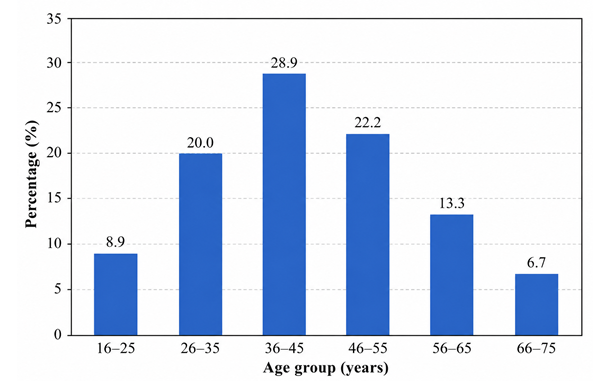
The highest proportion of patients belonged to the 36–45 years age group (13, 28.9%), followed by the 46–55 years age group (10, 22.2%) and the 26–35 years age group (9, 20.0%). The lowest frequency was observed among patients aged 66–75 years (3, 6.7%) [Table 1] [Graph 1].
Table 2. Distribution of ABO blood groups among patients with OPMDs and healthy controls
|
Blood Group |
OPMD Patients n (%) |
Controls n (%) |
p-value |
|
A |
18 (40.0) |
16 (35.6) |
0.662 |
|
B |
10 (22.2) |
12 (26.7) |
0.624 |
|
AB |
5 (11.1) |
3 (6.7) |
0.458 |
|
O |
12 (26.7) |
14 (31.1) |
0.648 |
|
Total |
45 (100) |
45 (100) |
Graph 2: Distribution of ABO blood groups among patients with OPMDs and healthy controls
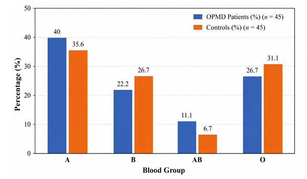
Blood group A was the most common blood group among patients with OPMDs (18, 40.0%), followed by blood groups O (12, 26.7%), B (10, 22.2%), and AB (5, 11.1%). A similar distribution was observed among healthy controls. Comparison of ABO blood group frequencies between the two groups showed no statistically significant association (p > 0.05) [Table 2] [Graph 2].
Table 3. Odds ratios showing the association between ABO blood groups and oral potentially malignant disorders (OPMDs)
|
Blood Group |
Odds Ratio (OR) |
95% Confidence Interval |
|
A |
1.21 |
0.54–2.72 |
|
B |
0.78 |
0.31–1.98 |
|
AB |
1.75 |
0.39–7.84 |
|
O |
0.81 |
0.34–1.95 |
OR, Odds Ratio; CI, Confidence Interval.
Individuals with blood group A showed a slightly higher likelihood of developing OPMDs (OR = 1.21; 95% CI: 0.54–2.72), whereas blood group B (OR = 0.78; 95% CI: 0.31–1.98) and blood group O (OR = 0.81; 95% CI: 0.34–1.95) showed no increased risk. Although blood group AB demonstrated a relatively higher odds ratio (OR = 1.75; 95% CI: 0.39–7.84), the wide confidence interval indicates imprecision due to the small number of participants. As all confidence intervals included 1.0, none of the observed associations were statistically significant [Table 3].
Table 4. Distribution of tobacco-related habits among patients with oral potentially malignant disorders (OPMDDs) (n = 45)
|
Habit |
Frequency (n) |
Percentage (%) |
|
Tobacco chewing |
31 |
68.9 |
|
Tobacco smoking |
5 |
11.1 |
|
Combination of habits* |
9 |
20.0 |
|
Total |
45 |
100.0 |
Combination of habits includes participants with more than one tobacco-related habit (e.g., tobacco chewing with smoking).
Graph 3: Distribution of tobacco-related habits among patients with oral potentially malignant disorders (OPMDDs) (n = 45)
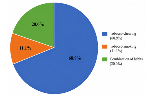
Tobacco chewing was the most common habit, reported by 31 (68.9%) patients, followed by combined tobacco habits in 9 (20.0%) patients. Tobacco smoking alone was reported by 5 (11.1%) patients. These findings indicate that smokeless tobacco use was the predominant habit among patients with OPMDs [Table 4] [Graph 3].
Table 5. Salivary secretor status among patients with oral potentially malignant disorders (OPMDs) and healthy controls
|
Group |
Non-secretors n (%) |
Secretors n (%) |
Total n (%) |
|
OPMDs |
39 (86.7) |
6 (13.3) |
45 (100) |
|
Controls |
8 (17.8) |
37 (82.2) |
45 (100) |
|
p-value |
<0.001 |
||
Graph 4: Salivary secretor status among patients with oral potentially malignant disorders (OPMDs) and healthy controls
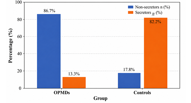
Among the 45 patients with OPMDs, 39 (86.7%) were non-secretors, whereas only 6 (13.3%) were secretors. In contrast, among the 45 healthy controls, 8 (17.8%) were non-secretors and 37 (82.2%) were secretors. The difference in secretor status between the two groups was highly statistically significant (p < 0.001), indicating a strong association between non-secretor status and oral potentially malignant disorders [Table 5] [Graph 4].
Table 6. Association between gender and salivary secretor status among patients with OPMDs and healthy controls
|
Gender |
OPMD Group |
Control Group |
||
|
Non-secretors n (%) |
Secretors n (%) |
Non-secretors n (%) |
Secretors n (%) |
|
|
Men |
31 (68.9) |
5 (11.1) |
5 (11.1) |
18 (40.0) |
|
Women |
8 (17.8) |
1 (2.2) |
3 (6.7) |
19 (42.2) |
|
Total |
39 (86.7) |
6 (13.3) |
8 (17.8) |
37 (82.2) |
|
p-value |
<0.001 |
|||
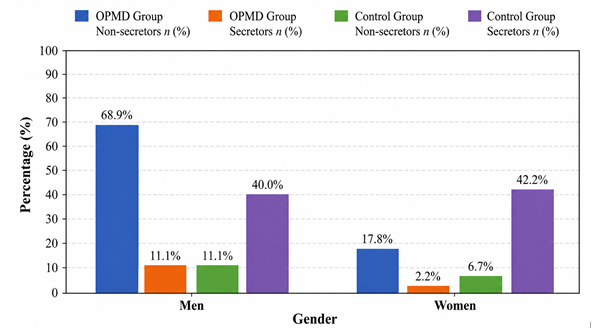
Graph 5: Association between gender and salivary secretor status among patients with OPMDs and healthy controls
In the case group, 31 (68.9%) men and 8 (17.8%) women were non-secretors, whereas 5 (11.1%) men and 1 (2.2%) woman were secretors. In the control group, 5 (11.1%) men and 3 (6.7%) women were non-secretors, while 18 (40.0%) men and 19 (42.2%) women were secretors. The distribution of secretor status differed significantly between the OPMD and control groups (p < 0.001), with non-secretor status being markedly more prevalent among patients with OPMDs irrespective of gender [Table 6] [Graph 5].
Table 7. Association between salivary secretor status and number of lesion sites in patients with OPMDs (n = 45)
|
Number of lesion sites |
Secretors n (%) |
Non-secretors n (%) |
|
Single site |
6 (13.3) |
24 (53.3) |
|
Multiple sites |
0 (0.0) |
15 (33.4) |
|
Total |
6 (13.3) |
39 (86.7) |
|
p-value |
0.098 |
|
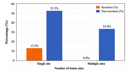
Graph 6: Association between salivary secretor status and number of lesion sites in patients with OPMDs (n = 45)
Among patients with single-site lesions, 24 (53.3%) were non-secretors and 6 (13.3%) were secretors. All patients with multiple-site lesions (15; 33.4%) were non-secretors, while no secretors had multiple-site involvement. Although multiple-site lesions were observed exclusively among non-secretors, the association between secretor status and the number of lesion sites did not reach statistical significance (p = 0.098) [Table 7] [Graph 6].
DISCUSSION:
The ABO blood group system is the most extensively studied erythrocyte antigen system, and its association with various diseases, including malignancies, diabetes mellitus, cardiovascular diseases, and oral disorders, has been widely investigated. Although several studies have suggested an association between ABO blood groups and oral cancer, the findings remain inconsistent across different populations.1-4
The present case–control study evaluated the association of ABO blood groups and salivary secretor status with oral potentially malignant disorders (OPMDs) in patients attending Government General Hospital, Kadapa. The results demonstrated a significant association between non-secretor status and OPMDs, whereas no significant association was observed between ABO blood groups and the occurrence of OPMDs.
In the present study, the highest proportion of patients with OPMDs belonged to the 36–45-year age group, reflecting the age at which cumulative exposure to tobacco and areca nut is sufficient to produce premalignant changes. Tobacco chewing was the predominant habit, accounting for 68.9% of cases, highlighting its role as the major etiological factor for OPMDs in the study population.5,6
Although blood group A was the most common blood group among patients with OPMDs, the difference in ABO blood group distribution between cases and controls was not statistically significant. Similar findings were reported by Moshaverinia et al., who found no association between ABO blood groups and oral lichen planus, and by Hallikeri et al., who observed no significant relationship between ABO blood groups and oral submucous fibrosis.7,8 In contrast, Tyagi et al., Jaleel and Nagarajappa, and Kumar et al. reported a higher prevalence of blood group A among patients with oral cancer or oral lichen planus.9-11 These discrepancies may be attributed to differences in ethnicity, geographic distribution, sample size, and study design.
The most significant finding of the present study was the strong association between non-secretor status and OPMDs. Among patients with OPMDs, 86.7% were non-secretors compared with only 17.8% of healthy controls, and this difference was highly significant (p < 0.001). Secretor status is genetically determined by the FUT2 (Se) gene, which regulates the secretion of ABO(H) blood group antigens into saliva and other body fluids. Individuals carrying the dominant Se allele express these antigens in secretions, whereas homozygous se/se individuals lack them and are classified as non-secretors.12,13
The absence of ABO(H) antigens in saliva may reduce mucosal protection, alter oral microbial colonization, and impair local immune defence mechanisms, thereby increasing susceptibility to oral mucosal disease. Previous studies by Lamey et al. suggested that non-secretor status is a genetically determined risk marker for candidal leukoplakia.14 Similarly, Vidas et al. demonstrated a higher prevalence of non-secretors among patients with oral precancerous lesions, with epithelial dysplasia occurring exclusively in non-secretors.15 Hallikeri et al. also reported that all patients with oral submucous fibrosis in their study were non-secretors, while Pourazar et al. observed a significantly higher frequency of non-secretors among patients with leukoplakia.8,16 The findings of the present study are in agreement with these reports and further support the role of non-secretor status as a host susceptibility factor for OPMDs.
The association between secretor status and the number of lesion sites was not statistically significant (p = 0.098), although all patients with multiple-site lesions were non-secretors. This trend suggests that non-secretor status may influence disease severity or extent; however, larger studies are required to confirm this observation.
However, conflicting evidence also exists. Cerovic et al. and Lamey et al. found no significant association between secretor status and oral cancer, indicating that secretor phenotype alone may not determine malignant transformation.17,18 These differences may reflect variations in study populations, disease spectrum, environmental exposures, and genetic background.
The present study has certain limitations. It was conducted at a single tertiary care centre with a relatively small sample size, limiting the generalizability of the findings. Molecular evaluation of the FUT2 gene was not performed, and patients were not followed longitudinally to assess malignant transformation. Future multicentre studies involving larger populations, molecular genetic analysis, and long-term follow-up are warranted to further clarify the role of ABO blood groups and secretor status in oral carcinogenesis.
In conclusion, the present study demonstrates that salivary non-secretor status is significantly associated with oral potentially malignant disorders and may serve as a useful host-related risk marker, whereas ABO blood group alone does not appear to be significantly associated with the occurrence of OPMDs. Assessment of salivary secretor status may aid in identifying individuals at increased risk for developing OPMDs and facilitate early preventive interventions.
CONCLUSION:
CONCLUSION
Oral submucous fibrosis (OSF) was the most common oral potentially malignant disorder (OPMD) observed in the present study, likely reflecting the high prevalence of chronic gutka and pan chewing in the study population. Although blood group A was more frequently observed among patients with OPMDs than other ABO blood groups, no statistically significant association was found between ABO blood group and OPMDs, indicating that ABO blood group alone is not a significant risk factor.
In contrast, a strong and statistically significant association was observed between salivary non-secretor status and OPMDs. The markedly higher prevalence of non-secretors among patients with OPMDs suggests that the inability to secrete ABO blood group antigens into saliva may represent an important host-related susceptibility factor. Assessment of salivary secretor status may therefore serve as a useful adjunctive biomarker for identifying individuals at increased risk of developing OPMDs. Further multicentre studies with larger sample sizes and molecular evaluation are recommended to validate these findings.
REFERENCES:
1. World Health Organization. Oral health. Geneva: World Health Organization; 2020.
2. Warnakulasuriya S. Global epidemiology of oral and oropharyngeal cancer. Oral Oncol. 2009;45(4-5):309-316.
3. Indian Council of Medical Research, National Centre for Disease Informatics and Research. National Cancer Registry Programme Report 2020. Bengaluru: ICMR-NCDIR; 2020.
4. Sankaranarayanan R, Ramadas K, Amarasinghe H, Subramanian S, Johnson N. Oral cancer: prevention, early detection, and treatment. In: Disease Control Priorities. 3rd ed. Vol. 3. Washington (DC): World Bank; 2015.
5. Gupta B, Johnson NW, Kumar N. Global epidemiology of head and neck cancers: a continuing challenge. Oncology. 2016;91(1):13-23.
6. Oral Submucous Fibrosis. Tilakaratne WM, Klinikowski MF, Saku T, Peters TJ, Warnakulasuriya S. Oral submucous fibrosis: review on aetiology and pathogenesis. Oral Oncol. 2006;42(6):561-568.
7. Moshaverinia M, Rakhshan V, Moshaverinia A, et al. ABO blood group and oral lichen planus: a case-control study. J Contemp Dent Pract. 2014;15(5):558-562.
8. Hallikeri K, Naikmasur VG, Guttal KS, Shodan M. Analysis of salivary secretor status in patients with oral submucous fibrosis. J Oral Maxillofac Pathol. 2012;16(3):359-363.
9. Tyagi SP, Maheshwari V, Tyagi N. ABO blood groups and oral cancer: a hospital-based study. Indian J Otolaryngol Head Neck Surg. 1988;40:20-23.
10. Jaleel BF, Nagarajappa R. Relationship between ABO blood groups and oral cancer. Indian J Dent Res. 2012;23(1):7-10.
11. Kumar S, Koneru A, Babu AM, et al. Association of ABO blood groups with oral lichen planus. J Clin Diagn Res. 2015;9(4):ZC40-ZC42.
12. Secretor Status. Henry S. Molecular diversity in the biosynthesis of ABO, H, Lewis and related histo-blood group antigens. Vox Sang. 1995;69(3):166-182.
13. Kelly RJ, Rouquier S, Giorgi D, Lennon GG, Lowe JB. Sequence and expression of a candidate for the human Secretor blood group α(1,2)-fucosyltransferase gene (FUT2). J Biol Chem. 1995;270(9):4640-4649.
14. Lamey PJ, Darwazeh AM, Muirhead J, Rennie JS, Samaranayake LP. Secretor status, candidal carriage and oral precancerous lesions. J Oral Pathol Med. 1991;20(3):103-105.
15. Vidas I, Grdović M, Brailo V, et al. Secretor status and oral precancerous lesions. Oral Dis. 1999;5(3):249-252.
16. Pourazar A, Cerović R, Furberg AS, et al. Salivary secretor status in patients with oral leukoplakia. J Oral Pathol Med. 2005;34(7):421-425.
17. Cerović R, Juretić M, Belušić-Gobić M, et al. Secretor status and oral cancer. Coll Antropol. 2001;25(Suppl):83-87.
18. Lamey PJ, Nolan A, Forsyth A. Secretor status and oral cancer in Sri Lankan patients. J Oral Pathol Med. 1994;23(6):271-273.