ROLE OF TWO-DIMENSIONAL ECHOCARDIOGRAPHY IN THE EARLY DETECTION OF LEFT VENTRICULAR DIASTOLIC DYSFUNCTION AMONG PATIENTS WITH HYPERTENSION: A HOSPITAL-BASED CROSS-SECTIONAL STUDY.
- Bajarang Lal Bansal , Professor & HOD, Department of Internal Medicine, Abhishek I Mishra Memorial Medical College and Hospital, Bhilai, Durg, Chhattisgarh, India.
- Ashok Kumar , Associate Professor, Department of General Medicine, Abhishek I Mishra Memorial Medical College and Hospital, Bhilai, Durg, Chhattisgarh, India.
- Chirag. C. Taunk , Senior Resident, Department of Respiratory Medicine, Shri Shankaracharya Institute of Medical Sciences, Bhilai, Durg, Chhattisgarh, India.
Article Information:
Abstract:
Background: Hypertension is a major risk factor for cardiovascular disease and causes progressive structural and functional changes in the myocardium. Left ventricular diastolic dysfunction (LVDD) is an early manifestation of hypertensive heart disease that often remains asymptomatic despite preserved systolic function. Early detection is essential to prevent progression to heart failure. Two-dimensional echocardiography is a reliable, non-invasive modality for identifying early diastolic dysfunction. Objective: To evaluate the role of two-dimensional echocardiography in the early detection of left ventricular diastolic dysfunction among patients with hypertension and to determine its association with selected clinical characteristics. Methods: This hospital-based cross-sectional study was conducted from December 2025 to May 2026 and included 120 adult patients with essential hypertension. All participants underwent clinical evaluation and two-dimensional echocardiography. Left ventricular systolic and diastolic functions were assessed using standard echocardiographic parameters including left ventricular ejection fraction, E/A ratio, E/e′ ratio, deceleration time, isovolumetric relaxation time, left atrial volume index, and left ventricular mass index. Statistical analysis was performed using SPSS version 26.0. Results: Left ventricular diastolic dysfunction was detected in 58.3% of patients. Increasing age, longer duration of hypertension, uncontrolled blood pressure, and left ventricular hypertrophy were significantly associated with diastolic dysfunction. Grade I diastolic dysfunction was the most common finding. Conclusion: Two-dimensional echocardiography is an effective non-invasive tool for the early detection of left ventricular diastolic dysfunction in hypertensive patients. Routine echocardiographic evaluation may facilitate timely diagnosis, improve risk stratification, and support early intervention to reduce cardiovascular complications.
Keywords:
Article :
INTRODUCTION:
Hypertension is one of the most common cardiovascular disorders worldwide and a major contributor to morbidity and mortality. Persistent elevation of blood pressure leads to structural and functional changes in the heart, including left ventricular hypertrophy, myocardial fibrosis, and impaired ventricular relaxation. These alterations often occur before the appearance of clinical symptoms and increase the risk of heart failure, arrhythmias, stroke, and other cardiovascular complications.[1]
Left ventricular diastolic dysfunction (LVDD) is considered an early manifestation of hypertensive heart disease and may develop despite preserved left ventricular systolic function. Because patients are frequently asymptomatic during the initial stages, early identification of diastolic dysfunction provides an opportunity for timely intervention and better blood pressure management, thereby reducing the likelihood of progression to heart failure with preserved ejection fraction.[2,3]
Two-dimensional echocardiography, together with Doppler and tissue Doppler imaging, is a reliable, non-invasive method for evaluating cardiac structure and diastolic function. Echocardiographic parameters such as the E/A ratio, E/e′ ratio, deceleration time, isovolumetric relaxation time, left atrial volume index, and left ventricular mass index are widely used for the assessment and grading of LVDD. Owing to its accuracy, availability, and cost-effectiveness, echocardiography has become an important investigation for the routine evaluation of hypertensive patients.[4]
Recent studies have demonstrated that advancing age, prolonged duration of hypertension, poor blood pressure control, and left ventricular hypertrophy are associated with a higher prevalence of left ventricular diastolic dysfunction and an increased risk of adverse cardiovascular outcomes.[5,6] Therefore, the present study was undertaken to evaluate the role of two-dimensional echocardiography in the early detection of left ventricular diastolic dysfunction among patients with hypertension and to determine its association with selected clinical characteristics.
Aim: To evaluate the role of two-dimensional echocardiography in the early detection of left ventricular diastolic dysfunction among patients with hypertension.
MATERIALS AND METHODS:
This hospital-based cross-sectional observational study was conducted in the Departments of Medicine and Cardiology of a tertiary care teaching hospital from December 2025 to May 2026. A total of 120 adult patients (≥18 years) with previously diagnosed or newly detected essential hypertension attending the outpatient department or admitted to the medicine wards during the study period were enrolled after fulfilling the eligibility criteria.
Inclusion Criteria
• Patients aged 18 years or above.
• Patients with previously diagnosed or newly detected essential hypertension.
• Patients willing to undergo two-dimensional echocardiographic evaluation.
• Patients who provided written informed consent.
Exclusion Criteria
• Patients with known coronary artery disease or previous myocardial infarction.
• Patients with moderate or severe valvular heart disease.
• Patients with congenital heart disease or cardiomyopathy.
• Patients with persistent atrial fibrillation or other significant arrhythmias.
• Patients with severe chronic kidney disease or inadequate echocardiographic windows.
Study Procedure
All enrolled patients underwent detailed clinical evaluation, blood pressure measurement, and transthoracic two-dimensional echocardiography. Left ventricular systolic and diastolic functions were evaluated using standard echocardiographic parameters, including left ventricular ejection fraction, transmitral E/A ratio, E/e′ ratio, deceleration time, isovolumetric relaxation time, left atrial volume index, and left ventricular mass index. Left ventricular diastolic dysfunction was graded according to contemporary echocardiographic recommendations.
Statistical Analysis
Statistical analysis was performed using SPSS version 26.0. Continuous variables were expressed as mean ± standard deviation, whereas categorical variables were presented as frequencies and percentages. Comparisons between groups were performed using the Independent Student's t-test for continuous variables and the Chi-square test or Fisher's exact test for categorical variables, as appropriate. A p-value of <0.05 was considered statistically significant.
RESULTS:
Table 1: Baseline demographic characteristics of the study participants (n=120)
|
Variable |
Frequency |
Percentage |
|
|
Age (years) |
18–40 |
20 |
16.7% |
|
41–50 |
30 |
25% |
|
|
51–60 |
42 |
35% |
|
|
>60 |
28 |
23.3% |
|
|
Gender |
Male |
68 |
56.7% |
|
Female |
52 |
43.3% |
|
The majority of participants belonged to the 51–60 years age group (35%). Male participants constituted 56.7% of the study population, indicating a slight male predominance.
Table 2: Echocardiographic findings among hypertensive patients (n=120)
|
Echocardiographic parameter |
Frequency |
Percentage |
|
Preserved LVEF |
111 |
92.5% |
|
Left ventricular hypertrophy |
67 |
55.8% |
|
Increased LV mass index |
62 |
51.7% |
|
Left atrial enlargement |
40 |
33.3% |
|
Left ventricular diastolic dysfunction |
70 |
58.3% |
Preserved left ventricular ejection fraction was observed in 92.5% of patients. Left ventricular hypertrophy, increased left ventricular mass index, and left atrial enlargement were identified in 55.8%, 51.7%, and 33.3% of patients, respectively. Overall, left ventricular diastolic dysfunction was detected in 58.3% of hypertensive patients.
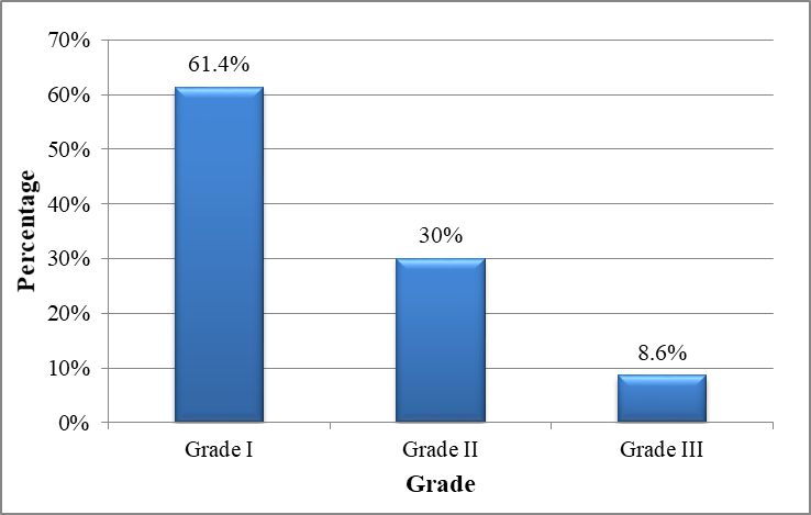
Figure 1: Distribution of Grades of Left Ventricular Diastolic Dysfunction Among Patients with LVDD (n=70)
Grade I diastolic dysfunction was the predominant finding (61.4%), followed by Grade II (30%) and Grade III (8.6%), indicating that impaired myocardial relaxation was the most common pattern of diastolic dysfunction.
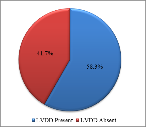
Figure 2: Prevalence of left ventricular diastolic dysfunction among hypertensive patients (n=120)
Left ventricular diastolic dysfunction was present in 58.3% of hypertensive patients, whereas 41.7% had normal diastolic function.
Table 3: Association between duration of hypertension and left ventricular diastolic dysfunction
|
Duration of hypertension |
LVDD Present |
LVDD Absent |
p-value |
|
<5 years (n=44) |
18 |
26 |
0.003 |
|
5–10 years (n=46) |
29 |
17 |
|
|
>10 years (n=30) |
23 |
7 |
The prevalence of left ventricular diastolic dysfunction increased significantly with increasing duration of hypertension (p=0.003), suggesting progressive impairment of diastolic function with prolonged disease duration.
Table 4: Association between blood pressure control and left ventricular diastolic dysfunction
|
Blood pressure status |
LVDD Present |
LVDD Absent |
p-value |
|
Controlled (n=46) |
18 |
28 |
0.001 |
|
Uncontrolled (n=74) |
52 |
22 |
Left ventricular diastolic dysfunction was significantly more frequent among patients with uncontrolled hypertension than among those with controlled blood pressure (p=0.001), highlighting the importance of adequate blood pressure control
DISCUSSION:
The present hospital-based cross-sectional study evaluated the role of two-dimensional echocardiography in the early detection of left ventricular diastolic dysfunction (LVDD) among patients with hypertension. Left ventricular diastolic dysfunction was detected in 58.3% of the study participants despite preserved left ventricular systolic function in the majority of patients. These findings indicate that diastolic dysfunction is an early manifestation of hypertensive heart disease and may be identified before the development of overt systolic dysfunction. Similar observations have been reported in previous studies, supporting the value of echocardiography as a reliable, non-invasive modality for detecting subclinical myocardial dysfunction in hypertensive individuals.[7,8]
Grade I diastolic dysfunction was the predominant abnormality, accounting for 61.4% of patients with LVDD, suggesting that impaired myocardial relaxation represents the earliest stage of diastolic dysfunction in hypertension. The present study also demonstrated a significant association between longer duration of hypertension and the presence of LVDD. Furthermore, uncontrolled blood pressure was significantly associated with diastolic dysfunction, highlighting the adverse effects of persistent pressure overload on myocardial structure and function. These findings are in agreement with previous reports showing that prolonged hypertension and inadequate blood pressure control contribute to progressive impairment of left ventricular diastolic function and increase cardiovascular risk.[9,10]
More than half of the study participants had left ventricular hypertrophy, and increased left ventricular mass index was frequently associated with diastolic dysfunction. Chronic hypertension promotes myocardial hypertrophy, interstitial fibrosis, and reduced ventricular compliance, resulting in impaired ventricular relaxation. Although left ventricular ejection fraction remained preserved in most patients, evidence of diastolic dysfunction was already present, emphasizing that abnormalities of ventricular relaxation generally precede systolic impairment in hypertensive heart disease.[11,12]
Two-dimensional echocardiography, together with Doppler and tissue Doppler imaging, provides a comprehensive and non-invasive assessment of left ventricular diastolic function. Echocardiographic parameters such as the E/A ratio, E/e′ ratio, left atrial volume index, and left ventricular mass index facilitate early detection of myocardial dysfunction and help identify patients at increased cardiovascular risk. The findings of the present study further support the routine use of two-dimensional echocardiography in hypertensive patients for early diagnosis, risk stratification, and timely therapeutic intervention, thereby reducing the likelihood of progression to symptomatic heart failure and other adverse cardiovascular outcomes.[2,13].
CONCLUSION:
Left ventricular diastolic dysfunction is a common subclinical cardiac abnormality among patients with hypertension and frequently develops despite preserved left ventricular systolic function. The present study demonstrated a significant association of diastolic dysfunction with increasing age, longer duration of hypertension, uncontrolled blood pressure, and left ventricular hypertrophy. Two-dimensional echocardiography proved to be an effective, non-invasive, and reliable modality for the early detection of these functional abnormalities. Incorporating routine echocardiographic assessment into the evaluation of hypertensive patients may facilitate early diagnosis, improve cardiovascular risk stratification, and enable timely therapeutic interventions to prevent progression to symptomatic heart failure and other adverse cardiovascular outcomes.
REFERENCES:
1. Williams B, Mancia G, Spiering W, Agabiti Rosei E, Azizi M, Burnier M, et al. 2018 ESC/ESH Guidelines for the management of arterial hypertension. Eur Heart J. 2018 Sep 1;39(33):3021-3104. doi: 10.1093/eurheartj/ehy339.
2. Nagueh SF, Smiseth OA, Appleton CP, Byrd BF 3rd, Dokainish H, Edvardsen T, et al. Recommendations for the Evaluation of Left Ventricular Diastolic Function by Echocardiography: An Update from the American Society of Echocardiography and the European Association of Cardiovascular Imaging. J Am Soc Echocardiogr. 2016 Apr;29(4):277-314. doi: 10.1016/j.echo.2016.01.011.
3. Whelton PK, Carey RM, Aronow WS, Casey DE Jr, Collins KJ, Dennison Himmelfarb C, et al. 2017 ACC/AHA/AAPA/ABC/ACPM/AGS/APhA/ASH/ASPC/NMA/PCNA Guideline for the Prevention, Detection, Evaluation, and Management of High Blood Pressure in Adults: A Report of the American College of Cardiology/American Heart Association Task Force on Clinical Practice Guidelines. Hypertension. 2018 Jun;71(6):e13-e115. doi: 10.1161/HYP.0000000000000065.
4. Redfield MM. Heart Failure with Preserved Ejection Fraction. N Engl J Med. 2016 Nov 10;375(19):1868-1877. doi: 10.1056/NEJMcp1511175.
5. Yancy CW, Jessup M, Bozkurt B, Butler J, Casey DE Jr, Colvin MM, et al. 2017 ACC/AHA/HFSA Focused Update of the 2013 ACCF/AHA Guideline for the Management of Heart Failure: A Report of the American College of Cardiology/American Heart Association Task Force on Clinical Practice Guidelines and the Heart Failure Society of America. Circulation. 2017 Aug 8;136(6):e137-e161. doi: 10.1161/CIR.0000000000000509.
6. Oh JK, Miranda WR, Bird JG, Kane GC, Nagueh SF. The 2016 Diastolic Function Guideline: Is it Already Time to Revisit or Revise Them? JACC Cardiovasc Imaging. 2020 Jan;13(1 Pt 2):327-335. doi: 10.1016/j.jcmg.2019.12.004.
7. Almeida JG, Fontes-Carvalho R, Sampaio F, Ribeiro J, Bettencourt P, Flachskampf FA, et al. Impact of the 2016 ASE/EACVI recommendations on the prevalence of diastolic dysfunction in the general population. Eur Heart J Cardiovasc Imaging. 2018 Apr 1;19(4):380-386. doi: 10.1093/ehjci/jex252.
8. van de Bovenkamp AA, Enait V, de Man FS, Oosterveer FTP, Bogaard HJ, Vonk Noordegraaf A, et al. Validation of the 2016 ASE/EACVI Guideline for Diastolic Dysfunction in Patients With Unexplained Dyspnea and a Preserved Left Ventricular Ejection Fraction. J Am Heart Assoc. 2021 Sep 21;10(18):e021165. doi: 10.1161/JAHA.121.021165.
9. Silbiger JJ. Pathophysiology and Echocardiographic Diagnosis of Left Ventricular Diastolic Dysfunction. J Am Soc Echocardiogr. 2019 Feb;32(2):216-232.e2. doi: 10.1016/j.echo.2018.11.011.
10. Pradhan A, Vohra S, Sethi R. A "20/20" Match: ACC/AHA 2017 versus ESC/ESH 2018 Guidelines for Management of Hypertension. Int J Angiol. 2021 Nov 22;30(4):243-248. doi: 10.1055/s-0041-1732433.
11. Unger T, Borghi C, Charchar F, Khan NA, Poulter NR, Prabhakaran D, et al. 2020 International Society of Hypertension Global Hypertension Practice Guidelines. Hypertension. 2020 Jun;75(6):1334-1357. doi: 10.1161/HYPERTENSIONAHA.120.15026.
12. Mancia G, Kreutz R, Brunström M, Burnier M, Grassi G, Januszewicz A, et al. 2023 ESH Guidelines for the management of arterial hypertension The Task Force for the management of arterial hypertension of the European Society of Hypertension: Endorsed by the International Society of Hypertension (ISH) and the European Renal Association (ERA). J Hypertens. 2023 Dec 1;41(12):1874-2071. doi: 10.1097/HJH.0000000000003480.
13. McDonagh TA, Metra M, Adamo M, Gardner RS, Baumbach A, Böhm M, et al. 2021 ESC Guidelines for the diagnosis and treatment of acute and chronic heart failure. Eur Heart J. 2021 Sep 21;42(36):3599-3726. doi: 10.1093/eurheartj/ehab368.