Diagnostic Utility of Cytology in Colorectal Lesions: A Comparative Study with Histopathology.
- Shraddha Singh , Junior Resident, BKLRMC, India.
- Rahul Yeshwant Sakpal , Professor, BKLWRMC, India.
- Amardeep Ajinath Patil , Associate Professor, BKLWRMC, India.
- Vijay Dhondiraj Dombale , Professsor and HOD, BKLWRMC, India..
Article Information:
Abstract:
Background: Colorectal cancer is a major cause of morbidity and mortality worldwide. Cytology offers a rapid, minimally invasive diagnostic approach, while histopathology remains the gold standard. Comparative evaluation of these methods is essential to establish cytology’s diagnostic utility. Materials and Methods: This prospective observational study included 72 patients with colorectal lesions at a tertiary care hospital. Colonoscopy-guided biopsies were subjected to cytological smear preparation and histopathological examination. Cytology findings were categorized as benign or malignant and compared with histopathology. Diagnostic performance was calculated in terms of sensitivity, specificity, positive predictive value (PPV), negative predictive value (NPV), and accuracy. Results: The majority of patients were aged >60 years (57%), with nearly equal gender distribution. Rectal specimens predominated (52.8%). Colonoscopy revealed circumferential polypoidal (27.8%) and ulcer-proliferative lesions (23.6%) as the most common findings. Cytology diagnosed 34% as malignant, while histopathology confirmed 25% malignancy. Correlation showed sensitivity of 83.3%, specificity of 81.5%, PPV of 60%, NPV of 93.6%, and overall accuracy of 81.9%. Conclusion: Cytology is a valuable adjunct diagnostic tool in colorectal lesions, particularly for rapid screening and ruling out malignancy. However, histopathology remains essential for definitive diagnosis and therapeutic planning.
Keywords:
Article :
INTRODUCTION:
Colorectal cancer is one of the leading causes of cancer-related morbidity and mortality globally, with its incidence rising steadily in both developed and developing countries¹. Early detection and accurate diagnosis are crucial for improving patient outcomes and guiding appropriate management strategies². Histopathology, the microscopic examination of tissue architecture, is considered the gold standard for diagnosing colorectal lesions. It provides detailed information about tissue organization, cellular morphology, and disease extent, making it indispensable for confirming malignancy and staging³. However, histopathology requires invasive sampling, longer processing times, and specialized laboratory facilities. Cytology, on the other hand, focuses on the study of individual cells obtained through exfoliative or fine-needle aspiration techniques. It is less invasive, cost-effective, and provides rapid preliminary results⁴. Cytology is particularly useful in screening and early detection, as well as in cases where surgical biopsy is not feasible⁵. Despite these advantages, cytology has limitations in sensitivity and specificity compared to histopathology, especially in differentiating borderline or atypical lesions⁶. Given these complementary roles, correlating cytological findings with histopathological results is essential to establish the diagnostic accuracy of cytology. Such comparative studies help determine the reliability of cytology as a screening tool, while reinforcing the necessity of histopathology for definitive diagnosis. The present study aims to evaluate the diagnostic utility of cytology in colorectal lesions by comparing its findings with histopathology, and to calculate sensitivity, specificity, predictive values, and accuracy.
Aim
To evaluate the diagnostic utility of cytology in colorectal lesions by comparing its findings with histopathological results.
Objectives
1. To analyze demographic characteristics, specimen distribution of colorectal lesions.
2. To document colonoscopic findings, cytological & histopathology (HPR) diagnoses in terms of benign versus malignant outcomes.
3. To compare cytology findings with histopathology (HPR) and calculate sensitivity, specificity, positive predictive value, negative predictive value, and diagnostic accuracy.
MATERIALS AND METHODS:
Study Design and Setting
This was a prospective observational study conducted in the Department of Pathology at BKL Walawalkar Rural Medical College & Hospital, Ratnagiri, Maharashtra. A total of 72 patients presenting with colorectal lesions were included.
Inclusion Criteria
• Patients undergoing colonoscopy with suspected colorectal lesions.
• Adequate cytology and histopathology specimens available for evaluation.
Exclusion Criteria
• Inadequate or poorly preserved samples.
• Patients with incomplete clinical or colonoscopic data.
Specimen Collection
Colonoscopy-guided biopsies were obtained from suspected lesions across different anatomical sites of the colon and rectum. Cytology smears were prepared from biopsy material, while tissue samples were processed for histopathological examination (HPR).
Cytology Evaluation
Smears were stained using hematoxylin and eosin (H&E) and Papanicolaou stains. Cytological findings were categorized as benign or malignant based on cellular morphology, nuclear atypia, and background features.
Histopathology Evaluation
Biopsy specimens were fixed in 10% buffered formalin, processed routinely, and stained with H&E. Histopathological diagnosis was considered the gold standard for comparison.
Colonoscopy Findings
Endoscopic observations were documented, including polypoidal growths, ulcerative lesions, thickened folds, and other morphological changes.
Statistical Analysis
Cytology results were compared with histopathology findings. Diagnostic performance was calculated in terms of sensitivity, specificity, positive predictive value (PPV), negative predictive value (NPV), and overall accuracy. Data were expressed as counts and percentages.
RESULTS:
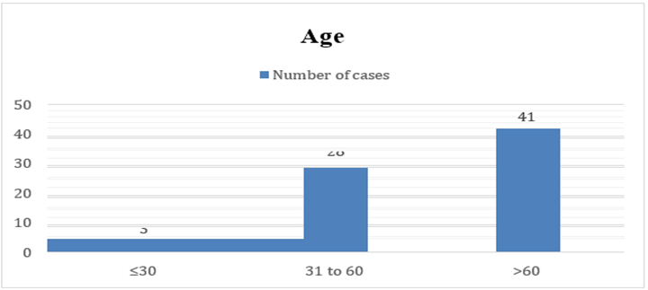
Graph 1: Age distribution
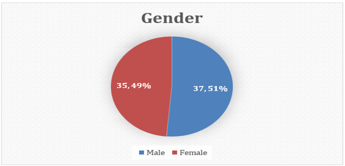
Graph 2: Gender distribution
The study included 72 cases, with the majority of patients aged above 60 years (57%), followed by those between 31–60 years (39%), while only 4% were younger than 30 years. Gender distribution was nearly equal, with males comprising 51% and females 49% of the cohort. This indicates that colorectal lesions were more common in older individuals, with no significant gender predominance.
Table 2: Specimen type
|
Sr No |
Specimen |
Number of cases n |
Percentage % |
|
1 |
Rectum |
38 |
52.8% |
|
2 |
Hepatic Flexure of Colon |
7 |
9.7% |
|
3 |
Sigmoid Colon |
7 |
9.7% |
|
4 |
Rectosigmoid Junction |
4 |
5.6% |
|
5 |
Splenic Flexure of Colon |
2 |
2.8% |
|
6 |
Transverse Colon |
2 |
2.8% |
|
7 |
Caecum |
4 |
5.6% |
|
8 |
Descending Colon |
4 |
5.6% |
|
9 |
Mid Transverse Colon |
2 |
2.8% |
|
10 |
Ascending Colon |
2 |
2.8% |
|
Total N (%) |
72 |
100 % |
|
Rectal specimens accounted for the largest proportion (52.8%), followed by specimens from the hepatic flexure and sigmoid colon (9.7% each). Other sites such as rectosigmoid junction, caecum, descending colon, and splenic flexure contributed smaller proportions ranging between 2.8% and 5.6%. This highlights that rectal involvement was predominant in the study population.
Table 3: Colonoscopy findings
|
Sr No |
Colonoscopy findings |
Number of cases n |
Percentage % |
|
1 |
Circumferential Polypoidal Lesion / Growth |
20 |
27.8% |
|
2 |
Ulcer proliferative Growth / Lesion |
17 |
23.6% |
|
3 |
Circumferential Lesion with Thickened Folds / Thickening |
8 |
11.1% |
|
4 |
Polypoidal Lesion / Growth (non- circumferential) |
6 |
8.3% |
|
5 |
Sessile / Pedunculated Polyp |
4 |
5.6% |
|
6 |
Cauliflower-like Growth |
2 |
2.8% |
|
7 |
Proliferative Growth (non-ulcerative) |
2 |
2.8% |
|
8 |
Ulcerated Lesion R/O Carcinoma |
2 |
2.8% |
|
9 |
Normal / Scope Non-Negotiable |
7 |
9.7% |
|
10 |
Mild Erythematous Edema |
4 |
5.6% |
|
Total N (%) |
72 |
100 % |
|
Circumferential polypoidal lesions were the most frequent colonoscopic finding (27.8%), followed by ulcer-proliferative growths (23.6%). Thickened circumferential lesions (11.1%) and non-circumferential polypoidal lesions (8.3%) were also observed. Sessile/pedunculated polyps and cauliflower-like growths were less common (5.6% and 2.8%, respectively). Notably, 9.7% of cases had normal or non-negotiable scopes, while mild erythematous edema was seen in 5.6%. Overall, proliferative and polypoidal growth patterns dominated the colonoscopic spectrum.
Table 4: Cytology findings
|
Sr No |
Cytology findings |
Number of cases n |
Percentage % |
|
1 |
Benign |
47 |
66 % |
|
2 |
Malignant |
25 |
34 % |
|
Total N (%) |
72 |
100 % |
|
Cytological evaluation revealed that 66% of cases were benign, while 34% were malignant. This suggests that cytology was able to identify a significant proportion of malignant lesions, though benign diagnoses were more frequent.
Table 5: HPR findings
|
Sr No |
HPR findings |
Number of cases n |
Percentage % |
|
1 |
Benign |
54 |
75 % |
|
2 |
Malignant |
18 |
25 % |
|
Total N (%) |
72 |
100 % |
|
Histopathological review showed that 75% of specimens were benign, whereas 25% were malignant. Compared to cytology, histopathology yielded a slightly lower malignant detection rate, reinforcing its role as the gold standard for confirmation.
Table 6: Cytology Vs HPR findings
|
Sr No |
Cytology findings |
HPR findings |
Total N (%) |
|
|
Benign n (%) |
Malignant n (%) |
|||
|
1 |
Benign n (%) |
44 (62 %) |
3 (4 %) |
47 (66 %) |
|
2 |
Malignant n (%) |
10 (13 %) |
15 (21 %) |
25 (34 %) |
|
Total N (%) |
54 (75 %) |
18 (25 %) |
72 100 %) |
|
|
Sensitivity = 83.3%, Specificity 81.5%, Positive Predictive Value (PPV) = 60.0% Negative Predictive Value (NPV) = 93.6%, Accuracy = 81.9% |
||||
Among cytologically benign cases (66%), histopathology confirmed benignity in 62% but identified malignancy in 4%. Conversely, of cytologically malignant cases (34%), histopathology confirmed malignancy in 21% and reclassified 13% as benign. The diagnostic performance of cytology showed sensitivity of 83.3%, specificity of 81.5%, positive predictive value of 60%, negative predictive value of 93.6%, and overall accuracy of 81.9%. This indicates that cytology is a reliable screening tool with high sensitivity and NPV, though histopathology remains essential for definitive diagnosis.
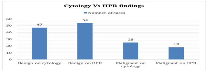
Graph 3: Cytology Vs HPR findings
Table 7: Histopathological Subtype
|
Sr No |
Subtype |
Number of cases n |
Percentage % |
|
1 |
Well-Differentiated Adenocarcinoma |
6 |
33.3% |
|
2 |
Moderately-Differentiated Adenocarcinoma |
4 |
22.2% |
|
3 |
Poorly-Differentiated Adenocarcinoma |
3 |
16.7% |
|
4 |
Mucinous Adenocarcinoma |
3 |
16.7% |
|
5 |
Signet Ring Cell Adenocarcinoma |
2 |
11.1% |
|
Total N (%) |
18 |
100% |
|
Histopathological subtyping of the 18 malignant colorectal lesions revealed that well-differentiated adenocarcinomas were the most frequent (33.3%), followed by moderately differentiated (22.2%) and poorly differentiated tumors (16.7%). Among the special variants, mucinous adenocarcinoma accounted for 16.7% and signet ring cell adenocarcinoma for 11.1%.
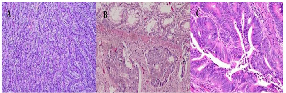
Figure a: adenocarcinoma A. Poorly differentiated B. Moderately differentiated C. Well differentiated.
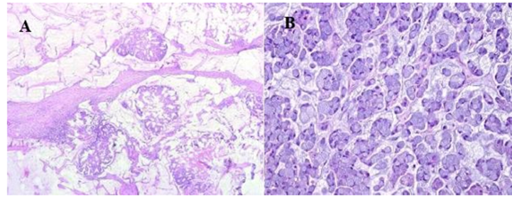
Figure b: A) Mucinous B)Signet ring cell
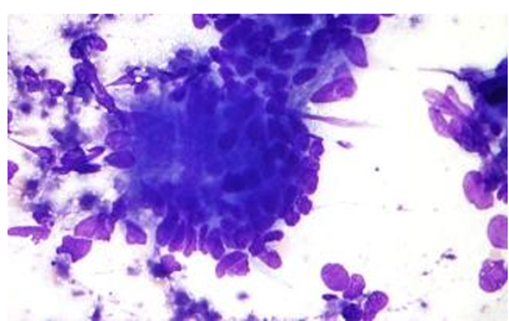
Figure c: Cytology - positive for malignancy
DISCUSSION:
The present study demonstrated that cytology achieved a sensitivity of 83.3%, specificity of 81.5%, and overall accuracy of 81.9% in diagnosing colorectal lesions when compared with histopathology. The high negative predictive value (93.6%) indicates that cytology is particularly reliable in ruling out malignancy, although its positive predictive value (60%) reflects limitations in differentiating borderline or atypical lesions.
Comparable studies have reported similar diagnostic performance. Patil et al. observed scrape cytology concordance with histopathology in 93.3% of large intestinal tumors, emphasizing its utility as a rapid intraoperative diagnostic tool⁷. Keihanian et al. found strong diagnostic concordance between cytology and histology in endoscopic ultrasound-guided fine-needle biopsy samples, supporting cytology’s role in gastrointestinal lesion evaluation⁸. Diaz-Mercedes et al. demonstrated that advanced cytology smear techniques improved lymph node staging in colorectal cancer, achieving sensitivity of 96.8% compared to conventional H&E analysis, thereby enhancing prognostic accuracy⁹. Similarly, Mitra and Dey reported cytology-histology correlation in gastrointestinal lesions with diagnostic accuracy exceeding 85%, reinforcing cytology’s role in early detection¹⁰.
The slightly lower malignant detection rate in cytology compared to histopathology in our study may be explained by sampling limitations and interpretative challenges. Cytology relies on exfoliated or aspirated cells, which may not capture deeper tissue architecture or invasive fronts of tumors. This can lead to false negatives in cases where malignant cells are sparsely distributed or obscured by inflammatory background. Conversely, false positives may occur when reactive atypia mimics malignancy, reducing specificity.
In addition to overall malignant detection, histopathological subtyping in our cohort revealed that well-differentiated adenocarcinomas constituted the largest proportion (33.3%), followed by moderately differentiated (22.2%) and poorly differentiated tumors (16.7%). Among the special variants, mucinous adenocarcinoma accounted for 16.7% and signet ring cell adenocarcinoma for 11.1%. This distribution highlights the predominance of differentiated adenocarcinomas in colorectal malignancies, while also underscoring the diagnostic challenges posed by mucinous and signet ring variants. The abundant extracellular mucin in mucinous lesions and the cytoplasmic vacuoles in signet ring cells often obscure nuclear details, which may contribute to interpretative difficulties in cytology and explain the reduced positive predictive value observed in our study.
Mechanistically, the high sensitivity and NPV observed in our study can be attributed to the fact that malignant colorectal lesions often shed atypical cells with distinct nuclear features, making them readily identifiable in cytology smears. However, the reduced PPV reflects the overlap between reactive epithelial changes and neoplastic atypia, which histopathology resolves by assessing tissue architecture and invasion patterns.
Taken together, our findings and those of other studies confirm that cytology is a valuable adjunct diagnostic tool, particularly for rapid screening and intraoperative consultation. Nonetheless, histopathology remains indispensable for definitive diagnosis, staging, and therapeutic planning.
CONCLUSION:
Cytology demonstrated high sensitivity (83.3%) and negative predictive value (93.6%) in diagnosing colorectal lesions, making it a reliable screening tool for ruling out malignancy. However, its lower positive predictive value (60%) reflects limitations in differentiating atypical or borderline lesions, necessitating histopathology for definitive diagnosis. The predominance of rectal involvement and polypoidal/ulcerative growth patterns in this cohort underscores the importance of early detection strategies. Overall, cytology serves as a rapid, cost-effective adjunct, but histopathology remains indispensable for confirmation, staging, and guiding treatment.
REFERENCES:
1. Sung H, Ferlay J, Siegel RL, Laversanne M, Soerjomataram I, Jemal A, et al. Global cancer statistics 2020: GLOBOCAN estimates of incidence and mortality worldwide for 36 cancers in 185 countries. CA Cancer J Clin. 2021;71(3):209-49.
2. Hamilton SR, Aaltonen LA. Pathology and genetics of tumours of the digestive system. Lyon: IARC Press; 2000.
3. Koss LG. Diagnostic cytology and its histopathologic bases. 5th ed. Philadelphia: Lippincott Williams & Wilkins; 2006.
4. Orell SR, Sterrett GF, Whitaker D. Fine needle aspiration cytology. 5th ed. London: Churchill Livingstone; 2012.
5. Mitra S, Dey P. Role of cytology in the diagnosis of gastrointestinal tract lesions with histopathological correlation. J Cytol. 2016;33(4):216-21.
6. Stewart CJ, Hillery S, Platell C, Murray D. Utility of fine-needle aspiration cytology in the diagnosis of colorectal carcinoma. Cytopathology. 2002;13(3):150-9.
7. Patil S, Kulkarni N, Manickyam S, Hulikeri SS. Scrape cytology – Diagnostic tool in large intestinal tumors. Indian J Pathol Oncol. 2023;10(4):382-6.
8. Keihanian T, Diaz L, Plafsky L, Shergill U, Satiya J, Abraham R, et al. Diagnostic concordance of cytology and histology in samples obtained via endoscopic ultrasound-guided fine-needle biopsy. Cureus. 2021;13(6):e15596.
9. Diaz-Mercedes S, Archilla I, Lahoz S, Rodrigo-Calvo MT, Lopez-Prades S, Tarragona J, et al. Cytology smears: An enhanced alternative method for colorectal cancer pN stage—A multicentre study. Cancers (Basel). 2022;14(24):6072.
10. Mitra S, Dey P. Role of cytology in the diagnosis of gastrointestinal tract lesions with histopathological correlation. J Cytol. 2016;33(4):216-21.