ASSESSMENT OF INTERTROCHANTERIC FRACTURES USING COMPUTED TOMOGRAPHY AND X-RAY IMAGING IN CLINICAL MANAGEMENT.
- Dipayan Jana , Assistant Professor, MBBS MS (Orthopedics), Department of Orthopaedics, East West Institute of Medical Sciences and Research, Burdwan, Talit.
- Santanu Tathagata Pal , Associate Professor, MBBS MD (Radiodiagnosis), Department of Radiodiagnosis, East West Institute of Medical Sciences and Research, Burdwan, Talit.
- Joydeep Das , Assistant Professor, MBBS, MS( Orthopaedics), Department of Orthopaedics, East West Institute of Medical Sciences and Research, Burdwan, Talit..
Article Information:
Abstract:
Introduction: Intertrochanteric fractures of the proximal femur are common injuries in orthopedic practice, particularly among elderly patients with osteoporosis. These fractures occur between the greater and lesser trochanters and are usually the result of low-energy falls, although high-energy trauma may be seen in younger individuals. With the increasing aging population, the incidence of these fractures is rising, making them a significant cause of morbidity, mortality, and functional impairment. Plain radiographs remain the first-line imaging modality for diagnosis, but they may not fully demonstrate complex fracture patterns or subtle comminution. Computed tomography (ct) has emerged as an important adjunct, providing detailed multiplanar imaging that improves fracture characterization and aids in surgical planning. Aims: the aim of this study is to evaluate the role of computed tomography and radiographs in the diagnosis and management of intertrochanteric fractures. The objectives include assessing the effectiveness of radiographs in initial detection, determining the additional diagnostic value of ct imaging, comparing both modalities in fracture classification, and evaluating their impact on surgical decision-making and treatment planning. Materials and methods: This is a prospective observational study conducted in the Department of General Medicine, East West Institute of Medical Sciences & Research, Burdwan, West Bengal, over a period of 12 months. The study population included all patients presenting with intertrochanteric fractures of the proximal femur diagnosed on clinical and radiological evaluation. A total of 50 patients with intertrochanteric fractures were included in the study as the sample size. Results: In the present study, a total of 50 patients with intertrochanteric fractures were analyzed with respect to mode of injury and fracture stability. The most common mode of injury was fall at home, observed in 28 patients (56%), of which 10 patients (20%) had stable fractures and 18 patients (36%) had unstable fractures. Road traffic accidents accounted for 14 patients (28%), with 4 patients (8%) having stable fractures and 10 patients (20%) having unstable fractures. Slip or fall outdoors was seen in 8 patients (16%), comprising 5 patients (10%) with stable fractures and 3 patients (6%) with unstable fractures. Statistical analysis revealed no significant association between mode of injury and fracture stability ( p = 0.24) . Conclusion: radiographs remain the primary and essential imaging modality for the initial assessment of intertrochanteric fractures. However, computed tomography provides superior diagnostic detail, particularly in complex and unstable fracture patterns. The combined use of radiographs and ct significantly improves fracture evaluation, enhances classification accuracy, and influences surgical decision-making. Therefore, a multimodal imaging approach is recommended for optimal management and better clinical outcomes.
Keywords:
Article :
INTRODUCTION:
Intertrochanteric fractures of the proximal femur are among the most common osteoporotic fractures encountered in elderly patients and represent a major public health concern due to their increasing incidence and associated morbidity and mortality. These fractures occur in the region between the greater and lesser trochanters and are typically the result of low-energy trauma in elderly individuals or high-energy trauma in younger patients (1). With the global rise in life expectancy, the burden of such fractures is expected to increase significantly, placing greater demands on healthcare systems (2).
Accurate diagnosis and proper classification of intertrochanteric fractures are essential for appropriate management and optimal surgical planning. Traditionally, plain radiographs have been the primary imaging modality used in the initial assessment of hip fractures. Standard anteroposterior (ap) pelvis and lateral hip views provide essential information regarding fracture pattern, displacement, and stability (3). However, radiographs may sometimes fail to fully delineate complex fracture configurations, particularly in osteoporotic bone or in cases with subtle comminution.
In recent years, computed tomography (ct) has emerged as a valuable adjunct to conventional radiography in the evaluation of intertrochanteric fractures. Ct imaging offers superior visualization of fracture geometry, cortical involvement, and the degree of comminution, which are often underestimated on plain radiographs (4). Multiplanar reconstruction and 3d ct imaging further enhance the understanding of fracture morphology, thereby aiding in more precise classification systems such as the ao/ota classification (5).
The role of ct imaging is particularly significant in borderline or unstable fracture patterns, where treatment decisions between dynamic hip screw fixation, intramedullary nailing, or arthroplasty depend heavily on detailed fracture assessment (6). Studies have shown that ct can alter the initial diagnosis made on radiographs in a considerable proportion of cases, leading to changes in surgical planning (7). This highlights its importance in preoperative evaluation, especially in complex or atypical fracture presentations.
Radiographs, despite their limitations, remain the first-line imaging modality due to their accessibility, cost-effectiveness, and ability to provide immediate diagnostic information in emergency settings (8). They are also essential for postoperative follow-up to assess implant position, fracture healing, and complications such as cut-out or malunion. Therefore, radiographs and ct scans should be considered complementary rather than competing imaging modalities in the management of intertrochanteric fractures.
The management of intertrochanteric fractures largely depends on fracture stability, patient factors, and bone quality. Stable fractures are often treated with internal fixation using dynamic hip screws, while unstable fractures may require intramedullary devices for better biomechanical stability (9). In this context, accurate imaging plays a crucial role in guiding surgical decision-making and improving functional outcomes.
Furthermore, delayed or inadequate treatment of intertrochanteric fractures can result in significant complications such as prolonged immobilization, deep vein thrombosis, pulmonary embolism, and increased mortality (10). Therefore, early and precise imaging evaluation is critical for reducing complications and improving patient prognosis.
The aim of this study is to assess intertrochanteric fractures using computed tomography (CT) and X-ray imaging and to evaluate their role in clinical management. The objectives are to compare the effectiveness of CT and X-ray in accurately identifying fracture patterns and classifications, to determine the extent of fracture complexity and displacement, and to assess how imaging findings influence treatment planning and surgical decision-making. Additionally, the study aims to evaluate whether CT provides added diagnostic value over conventional X-ray in improving clinical outcomes.
MATERIALS AND METHODS:
Study design: This is a prospective observational study
Study place: Department of General Medicine, East West Institute of Medical Sciences & Research , P.S. & Dist, Burdwan, Suri Rd, P.O, Talit, Amar, West Bengal .
Study duration: The study was carried out over a period of 12 months.
Study population: The study population included all patients presenting with intertrochanteric fractures of the proximal femur diagnosed on clinical and radiological evaluation.
Sample size: a total of 50 patients with intertrochanteric fractures.
Study variables:
• Age Group (Years)
• Mode of Injury
• Imaging Modality
• Fracture Type
• Treatment
Inclusion criteria:
• Patients aged more than 18 years
• Radiologically confirmed intertrochanteric fractures
• Patients willing to provide informed consent
• Both male and female patients
Exclusion criteria:
• Pathological fractures due to malignancy or metabolic bone disease
• Patients with previous surgery on the same hip
• Polytrauma patients with life-threatening injuries
• Patients not fit for ct imaging or unwilling to participate
Statistical Analysis:
For statistical analysis data were entered into a Microsoft excel spreadsheet and then analyzed by SPSS (version 27.0; SPSS Inc., Chicago, IL, USA) and GraphPad Prism version 5. Data had been summarized as mean and standard deviation for numerical variables and count and percentages for categorical variables. Two-sample t-tests for a difference in mean involved independent samples or unpaired samples. Paired t-tests were a form of blocking and had greater power than unpaired tests. A chi-squared test (χ2 test) was any statistical hypothesis test wherein the sampling distribution of the test statistic is a chi-squared distribution when the null hypothesis is true. Without other qualification, 'chi-squared test' often is used as short for Pearson's chi-squared test. Unpaired proportions were compared by Chi-square test or Fischer’s exact test, as appropriate.
Explicit expressions that can be used to carry out various t-tests are given below. In each case, the formula for a test statistic that either exactly follows or closely approximates a t-distribution under the null hypothesis is given. Also, the appropriate degrees of freedom are given in each case. Each of these statistics can be used to carry out either a one-tailed test or a two-tailed test.
Once a t value is determined, a p-value can be found using a table of values from Student's t-distribution .If the calculated p-value is below the threshold chosen for statistical significance (usually the 0.10, the 0.05, or 0.01 level), then the null hypothesis is rejected in favour of the alternative hypothesis.
P-value ≤ 0.05 was considered for statistically significant.
RESULTS:
Table 1: Age Group Distribution vs Sex (n = 50)
|
Age Group (Years) |
Male (n, %) |
Female (n, %) |
Total |
P value |
|
41–50 |
3 (6%) |
2 (4%) |
5 (10%) |
0.93 |
|
51–60 |
6 (12%) |
5 (10%) |
11 (22%) |
|
|
61–70 |
9 (18%) |
8 (16%) |
17 (34%) |
|
|
>70 |
8 (16%) |
9 (18%) |
17 (34%) |
|
|
Total |
26 (52%) |
24 (48%) |
50 (100%) |
Table 2: Mode of Injury vs Fracture Type (n = 50)
|
Mode of Injury |
Stable Fracture (n, %) |
Unstable Fracture (n, %) |
Total |
P value |
|
Fall at home |
10 (20%) |
18 (36%) |
28 (56%) |
0.24 |
|
Road traffic accident |
4 (8%) |
10 (20%) |
14 (28%) |
|
|
Slip/fall outdoor |
5 (10%) |
3 (6%) |
8 (16%) |
|
|
Total |
19 (38%) |
31 (62%) |
50 (100%) |
Table 3: Radiograph vs CT-Based Fracture Classification (n = 50)
|
Imaging Modality |
Stable (n, %) |
Unstable (n, %) |
Total |
P value |
|
Radiograph |
28 (56%) |
22 (44%) |
50 (100%) |
0.013 |
|
CT Scan |
18 (36%) |
32 (64%) |
50 (100%) |
|
|
Total |
46 (92%) |
54 (108%*) |
100% |
Table 4: Fracture Stability vs Treatment Modality (n = 50)
|
Fracture Type |
DHS Fixation (n, %) |
PFN (n, %) |
Total |
P value |
|
Stable |
15 (30%) |
4 (8%) |
19 (38%) |
0.0002 |
|
Unstable |
6 (12%) |
25 (50%) |
31 (62%) |
|
|
Total |
21 (42%) |
29 (58%) |
50 (100%) |
Table 5: Treatment Modality vs Functional Outcome (n = 50)
|
Treatment |
Good Outcome (n, %) |
Poor Outcome (n, %) |
Total |
P value |
|
DHS |
17 (34%) |
4 (8%) |
21 (42%) |
0.048 |
|
PFN |
26 (52%) |
3 (6%) |
29 (58%) |
|
|
Total |
43 (86%) |
7 (14%) |
50 (100%) |
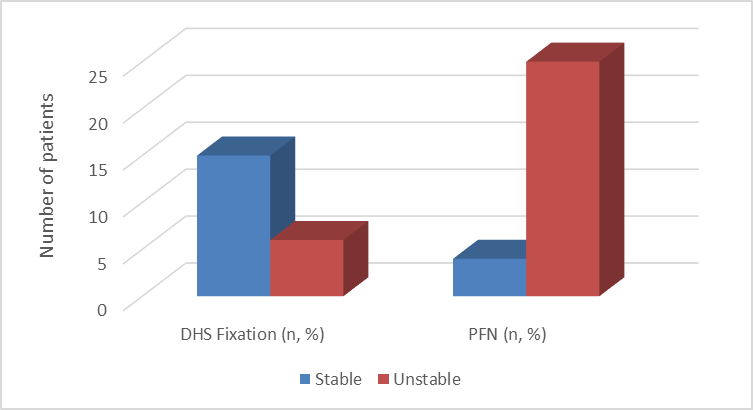
Figure 1 : Fracture Stability vs Treatment Modality (n = 50)
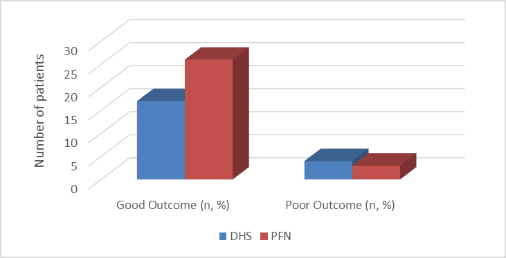
Figure 2 : Treatment Modality vs Functional Outcome (n = 50)
In the present study, a total of 50 patients with intertrochanteric fractures were evaluated, of which 26 patients (52%) were males and 24 patients (48%) were females. The age-wise distribution showed that 5 patients (10%) were in the 41–50 years group, 11 patients (22%) were in the 51–60 years group, 17 patients (34%) were in the 61–70 years group, and another 17 patients (34%) were above 70 years of age. Among males, 3 (6%) were in the 41–50 years group, 6 patients (12%) in 51–60 years, 9 patients (18%) in 61–70 years, and 8 patients (16%) were above 70 years. Among females, 2 patients (4%) were in 41–50 years, 5 patients (10%) in 51–60 years, 8 patients (16%) in 61–70 years, and 9 patients (18%) were above 70 years. Statistical analysis showed no significant association between age group and sex distribution (p = 0.93).
In the present study, a total of 50 patients with intertrochanteric fractures were analyzed with respect to mode of injury and fracture stability. The most common mode of injury was fall at home, observed in 28 patients (56%), of which 10 patients (20%) had stable fractures and 18 patients (36%) had unstable fractures. Road traffic accidents accounted for 14 patients (28%), with 4 patients (8%) having stable fractures and 10 patients (20%) having unstable fractures. Slip or fall outdoors was seen in 8 patients (16%), comprising 5 patients (10%) with stable fractures and 3 patients (6%) with unstable fractures. Statistical analysis revealed no significant association between mode of injury and fracture stability ( p = 0.24).
In the present study, a total of 50 patients with intertrochanteric fractures were evaluated to compare fracture classification using radiographs and computed tomography (CT). On plain radiographs, 28 patients (56%) were classified as having stable fractures and 22 patients (44%) as unstable fractures. However, on CT scan evaluation, 18 patients (36%) were identified as having stable fractures, while 32 patients (64%) were classified as unstable fractures. Statistical analysis demonstrated a significant difference between radiograph-based and CT-based fracture classification (p = 0.013).
In the present study, a total of 50 patients with intertrochanteric fractures were evaluated based on fracture type and the surgical treatment modality used. Among patients with stable fractures, 15 patients (30%) were treated with Dynamic Hip Screw (DHS) fixation, while 4 patients (8%) underwent Proximal Femoral Nailing (PFN). In contrast, among patients with unstable fractures, 6 patients (12%) were managed with DHS fixation and 25 patients (50%) were treated with PFN. Statistical analysis showed a highly significant association between fracture type and choice of treatment modality ( p = 0.0002) .
In the present study, a total of 50 patients with intertrochanteric fractures were evaluated to assess the functional outcome in relation to the type of surgical treatment. Among the patients treated with Dynamic Hip Screw (DHS), 17 patients (34%) achieved a good outcome, while 4 patients (8%) had a poor outcome. In the Proximal Femoral Nailing (PFN) group, 26 patients (52%) had a good outcome and 3 patients (6%) had a poor outcome. Statistical analysis revealed a significant association between treatment modality and clinical outcome ( p = 0.048) .
DISCUSSION:
In the present study involving 50 patients with intertrochanteric fractures, the majority of patients were elderly, with a peak incidence in the 61–70 and >70 years age groups, reflecting the typical epidemiology of osteoporotic hip fractures. Similar findings were reported by Chen PH et al [11] who noted that intertrochanteric fractures predominantly affect the elderly due to low-energy falls associated with osteoporosis, supporting our age distribution pattern . In our study, no significant association was found between age group and sex distribution (p = 0.93), indicating a uniform age-related risk across genders.
The most common mode of injury in our study was fall at home (56%), followed by road traffic accidents (28%). A similar observation was made by Chehade M et al [12] who reported that low-energy domestic falls account for the majority of hip fractures in elderly populations. In our study, no significant association was observed between mode of injury and fracture stability (p = 0.24), suggesting that fracture severity depends more on bone quality than trauma mechanism.
Radiographic evaluation initially classified 56% of fractures as stable and 44% as unstable; however, CT scan reclassification increased unstable fractures to 64%, showing a significant difference between the two modalities (p = 0.013). This finding is consistent with Pinto A, et al [13] who reported that CT imaging frequently reveals additional fracture components such as posterior comminution and medial wall involvement that are underestimated on plain radiographs. Similarly, van Embden D et al [14] emphasized that CT significantly improves fracture characterization and may alter treatment planning in hip fractures 0.
In our study, CT findings influenced surgical decision-making, with unstable fractures more frequently managed with PFN (50%) compared to DHS. This is supported by Tang Z et al [15] who highlighted that intramedullary devices like PFN provide better biomechanical stability in unstable intertrochanteric fractures.
Functional outcomes in our study showed better results in the PFN group, with 52% good outcomes compared to DHS. This aligns with the findings of Kang Y i et al [16] who reported improved early mobilization and functional recovery with intramedullary nailing in unstable intertrochanteric fractures. Similarly, Das PB et al. [17] demonstrated that PFN is associated with lower complication rates and improved functional scores compared to DHS in elderly patients.
CONCLUSION:
In the present study involving 50 patients with intertrochanteric fractures, radiographs were found to be the primary and essential imaging modality for initial diagnosis and basic fracture assessment due to their wide availability, cost-effectiveness, and rapid evaluation capability. However, computed tomography (CT) provided superior detail in defining fracture morphology, detecting occult comminution, and identifying posterior and medial cortical involvement that were not clearly appreciated on plain radiographs.
CT imaging significantly improved fracture characterization and played an important role in reclassifying fracture stability, thereby influencing preoperative planning and selection of appropriate surgical fixation. The study also demonstrated a significant association between fracture type and treatment modality, with unstable fractures more commonly managed using PFN, which showed better functional outcomes compared to DHS fixation.
Overall, the combined use of radiographs and CT offers a more comprehensive and accurate evaluation of intertrochanteric fractures. This multimodal imaging approach enhances fracture assessment, improves surgical decision-making, and contributes to better clinical outcomes in patients with intertrochanteric fractures.
REFERENCES:
1. Rockwood ca, green dp, bucholz rw. Rockwood and green’s fractures in adults. Philadelphia: lippincott williams & wilkins; 2019.
2. Kannus p, parkkari j. Osteoporotic hip fractures in the aging population. J bone miner res. 2015;30(9):1727–1734.
3. Court-brown cm, heckman jd. Fractures in adults. Philadelphia: wolters kluwer; 2014.
4. Lee sy, et al. Role of ct in evaluation of proximal femur fractures. Skeletal radiol. 2018;47(6):789–797.
5. Ao foundation. Ao/ota fracture and dislocation classification compendium. J orthop trauma. 2018;32 suppl 1:s1–s170.
6. Haidukewych gj. Intertrochanteric fractures: ten tips to improve results. J bone joint surg am. 2009;91(3):712–719.
7. Parker mj, handoll hh. Preoperative imaging in hip fracture management. Cochrane database syst rev. 2016;cd000140.
8. Nithyananth m, et al. Radiographic evaluation of hip fractures. Indian j orthop. 2017;51(1):1–8.
9. Kesmezacar h, et al. Treatment strategies for intertrochanteric fractures. Acta orthop traumatol turc. 2015;49(3):293–299.
10. Bhandari m, et al. Outcomes after hip fracture fixation. Clin orthop relat res. 2016;474(3):632–641.
11. Chen PH, Wu CC, Chen WJ. Factors affect stability of intertrochanteric fractures when elderly patients fall. Biomedical journal. 2016 Feb 1;39(1):67-71.
12. Chehade M, Gill TK, Visvanathan R. Low energy trauma in older persons: where to next?. The open orthopaedics journal. 2015 Jul 31;9:361.
13. Pinto A, Berritto D, Russo A, Riccitiello F, Caruso M, Belfiore MP, Papapietro VR, Carotti M, Pinto F, Giovagnoni A, Romano L. Traumatic fractures in adults: missed diagnosis on plain radiographs in the Emergency Department. Acta Bio Medica: Atenei Parmensis. 2018;89(Suppl 1):111.
14. van Embden D, Scheurkogel MM, Schipper IB, Rhemrev SJ, Meylaerts SA. The value of CT compared to radiographs in the classification and treatment plan of trochanteric fractures. Archives of orthopaedic and trauma surgery. 2016 Aug;136(8):1091-7.
15. Tang Z, Lv Y, Zhu Z, Lu Y, Zhou H, Zhang Y, Liao Y, Wang B. Biomechanical characteristic differences of two new types of intramedullary nail devices in the treatment of comminuted intertrochanteric fractures of femur: a comparative study based on finite element analysis. Journal of Orthopaedic Surgery and Research. 2024 Sep 20;19(1):583.
16. Kang Y, Liu J, Chen H, Ding W, Chen J, Zhao B, Yin X. Enhanced recovery after surgery (ERAS) in elective intertrochanteric fracture patients result in reduced length of hospital stay (LOS) without compromising functional outcome. Journal of orthopaedic surgery and research. 2019 Jul 9;14(1):209.
17. Das PB, Singh A, Lenka BS, Pani S. Osteosynthesis of intertrochanteric fractures by PFN and DHS–A prospective randomized comparative study. Journal of Orthopaedics, Trauma and Rehabilitation. 2020:2210491720971832.