ROLE OF ULTRASOUND IN EVALUATION OF BREAST LESIONS AND ITS FNAC CORRELATION.
- Miryala Vamshi Krishna , Post graduate resident, Department of General Surgery, Mamata Medical College, Khammam, Telangana, India.
- Gurram Laxmikanth , Associate professor, Department of General Surgery, Mamata Medical College, Khammam, Telangana, India.
- Suresh Clement , Professor, Department of General Surgery, Mamata Medical College, Khammam, Telangana, India.
- Polnati Swamikumar , Assistant professor, Department of General Surgery, Mamata Medical College, Khammam, Telangana, India.
Article Information:
Abstract:
Background: Diagnosis of breast cancer has been widely improved since the development of high-resolution ultrasound. Ultrasound is an indispensable tool in breast imaging. FNAC is relevant and important for pre-operative pathological evaluation in management of breast cancer. Present study was aimed to study ultrasound guided evaluation of breast lesions followed by correlation with findings of FNAC (cyto-pathological correlation). Materials and Methods: Present study was a single centre prospective and observational study conducted among females of age > 18 year, complaining of palpable breast lump or having suspicious lesions on mammography. Malignancy was predicted by using BIRADS score interpreted by using ultrasound. Results: In present study 233 women were included. Majority of them were from 31-60 years age group, with mean age of 47.22±11.43 years. Common indication for ultrasound was lump (71.2%) followed by pain (16.3%), screening (7.7%) and nipple discharge (4.7%). FNAC report was malignant (56.7%) in majority of cases as compared to benign (43.3%). Sensitivity and Specificity of BIRADS ≥ 4b was highest (90.9% &89.1% respectively) followed by BIRADS ≥ 4a (88.6&75.3% respectively) and BIRADS ≥4c (71.2% &95.1% respectively. No statistically significant difference was noted between oval and round shape with (P-O.859). Benign lesions on FNAC are commonly seen with circumscribed margin (64.4%) while malignant lesions on FNAC are commonly seen with micro-lobulated margin (57.6%) association was statistically significant (P<0.001). Conclusion: Using integrated approachby using imaging and FNAC (cytological method) procedure is the best way in addressing the diagnostic needs of patients with breast disease.
Keywords:
Article :
INTRODUCTION:
Breast lesions are among the most common clinical conditions encountered in women and encompass a wide spectrum of benign and malignant disorders. Breast cancer remains the most frequently diagnosed malignancy among women worldwide, making early detection and accurate diagnosis essential for improving prognosis and reducing mortality. Clinical examination alone is often insufficient to differentiate benign from malignant lesions, necessitating the use of imaging and cytological evaluation as part of the standard triple assessment approach [1].
Ultrasonography (USG) has become an indispensable imaging modality in the evaluation of breast lesions because it is non-invasive, radiation-free, cost-effective, and highly effective in differentiating cystic from solid masses. It is particularly useful in younger women with dense breast tissue and serves as an excellent tool for assessing lesion morphology, margins, vascularity, and axillary lymph nodes. The use of the Breast Imaging Reporting and Data System (BI-RADS) has standardized ultrasound reporting and improved diagnostic accuracy and patient management [2].
Fine-needle aspiration cytology (FNAC) is a simple, minimally invasive, and economical diagnostic procedure that provides rapid cytological diagnosis. Correlation of ultrasound findings with FNAC enhances diagnostic confidence, facilitates early treatment planning, and reduces unnecessary surgical biopsies. Recent studies have demonstrated a strong association between higher BI-RADS categories and malignant cytological findings, highlighting the complementary role of ultrasound and FNAC in the evaluation of breast lesions [3].
Despite advances in breast imaging, variations in ultrasound interpretation and cytological reporting continue to exist. Most recent studies have focused on correlating ultrasound findings with histopathology, whereas relatively fewer have specifically evaluated the concordance between ultrasonographic findings and FNAC, particularly in resource-limited settings where FNAC remains widely used. Therefore, the present study aims to evaluate the role of ultrasonography in the assessment of breast lesions and correlate its findings with FNAC to determine its diagnostic accuracy and clinical utility in the early diagnosis and management of breast diseases [4].
MATERIALS AND METHODS:
Present study was single-center, prospective, observational study, conducted in department of General surgery, Mamata Medical College&Hospital ,hammam,Telangana , India.over a period of two years from October 2023 to October 2025Study approval was obtained from institutional ethical committee.
Inclusion criteria
• Females, age > 18 years, complaining of palpable breast lesions, OR had suspicious lesions on mammography, willing to participate in present study.
Exclusion criteria
• All patients who did not give consent to be part of this STUDY.
• Pregnant and lactating patients.
• Patients with inflammatory breast lesions
Methodology
Study was explained to patients in local language & written consent was taken for participation & study. Sample size was calculated by considering Sensitivity of BIRADS score. The minimum required sample was 232. Demographic data (age and address) along with complaints, detailed history of previous operation, similar complaints in past, family history of breast cancer were noted.
Using Philips Affinity 70 and Philip's HDXE 11 ultrasound machines, the USG was performed with the patient lying in supine position. The high frequency linear probe (Transducer 5-7 MHz) had been used to image the breast tissues. Both the breasts were exposed and the transducer was swept in radial and anti-radial direction to look for abnormalities.
FNAC Fine needle aspiration cytology (FNAC) of the lesion was performed under ultrasound guidance. The skin disinfection done with the disinfectant. The needle was inserted near one of the side of the transducer and then it was advanced along a trajectory which is lying parallel to the long axis of the transducer. Until the lesion was penetrated, the needle was visualized on the monitor in real time.
Aspiration was applied and the tip was moved in various directions to collect multiple samples. When the needle was being withdrawn, no aspiration applied. The collected specimen then sent for histopathological examination which was examined by the pathologist.
BIRAD score interpreted by USG was used for prediction of malignancy. The accuracy of this prediction at various cut off values of BIRAD score was estimated by calculating sensitivity (true positive rate) specificity (true negative rate), Positive predictive value (probability that the disease is present when the test is positive) and Negative predictive value (probability that the disease is not present when the test is negative).
A receiver operating characteristic curve, or ROC curve, which is a graphical plot that illustrates the diagnostic ability of a binary classifier system as its discrimination threshold is varied was plotted and Area under the curve was calculated for each cut off value of BIRADS Score.
Statistical Analysis
Data was collected and compiled using Microsoft Excel, analysed using SPSS 23.0 version. Frequency, percentage, means and standard deviations (SD) was calculated for the continuous variables, while ratios and proportions were calculated for the categorical variables.
Difference of proportions between qualitative variables were tested using chi- square test or Fisher exact test as applicable. P value less than 0.5 was considered as statistically significant.
RESULTS:
Table 1. Age Distribution of the Study Participants (n = 233)
|
Age Group (Years) |
Number of Patients (n) |
Percentage (%) |
|
<30 |
23 |
9.87 |
|
31–60 |
175 |
75.11 |
|
61–90 |
35 |
15.02 |
|
Total |
233 |
100.00 |
|
Mean Age ± SD (Years) |
47.22 ± 11.43 |
— |
Table 1 presents the age distribution of the 233 study participants. The majority of the participants (75.11%) belonged to the 31–60 years age group, indicating that middle-aged adults constituted the largest proportion of the study population. Participants aged 61–90 years accounted for 15.02%, while those younger than 30 years represented only 9.87% of the total sample. The mean age of the participants was 47.22 ± 11.43 years, suggesting that the study population predominantly consisted of middle-aged individuals with a moderate variation in age. This age distribution is appropriate for studies involving adult clinical populations, where the prevalence of chronic diseases and associated complications is generally higher during middle age.
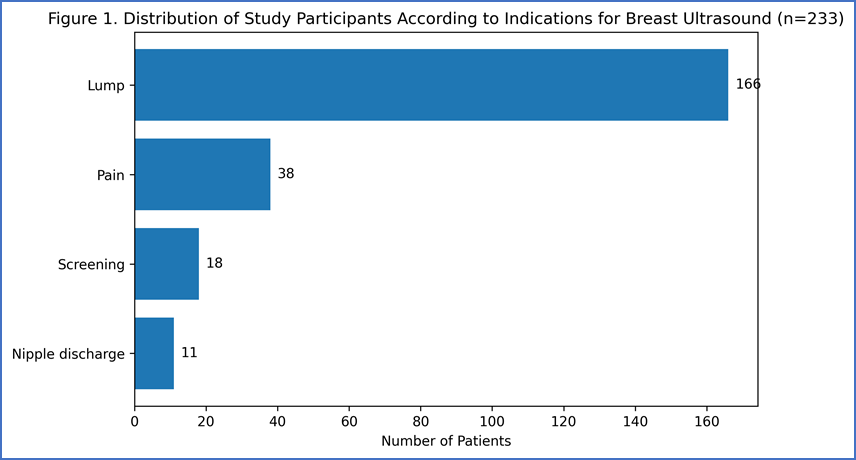
Figure 1. Distribution of Study Participants According to Indications for Breast Ultrasound (n = 233)
Figure 1 shows the distribution of study participants based on the indication for undergoing breast ultrasound examination. The most common indication was the presence of a breast lump, accounting for 166 (71.2%) patients, making it the predominant reason for imaging. Breast pain was the second most common indication, observed in 38 (16.3%) patients. Screening examinations were performed in 18 (7.7%) patients, while 11 (4.7%) patients underwent ultrasound due to nipple discharge. These findings indicate that palpable breast lumps constituted the primary clinical presentation prompting breast ultrasound evaluation in the study population.
Table 2. Distribution of Study Participants According to Pathological Characteristics (n = 233)
|
Pathological Characteristic |
Number of Patients (n) |
Percentage (%) |
|
Skin Changes |
||
|
Normal |
94 |
40.3 |
|
Skin thickening |
72 |
30.9 |
|
Skin retraction |
67 |
28.8 |
|
Total |
233 |
100.0 |
|
Invasion of Lesion |
||
|
No invasion |
138 |
59.2 |
Table 3 summarizes the pathological characteristics observed among the study participants. Regarding skin changes, 40.3% of patients had normal skin appearance, while 30.9% exhibited skin thickening and 28.8% showed skin retraction. These findings indicate that skin abnormalities were present in nearly 60% of the study population, suggesting advanced local disease in a substantial proportion of patients. With respect to lesion invasion, 138 (59.2%) patients showed no evidence of invasion. The remaining invasion-related findings should be interpreted after including the complete dataset.
Table 3. Correlation Between Ultrasonography (USG) and Fine Needle Aspiration Cytology (FNAC) Findings with Distribution of BI-RADS Categories (n = 233)
|
Variable |
Number of Patients (n) |
Percentage (%) |
|
USG–FNAC Correlation |
||
|
Concordance |
222 |
95.3 |
|
Discordance |
11 |
4.7 |
|
Total |
233 |
100.0 |
|
BI-RADS Category |
||
|
BI-RADS I (Negative) |
7 |
3.00 |
|
BI-RADS II (Benign) |
46 |
19.74 |
|
BI-RADS III (Probably Benign) |
31 |
13.30 |
|
BI-RADS IVa (Low Suspicion of Malignancy) |
18 |
7.73 |
|
BI-RADS IVb (Moderate Suspicion of Malignancy) |
32 |
13.73 |
|
BI-RADS IVc (High Suspicion of Malignancy) |
55 |
23.60 |
|
BI-RADS V (Highly Suggestive of Malignancy) |
44 |
18.88 |
|
Total |
233 |
100.0 |
Table 3 illustrates the correlation between ultrasonography (USG) and fine needle aspiration cytology (FNAC) findings, along with the distribution of BI-RADS categories among the study participants. A high degree of agreement was observed between USG and FNAC, with 222 (95.3%) cases demonstrating concordance and only 11 (4.7%) showing discordance, indicating excellent diagnostic correlation. Regarding BI-RADS classification, BI-RADS IVc (23.60%) was the most frequently assigned category, followed by BI-RADS II (19.74%) and BI-RADS V (18.88%). Lower proportions of patients were categorized as BI-RADS I (3.00%), BI-RADS III (13.30%), BI-RADS IVa (7.73%), and BI-RADS IVb (13.73%). Overall, a considerable proportion of lesions fell into higher BI-RADS categories (IV and V), reflecting a high prevalence of suspicious or malignant breast lesions within the study population.
Table 4. Sensitivity and Specificity of Different BI-RADS Thresholds for Predicting Malignancy
|
BI-RADS Threshold |
Sensitivity (%) |
Specificity (%) |
|
BI-RADS ≥ III |
93.9 |
44.6 |
|
BI-RADS ≥ IVa |
88.6 |
75.3 |
|
BI-RADS ≥ IVb |
90.9 |
89.1 |
|
BI-RADS ≥ IVc |
71.2 |
95.1 |
|
BI-RADS ≥ V |
32.6 |
99.0 |
Table 4 presents the diagnostic performance of different BI-RADS thresholds in predicting breast malignancy. BI-RADS ≥ IVb demonstrated the best overall diagnostic accuracy, with a high sensitivity of 90.9% and specificity of 89.1%, indicating an optimal balance between correctly identifying malignant lesions and excluding benign ones. BI-RADS ≥ IVa also showed high sensitivity (88.6%) but comparatively lower specificity (75.3%), making it useful as a screening threshold. In contrast, BI-RADS ≥ IVc exhibited lower sensitivity (71.2%) but a markedly higher specificity (95.1%), reflecting greater confidence in identifying malignancy. BI-RADS ≥ III had the highest sensitivity (93.9%) but poor specificity (44.6%), whereas BI-RADS ≥ V demonstrated the highest specificity (99.0%) with a substantially lower sensitivity (32.6%). These findings suggest that BI-RADS ≥ IVb provides the most favorable balance between sensitivity and specificity for predicting breast malignancy in the present study.
Table 5. Distribution of Echogenicity According to FNAC Diagnosis (n = 233)
|
Echogenicity |
Benign (n = 101) |
Malignant (n = 132) |
p-value |
|
Anechoic |
0 (0.0%) |
22 (16.7%) |
|
|
Hypoechoic |
69 (68.3%) |
73 (55.3%) |
|
|
Hyperechoic |
17 (16.8%) |
24 (18.2%) |
|
|
Isoechoic |
15 (14.9%) |
6 (4.5%) |
|
|
Complex solid and cystic lesion |
0 (0.0%) |
7 (5.3%) |
< 0.001 |
Table 5 presents the distribution of ultrasound echogenicity patterns according to FNAC diagnosis. Hypoechoic lesions were the most common finding in both benign (68.3%) and malignant (55.3%) groups. Anechoic lesions and complex solid-cystic lesions were observed exclusively in the malignant group, accounting for 16.7% and 5.3% of malignant lesions, respectively. Isoechoic lesions were more frequently associated with benign lesions (14.9%) than malignant lesions (4.5%), while hyperechoic lesions showed a comparable distribution in both groups. The association between echogenicity pattern and FNAC diagnosis was highly statistically significant (p < 0.001), indicating that ultrasound echogenicity is an important parameter in differentiating benign from malignant breast lesions.
DISCUSSION:
The general role of diagnostic breast ultrasound is to make a non-invasive diagnosis in patients who have clinical or mammographic abnormalities than could be achieved with mammography and clinical findings alone. The use of breast ultrasound in patients (those who have clinical or mammographic findings that are not clearly malignant) should increase the certainty of a benign diagnosis in many patients and should increase the suspicion of carcinoma in a small number of patients [4]
Mammography is capable only of showing four different densities (air, water, fat, and calcium) and can further distinguish between different water-density tissues only by differences in thickness and compressibility and by whether the tissues contain some fatty or calcium density. Ultrasound, on the other hand, can distinguish among many different types of normal breast tissue. Like mammography, ultrasound can identify air, fat, calcium densities. However, unlike mammography, ultrasound can also differentiate among different types of normal water-density tissues by echogenicity as well as thickness and elasticity [5]. High resolution ultrasonography is a useful diagnostic modality for evaluation of various breast lumps. It is sensitive for microcalcifications, intraductal lesions and has good sensitivity, specificity, positive and negative predictive values for diagnosing breast lesions.
Ultrasound is cheap, easily available, with no risk of ionising radiation. So, it is useful investigation for follow up of indicated lesions. The FNAC of breast is cheap, less invasive, safe and highly accurate method for diagnosis of breast lesions. Many benign conditions, infections, degenerative conditions are easily detected by FNAC. So, no further invasive investigations can be avoided.
Almost 55.3% of hypoechoic lesions were malignant on fine needle aspiration cytology followed by hyperechoic and solid cystic lesions. Some benign lesions also were hypoechoic but they lacked other characteristic features like irregular or spiculated margins, skin invasion and posterior features typical of malignancy.
In our study, all the lesions which had a longitudinal versus anteroposterior diameter ratio of more than one was came to be benign on FNAC. The lesions those not fulfilling the criteria were almost equally either benign or malignant. From this study we can conclude that a lesion having a ratio of longitudinal to AP diameter of more than 1 were benign. These findings are similar to a study done by Berns et al [6] a round lesion with well- circumscribed margins, a longitudinal to AP diameter ratio which was greater than or equal to one with no hypoechogenicity when seen were most probably considered benign lesion on ultrasonography. Most of the cases that were given a grade of BI-RADS 5 had positive malignant ipsilateral lymph adenopathy which were enlarged rounded with loss of fatty hilum. In our study, the positive predictive value was 97.7 % and the negative predictive value was 52.9 % on ultrasonography.
In 95.3 % cases there was concordance of findings between ultrasonography and FNAC findings. It was concluded from the cases which we labelled BIRADS IV or V and these cases which came malignant on FNAC. The discordance is seen in around 4.7 % cases. These were the cases which showed the benign features on ultrasound like well circumscribed margins without significant internal vascularity, no obvious underlying chest wall invasion. Specificity of detecting BIRADS V lesion were turned to be highest i.e., 99 % on ultrasound, with the lesions showing hyperechoic foci i.e., microcalcifications, micro-lobulated margins, obvious underlying pectoralis invasion and overlying skin invasion.
Chaitanya et al. [7] noted that most of the cases 35 (63.6 %) were in the upper outer quadrant of the breast. There were (BI-RADS) 45 (81%) lumps were reported as benign lesions and 07 (12.7 %) as malignant and 3 (5 %) were reported as features suggestive or suspicious for malignancy. Similar findings were noted in present study.
Hiral Hapani et al,.[8] noted that 52% cases showed circumscribed margins (all were benign) followed by 12% Spiculated margins. Among the 64% cases appearing hypoechoic, 76.56% came benign and remaining 23.43% were reveal malignant. 51% of benign lesions showed posterior enhancement while 70% of malignant lesions showed posterior acoustic shadowing. Jahan et al, [9] sensitivity was proven to be 80.0%, specificity 96%, positive predictive value (PPV) (88.89%), negative predictive value 94.12% and the accuracy was 93.02% and comparable to other study. In diagnosis of benign breast lesion by ultrasound, sensitivity was about 96 %, specificity of 80%, positive predictive value (94.12%), negative predictive value (NPV)88.89% and accuracy was 93.02%.
USG is an appropriate imaging method for reliable diagnosis of diagnosis of palpable breast mass [10, 11]. Imaging with ultrasonography combined with FNAC yielded the best results showed the sensitivity of 94% for benign lesions and 96% for malignant lesions, while the specificity was 97% for benign lesions and 92% for malignant lesions. The detection of malignancy was proven to be more accurate when both the modalities were combined in our study group. Our results were slightly different from other studies which evaluated the sensitivity of radiological grading in predicting malignancy. It is concluded that imaging with ultrasonography and FNAC yielded a better result than with either modality in helping in more accurate characterization of lesions in the breast and to categorize breast lesions as benign and malignant.
CONCLUSION:
Using integrated approach employing imaging and FNAC (cytological method) procedure is the best way to in addressing the diagnostic needs of patients with breast diseases. Ultrasonography BIRADS combined with FNAC gives the excellent results. Hence Ultrasonography can safely be used as the first line investigation for evaluation of breast lump.
FNAC correlation of ultrasound findings revealed a high degree of sensitivity and specificity in differentiating benign from malignant lesions and obviated need for more invasive procedures like core biopsy and investigations like MRI.
REFERENCES:
1. American College of Radiology. [Internet] Philadelphia:ACR Breast Imaging Reporting and Data System (BI-RADS) [cited August 20, 2015].
2. Berg WA, Gutierrez L, Nessaiver MS, Carter WB, Bhargavan M, Lewis RS, Ioffe OB. Diagnostic accuracy of mammography, clinical examination, US and MR imaging in preoperative assessment of breast cancer. Radiology 2004; 233:830–49.
3. Kriege M, Brekelmans Ct, Boetes C, Besnard PE, Zonderland HM, Obdeijn IM, et al . Efficacy of MRI and mammography for breast – cancer screening in women with a familial or genetic predisposition. N Engl J Med. 2004;351:427-37.
4. Ferlay J, Soerjomataram I, Ervik M, Dikshit R, Eser S, Mathers C, GLOBOCAN 2012 v1.0, Cancer Incidence and Mortality Worldwide: IARC CancerBase No. 11 [Internet]. [Lyon, France]: International Agency for Research on Cancer; 2013 [cited 2015 June 21].
5. Etta D. Pisano, Martin J. Yaffe. Digital Mammography. Radiology 2005; 234:353–62
6. Berns EA, Hendrick RE, Cutter GR. Performance comparison of full-field digital mammography to screen-film mammography in clinical practice. Med Phys 2002;29:830–4
7. Chaitanya INVL, Prabhala S, Annapurna S, et al. Comparison of histopathologic findings with BIRADS Score in trucut biopsies of breast lesions. Indian Journal of Pathology: Research and Practice 2020;9(Pt 1):35- 41.
8. Marri RK, Sushmita P. Evaluation of breast lumps by ultrasound and its correlation with fnac findings in a teaching hospital in Telangana. J Evid Based Med Healthc 2020; 7(47), 2772-2776.
9. Hiral Hapani, Bhargav Patel,. Anjana Trivedi, Ultrasonographic evaluation of breast pathologies with FNAC correlation, International Journal of Scientific Research Volume- 7, Issue-12, December-2018
10. Jahan AB, Ahmed MU, Begum M, Hossain MM, Rahman MM, Sarwar JM, et al. Ultrasonographic evaluation of palpable breast mass and correlation with histopathology. Mymensingh Med J. 2017;26:223–9.
11. Tamaki K, Sasano H, Ishida T, et al. The correlation between ultrasonographic findings and pathologic features in breast disorders. Jpn J Clin Oncol 2010;40(10)905-912.