ARTIFICIAL INTELLIGENCE-ASSISTED MRI INTERPRETATION FOR EARLY DETECTION AND CHARACTERIZATION OF BRAIN TUMORS: A PROSPECTIVE DIAGNOSTIC ACCURACY STUDY.
- Shubhankar , Assistant Professor, Department of Radiodiagnosis, Ram Krishna Medical College Hospital and Research Centre, Bhopal, Madhya Pradesh India.
- Mayank Kumar Ujjaliya , Assistant Professor, Department of Radiodiagnosis, Ram Krishna Medical College Hospital and Research Centre, Bhopal, Madhya Pradesh India.
- Vinod Parmar , Assistant Professor, Department of Radiodiagnosis, Ram Krishna Medical College Hospital and Research Centre, Bhopal, Madhya Pradesh India.
Article Information:
Abstract:
Background: Early and accurate diagnosis of brain tumors is essential for optimizing treatment planning and improving patient outcomes. Although magnetic resonance imaging (MRI) is the standard imaging modality for brain tumor evaluation, diagnostic performance may be limited by overlapping imaging characteristics and inter-observer variability. Artificial intelligence (AI)-assisted MRI interpretation has emerged as a promising approach to enhance diagnostic accuracy and efficiency. Aim: To evaluate the diagnostic performance of AI-assisted MRI for the early detection and characterization of brain tumors compared with conventional radiologist interpretation. Materials and Methods: This prospective diagnostic accuracy study included 180 adult patients with suspected intracranial tumors who underwent 1.5 T brain MRI at a tertiary care teaching hospital between October 2024 and March 2026. MRI examinations were independently interpreted by experienced neuroradiologists before and after AI assistance using a validated deep learning-based platform. Histopathological examination or multidisciplinary consensus served as the reference standard. Diagnostic accuracy, sensitivity, specificity, positive predictive value (PPV), negative predictive value (NPV), receiver operating characteristic (ROC) curve analysis, Cohen's kappa coefficient, and reporting time were evaluated. Results: The mean age of participants was 49.8 ± 14.2 years, with males accounting for 56.7% of the study population. Glioma was the most common tumor (37.8%). AI-assisted MRI significantly improved diagnostic accuracy from 86.1% to 95.0% (p=0.008), with sensitivity increasing from 88.2% to 96.1% and specificity from 82.4% to 92.6%. PPV and NPV improved to 96.8% and 91.2%, respectively. AI-assisted interpretation demonstrated almost perfect agreement with the reference diagnosis (κ=0.92) compared with radiologist interpretation alone (κ=0.79). ROC analysis showed superior performance for AI-assisted MRI (AUC=0.964; 95% CI: 0.937–0.991) compared with conventional MRI (AUC=0.885; 95% CI: 0.831–0.938). Mean reporting time was significantly reduced from 11.8 ± 2.6 to 7.3 ± 1.8 minutes (p<0.001). Conclusion: AI-assisted MRI significantly enhances the early detection and characterization of brain tumors by improving diagnostic accuracy, agreement with reference diagnosis, and reporting efficiency. Integration of AI into routine neuroimaging practice may support radiologists in achieving faster, more consistent, and more accurate clinical decision-making.
Keywords:
Article :
INTRODUCTION:
Brain tumors comprise a diverse group of primary and secondary intracranial neoplasms that are associated with substantial morbidity and mortality worldwide. Despite advances in neurosurgery, radiotherapy, and molecularly targeted therapies, patient prognosis remains closely linked to the timing of diagnosis and accurate characterization of tumor type, location, and biological behavior. Early identification enables prompt intervention, facilitates optimal surgical planning, and improves overall clinical outcomes, particularly for aggressive tumors such as gliomas. Magnetic resonance imaging (MRI) remains the imaging modality of choice because of its superior soft-tissue contrast and ability to provide detailed anatomical and functional information without ionizing radiation [1, 2].
Interpretation of brain MRI, however, is often challenging due to overlapping imaging features among different tumor types, intratumoral heterogeneity, and inter-observer variability. Diagnostic accuracy may also be influenced by radiologist experience, imaging protocols, and increasing clinical workload. These limitations can delay diagnosis or lead to inconsistent lesion characterization, emphasizing the need for reliable decision-support tools that enhance radiological assessment while maintaining clinical efficiency [3].
Recent developments in artificial intelligence (AI), particularly deep learning and convolutional neural network (CNN)-based algorithms, have transformed medical image analysis. AI-assisted MRI interpretation has demonstrated promising performance in automated tumor detection, segmentation, and classification by identifying complex imaging patterns that may not be readily apparent to human observers. Meta-analyses have reported high pooled sensitivity and diagnostic accuracy for AI models in brain tumor detection, suggesting their potential to complement radiologist interpretation rather than replace it. Nevertheless, concerns regarding model generalizability, external validation, explainability, and integration into routine clinical practice remain important challenges before widespread implementation [4]. Although numerous retrospective studies and algorithm-development reports have demonstrated encouraging diagnostic performance, prospective clinical evidence evaluating AI-assisted MRI interpretation under real-world conditions remains limited. Further validation is needed to determine whether AI assistance can improve diagnostic accuracy, reduce interpretation variability, and support earlier detection of brain tumors across diverse patient populations [5, 6]. Therefore, the present prospective diagnostic accuracy study was designed to evaluate the performance of AI-assisted MRI interpretation for the early detection and characterization of brain tumors by comparing AI-assisted assessment with the reference standard of histopathological diagnosis and/or expert multidisciplinary consensus. The study also aims to determine the diagnostic accuracy, sensitivity, specificity, and predictive values of AI-assisted MRI in routine clinical practice.
MATERIALS AND METHODS:
Study Design and Setting: A prospective diagnostic accuracy study was conducted in the Department of Radiodiagnosis in collaboration with the Departments of Neurosurgery and Pathology at a tertiary care teaching hospital in central India, over a period of 18 months from October 2024 to March 2026.
Study Population: A total of 180 consecutive patients aged 18 years or older presenting with clinical features suggestive of an intracranial space-occupying lesion and referred for contrast-enhanced brain MRI were screened for eligibility.
Inclusion criteria: Patients with newly detected intracranial lesions who subsequently underwent histopathological evaluation or had a definitive diagnosis established by multidisciplinary consensus were included.
Exclusion criteria: Patients with previously treated brain tumors, contraindications to MRI or gadolinium-based contrast agents, severe motion-related image degradation, traumatic brain lesions, infectious or inflammatory intracranial diseases, vascular malformations, or incomplete clinical or imaging data were excluded.
MRI Acquisition Protocol and AI-Assisted MRI Interpretation: All examinations were performed using a 1.5-T MRI scanner (Siemens Magnetom Essenza) according to the institutional brain tumor imaging protocol. MRI datasets were analyzed using a validated deep learning–based AI platform incorporating convolutional neural network algorithms for automated lesion detection, segmentation, and tumor characterization. The AI system generated probability maps and classified lesions according to tumor type and likelihood of malignancy. Two experienced neuroradiologists independently interpreted all MRI examinations. Initially, images were reviewed without AI assistance, followed by a second assessment with AI-generated outputs after an appropriate washout period to minimize recall bias. The radiologists were blinded to histopathological findings during image interpretation.
Reference Standard: The reference standard for diagnosis was histopathological examination of surgically resected or biopsied tissue according to the latest World Health Organization (WHO) Classification of Central Nervous System Tumors [7]. For patients who did not undergo surgery, the final diagnosis was established through multidisciplinary consensus based on imaging findings, clinical presentation, and follow-up of at least six months.
Outcome Measures: The primary outcome was the diagnostic accuracy of AI-assisted MRI for detecting and characterizing brain tumors. Secondary outcomes included sensitivity, specificity, positive predictive value, negative predictive value, overall accuracy, and area under the receiver operating characteristic (ROC) curve. Agreement between AI-assisted interpretation and conventional radiologist assessment was evaluated using Cohen's kappa coefficient.
Statistical Analysis: Data were analyzed using IBM SPSS Statistics version 27.0 (IBM Corp., Armonk, NY, USA). Continuous variables were expressed as mean ± standard deviation or median (interquartile range), whereas categorical variables were presented as frequencies and percentages. Diagnostic performance indices with 95% confidence intervals were calculated using the reference standard. ROC curve analysis was performed to assess discriminative performance. Interobserver agreement was evaluated using Cohen's kappa coefficient. Comparisons between diagnostic methods were performed using McNemar's test for paired proportions. A two-sided p-value <0.05 was considered statistically significant.
RESULTS:
A total of 180 patients were included, with a mean age of 49.8 ± 14.2 years. The largest proportion belonged to the 41–60 years age group (45.0%). Males constituted 56.7% of the study population. Headache was the most common presenting symptom (76.7%) [Table: 1].
Table 1: Baseline demographic and clinical characteristics of the study population (N = 180)
|
Variable |
Frequency (n) |
Percentage (%) |
|
|
Age (years) |
18–40 |
54 |
30.0 |
|
41–60 |
81 |
45.0 |
|
|
>60 |
45 |
25.0 |
|
|
Mean ± SD |
49.8 ± 14.2 |
- |
|
|
Gender |
Male |
102 |
56.7 |
|
Female |
78 |
43.3 |
|
|
Clinical presentation |
Presenting headache |
138 |
76.7 |
|
Seizure |
61 |
33.9 |
|
|
Focal neurological deficit |
58 |
32.2 |
|
|
Vomiting |
42 |
23.3 |
|
|
Visual symptoms |
37 |
20.6 |
|
Glioma was the most frequently diagnosed brain tumor (37.8%), followed by meningioma (21.7%) and metastatic tumors (13.3%) [Graph: 1].
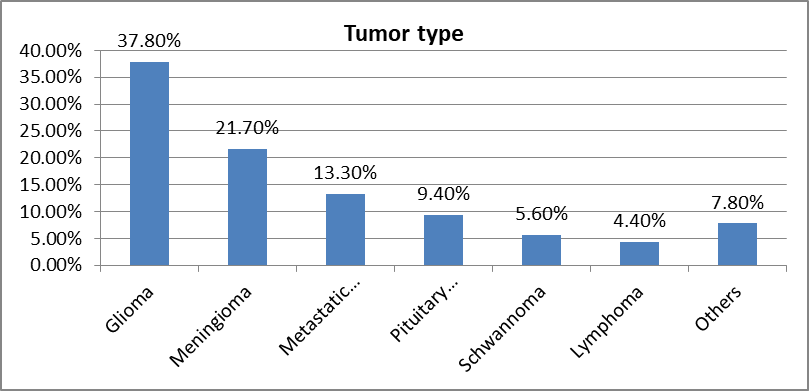
Graph 1: Histopathological diagnosis according to the Reference standard
AI-assisted MRI demonstrated superior diagnostic performance compared with conventional MRI. Overall diagnostic accuracy increased from 86.1% to 95.0%, with the improvement being statistically significant (McNemar's test, p = 0.008).
Table 2: Diagnostic performance of conventional MRI versus AI-assisted MRI
|
Parameter |
Conventional MRI |
AI-assisted MRI |
|
Sensitivity |
88.2% |
96.1% |
|
Specificity |
82.4% |
92.6% |
|
Positive Predictive Value |
90.0% |
96.8% |
|
Negative Predictive Value |
79.7% |
91.2% |
|
Overall Accuracy |
86.1% |
95.0% |
|
95% CI |
80.6–90.1 |
91.8–97.4 |
|
McNemar p-value |
- |
0.008 |
The confusion matrix demonstrated excellent classification performance of the AI-assisted MRI model, indicating a low rate of diagnostic errors [Graph: 2].
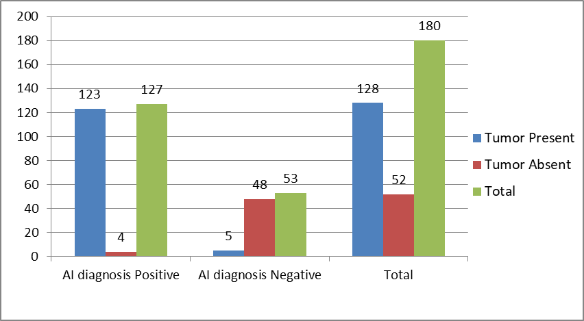
Graph 2: Confusion matrix for AI-assisted MRI
AI assistance significantly improved diagnostic accuracy compared with radiologist interpretation alone. Correct diagnoses increased from 86.1% to 95.0%, while incorrect diagnoses decreased from 13.9% to 5.0% (p = 0.008). Furthermore, AI significantly reduced the mean reporting time (p < 0.001) [Table: 3].
Table 3: Comparison between radiologist-alone and AI-assisted interpretation
|
Parameter |
Radiologist Alone |
AI-assisted |
p-value |
|
Correct diagnosis |
155 (86.1%) |
171 (95.0%) |
0.008 |
|
Incorrect diagnosis |
25 (13.9%) |
9 (5.0%) |
- |
|
Mean reporting time (minutes) |
11.8 ±2.6 |
7.3 ±1.8 |
<0.001 |
AI-assisted MRI showed almost perfect agreement with the reference diagnosis (Cohen's κ = 0.92), compared with substantial agreement for radiologist interpretation alone (κ = 0.79), indicating greater diagnostic consistency with AI assistance [Table: 4]..
Table 4: Agreement with reference diagnosis
|
Comparison |
Cohen's κ |
Interpretation |
|
Radiologist alone |
0.79 |
Substantial |
|
AI-assisted MRI |
0.92 |
Almost perfect |
Receiver operating characteristic (ROC) analysis demonstrated excellent discriminative ability for both diagnostic approaches. However, AI-assisted MRI achieved a significantly higher AUC of 0.964 compared with 0.885 for conventional MRI. Both models were statistically significant (p < 0.001), with AI-assisted MRI exhibiting superior overall diagnostic performance.
Table 5: Area under curve ROC analysis
|
Model |
AUC |
95% CI |
p-value |
|
Conventional MRI |
0.885 |
0.831–0.938 |
<0.001 |
|
AI-assisted MRI |
0.964 |
0.937–0.991 |
<0.001 |
DISCUSSION:
The present prospective diagnostic accuracy study demonstrated that AI-assisted MRI significantly improved the detection and characterization of brain tumors compared with conventional radiologist interpretation. AI assistance increased overall diagnostic accuracy from 86.1% to 95.0%, with significant improvements in sensitivity, specificity, positive predictive value, and negative predictive value. Furthermore, AI-assisted interpretation reduced reporting time while achieving almost perfect agreement with the reference diagnosis (κ=0.92) and an excellent ROC performance (AUC=0.964). These findings support the growing role of artificial intelligence as an effective adjunct to neuroradiologists in routine clinical practice.
The overall diagnostic accuracy of 95% observed in the present study is consistent with recent clinical evidence demonstrating that deep learning algorithms substantially improve MRI-based brain tumor diagnosis. In a multicenter evaluation, Bash et al [8] reported that AI-assisted image interpretation improved lesion detection and diagnostic confidence while maintaining high sensitivity across multiple neurological disorders. Likewise, Khalighi et al [9] highlighted that AI systems enhance tumor classification by integrating complex imaging features beyond human visual assessment, thereby improving diagnostic precision in neuro-oncology. These findings collectively suggest that AI functions best as a clinical decision-support tool rather than a replacement for radiologists.
The higher sensitivity (96.1%) and specificity (92.6%) achieved with AI-assisted MRI indicate that automated algorithms effectively reduce both false-negative and false-positive diagnoses. Early identification of malignant intracranial lesions is particularly important because delayed diagnosis may adversely affect surgical planning and patient survival. Deep learning models can detect subtle spatial and textural imaging characteristics that may be overlooked during routine visual interpretation, especially in infiltrative gliomas or small lesions with atypical imaging appearances. Such capabilities are particularly valuable in high-volume radiology departments where diagnostic workload is increasing.
Another important finding was the significant reduction in reporting time from 11.8 to 7.3 minutes following AI assistance. Similar observations have been reported by van Leeuwen et al [10], who demonstrated that AI-assisted radiology workflows improve reporting efficiency without compromising diagnostic accuracy. Reduced interpretation time can facilitate faster clinical decision-making, optimize workflow efficiency, and decrease radiologist fatigue, particularly in tertiary care centers managing large numbers of neuroimaging examinations.
The present study also demonstrated excellent agreement between AI-assisted MRI and the reference diagnosis (κ=0.92), compared with substantial agreement for conventional radiologist interpretation (κ=0.79). High inter-method agreement indicates consistent lesion characterization and reduced observer variability. Inter-observer variability remains a recognized limitation of conventional MRI interpretation because diagnostic performance is influenced by reader experience and institutional expertise. AI algorithms provide standardized image analysis, thereby improving reproducibility across different clinical settings [11].
Receiver operating characteristic analysis further confirmed the superior discriminative performance of AI-assisted MRI, with an AUC of 0.964, substantially higher than that of conventional MRI (0.885). Similar diagnostic performance has been reported in recent systematic reviews evaluating machine learning models for brain tumor diagnosis. Meta-analytic evidence indicates that modern deep learning architectures consistently achieve AUC values exceeding 0.95 when validated using multiparametric MRI datasets. The excellent ROC performance observed in the present study therefore supports the reliability of AI-assisted MRI for distinguishing neoplastic from non-neoplastic lesions and for tumor characterization [12].
The findings of this study have important clinical implications. AI-assisted MRI has the potential to enhance diagnostic confidence, reduce interpretation variability, accelerate reporting, and facilitate earlier therapeutic decision-making. Rather than replacing radiologists, AI should be viewed as an intelligent assistive technology capable of improving consistency and efficiency while allowing clinicians to focus on complex diagnostic challenges. Integration of AI into routine neuroimaging workflows may be particularly beneficial in resource-limited settings where experienced neuroradiologists are scarce [13].
CONCLUSION:
AI-assisted MRI significantly improved the early detection and characterization of brain tumors by enhancing diagnostic accuracy, sensitivity, specificity, and interobserver agreement while substantially reducing reporting time compared with conventional MRI interpretation. These findings support the integration of AI as a reliable decision-support tool to complement radiologist expertise, improve diagnostic consistency, and facilitate timely clinical decision-making. Further multicenter studies with larger cohorts are warranted to validate these results and establish the broader clinical utility of AI-assisted neuroimaging.
REFERENCES:
1. Dorfner FJ, Patel JB, Kalpathy-Cramer J, Gerstner ER, Bridge CP, et al. A review of deep learning for brain tumor analysis in MRI. NPJ Precis Oncol. 2025;9:2.
2. Khalighi S, Reddy K, Midya A, Pandav KB, Madabhushi A, Abedalthagafi M. Artificial intelligence in neuro-oncology: advances and challenges in brain tumor diagnosis, prognosis, and precision treatment. NPJ Precis Oncol. 2024;8:80.
3. Wang TW, Shiao YC, Hong JS, Lee WK, Hsu MS, Cheng HM, et al. Artificial intelligence detection and segmentation models: a systematic review and meta-analysis of brain tumors in magnetic resonance imaging. Mayo Clin Proc Digit Health. 2024;2(1):75-91. doi:10.1016/j.mcpdig.2024.01.002.
4. Al-Rumaihi A, et al. Performance evaluation of artificial intelligence techniques in the diagnosis of brain tumors: a systematic review and meta-analysis. Diagnostics (Basel). 2025. doi:10.3390/diagnostics15151766.
5. Abidin ZU, Naqvi RA, Haider A, Kim HS, Jeong D, Lee SW. Recent deep learning-based brain tumor segmentation models using multi-modality magnetic resonance imaging: a prospective survey. Front Bioeng Biotechnol. 2024;12:1392807. doi:10.3389/fbioe.2024.1392807
6. Dorfner FJ, Patel JB, Kalpathy-Cramer J, Gerstner ER, Bridge CP. A review of deep learning for brain tumor analysis in MRI. NPJ Precision Oncology. 2025;9(1):2. doi:10.1038/s41698-024-00789-2. PMID: 39753730.
7. WHO Classification of Tumours Editorial Board. Central Nervous System Tumours. 5th ed. WHO Classification of Tumours, Vol. 6. Lyon, France: International Agency for Research on Cancer; 2021. ISBN: 978-92-832-4508-7. Available from: https://publications.iarc.who.int/Book-And-Report-Series/Who-Classification-Of-Tumours/Central-Nervous-System-Tumours-2021
8. Bash S, Wang H, Varadan V, et al. Artificial intelligence applications in neuroradiology. Radiology. 2024;310(2):e230965. doi:10.1148/radiol.230965.
9. Khalighi S, Reddy K, Midya A, Pandav KB, Madabhushi A, Abedalthagafi M. Artificial intelligence in neuro-oncology: advances and challenges in brain tumor diagnosis, prognosis, and precision treatment. NPJ Precision Oncology. 2024;8:80. doi:10.1038/s41698-024-00595-w.
10. Van Leeuwen KG, Schalekamp S, Rutten MJCM, et al. Artificial intelligence in radiology: 100 commercially available products and their scientific evidence. Eur Radiol. 2021; 31:3797–3804. doi:10.1007/s00330-021-07892-z.
11. Louis DN, Weller M, Aldape K, et al. Glioma. Nat Rev Dis Primers. 2021; 7:74. doi:10.1038/s41572-021-00289-0.
12. Ostrom QT, Price M, Ryan K, et al. CBTRUS Statistical Report: Primary Brain and Other Central Nervous System Tumors Diagnosed in the United States, 2017–2021. Neuro Oncol. 2024; 26(Suppl 5):v1-v99. doi:10.1093/neuonc/noae190.
13. Topol EJ. High-performance medicine: the convergence of human and artificial intelligence. Nat Med. 2019; 25:44–56. doi:10.1038/s41591-018-0300-7.