Role of Ultrasound Elastography in the Evaluation of Diffuse Liver Diseases: A Non-Invasive Diagnostic Approach.
- Vaibhav Mehta. , Consultant Radiologist, Keshavam Imaging Centre, Gandhinagar.
Article Information:
Abstract:
Background: Diffuse liver diseases (DLDs), including hepatic steatosis, hepatitis, cirrhosis, hemochromatosis, amyloidosis, and Wilson's disease, are major causes of chronic liver-related morbidity and mortality. Accurate assessment of hepatic fibrosis is essential for determining disease severity, guiding treatment, and predicting prognosis. Although liver biopsy remains the reference standard, it is invasive and associated with sampling errors and potential complications. Ultrasound shear wave elastography (SWE) has emerged as a reliable, non-invasive technique for quantitative assessment of liver stiffness and hepatic fibrosis. Objectives: To evaluate the role of ultrasound shear wave elastography in the assessment of diffuse liver diseases and to determine its utility in staging hepatic fibrosis. Materials and Methods: This prospective observational study included 50 patients with diffuse liver diseases who underwent conventional ultrasonography and shear wave elastography at the Department of Radiodiagnosis, Smt. N.H.L. Municipal Medical College and SVP Hospital, Ahmedabad, between August 2018 and April 2020. Liver stiffness measurements were obtained from the right hepatic lobe using Sound Touch Elastography (STE) and Sound Touch Quantification (STQ). Fibrosis staging was performed according to the Castera elastography criteria. Results: The study included 30 males and 20 females with a mean age of 58.8 years. Hepatic steatosis was the most common diffuse liver disease (32%), followed by alcoholic hepatitis (26%), viral hepatitis (24%), and cirrhosis (12%). The highest mean liver stiffness value was observed in cirrhosis (15.08 kPa). According to the Castera fibrosis staging system, 48% of patients had significant fibrosis (F2), 30% had severe fibrosis (F3), 12% had cirrhosis (F4), and 10% had absent or mild fibrosis (F0–F1). Overall, 78% of patients demonstrated significant to advanced fibrosis (F2–F4). Conclusion: Ultrasound shear wave elastography is an accurate, reproducible, and non-invasive modality for evaluating diffuse liver diseases and staging hepatic fibrosis. It provides quantitative assessment of liver stiffness, facilitates early detection of fibrosis—particularly in hepatic steatosis—and reduces reliance on invasive liver biopsy. When combined with conventional ultrasonography, shear wave elastography enhances diagnostic accuracy and is valuable for disease staging, treatment planning, and follow-up of patients with chronic liver disease.
Keywords:
Article :
INTRODUCTION:
Diffuse liver diseases (DLDs) comprise a diverse group of hepatic disorders characterized by widespread involvement of the liver parenchyma rather than focal lesions. These include hepatic steatosis, viral and alcoholic hepatitis, cirrhosis, hemochromatosis, hemosiderosis, amyloidosis, Wilson's disease, and granulomatous diseases. Collectively, these conditions account for a substantial proportion of chronic liver disease worldwide and are associated with significant morbidity and mortality due to progressive fibrosis, portal hypertension, liver failure, and hepatocellular carcinoma (HCC). Early diagnosis and accurate assessment of disease severity are essential for guiding treatment and improving patient outcomes. (1–3)
Conventional ultrasonography (USG) is the first-line imaging modality for the evaluation of diffuse liver diseases because it is widely available, inexpensive, non-invasive, and free from ionizing radiation. It provides valuable information regarding liver size, parenchymal echotexture, surface nodularity, vascular anatomy, and complications such as portal hypertension. Fatty liver typically appears as diffuse increased hepatic echogenicity with posterior acoustic attenuation and poor visualization of intrahepatic vessels and the diaphragm. Acute viral hepatitis may demonstrate hepatomegaly with decreased hepatic echogenicity, whereas alcoholic hepatitis often exhibits increased echogenicity secondary to concomitant fatty infiltration. Cirrhosis is characterized by heterogeneous echotexture, irregular liver contour, altered lobar morphology, regenerative nodules, splenomegaly, and Doppler evidence of portal hypertension. However, conventional ultrasonography has limited sensitivity for detecting early fibrosis and often demonstrates nonspecific findings in disorders such as hemochromatosis, amyloidosis, and Wilson's disease. (4–6).
Hepatic fibrosis represents the common pathological pathway in most chronic diffuse liver diseases irrespective of etiology. Progressive fibrosis ultimately culminates in cirrhosis and its associated complications. Liver biopsy has traditionally been regarded as the reference standard for staging hepatic fibrosis; however, it is invasive, susceptible to sampling error and interobserver variability, and carries a risk of complications, making it unsuitable for serial monitoring. Consequently, there has been increasing interest in reliable non-invasive techniques for the assessment of liver fibrosis. (7,8).
Ultrasound elastography has emerged as a valuable non-invasive imaging modality that quantitatively measures liver stiffness, which closely correlates with the degree of hepatic fibrosis. Techniques such as transient elastography (TE), point shear-wave elastography (pSWE), and two-dimensional shear-wave elastography (2D-SWE) have demonstrated excellent diagnostic performance for staging liver fibrosis in chronic liver diseases. In addition to fibrosis assessment, elastography plays an important role in disease monitoring, evaluation of treatment response, prediction of portal hypertension, and risk stratification. Its integration into routine ultrasonography has significantly enhanced the diagnostic capability of hepatic imaging while reducing the need for liver biopsy. (8–11).
Although ultrasound elastography has become increasingly incorporated into routine hepatology practice, its role across the entire spectrum of diffuse liver diseases continues to evolve. Further evaluation of its diagnostic accuracy and clinical utility in comparison with conventional ultrasonography is warranted, particularly in heterogeneous patient populations.
Therefore, the present study aims to evaluate the role of ultrasound elastography in the assessment of diffuse liver diseases and to determine its effectiveness in detecting and staging hepatic fibrosis while correlating elastographic findings with conventional ultrasonographic features.
MATERIALS AND METHODS:
Study Design and Setting
A prospective observational study was conducted in the Department of Radiodiagnosis at Smt. N.H.L. Municipal Medical College and Sheth Vadilal Sarabhai (SVP) Hospital, Ahmedabad, Gujarat, India, over a period of August 2018 to April 2020. The study aimed to evaluate the role of ultrasound elastography in patients with diffuse liver diseases.
Ethical Approval
The study protocol was reviewed and approved by the Institutional Ethics Committee (IEC) of Smt. N.H.L. Municipal Medical College and SVP Hospital before commencement of the study. Written informed consent was obtained from all participants prior to enrollment.
Study Population
A total of 50 consecutive patients with clinically or radiologically suspected diffuse liver disease were included in the study. No selection bias regarding age or gender was applied. The study population comprised 30 males (60%) and 20 females (40%), with an age range of 24–78 years (mean age: 58.8 years).
Ultrasound Examination
All participants underwent conventional ultrasonography followed by ultrasound elastography using a standardized scanning protocol. The liver was evaluated for size, echotexture, echogenicity, surface nodularity, vascular architecture, and associated findings suggestive of diffuse liver disease.
Ultrasound Elastography Technique
Shear wave elastography was performed using Sound Touch Elastography (STE) and Sound Touch Quantification (STQ).
For elastography, the image depth was adjusted to approximately 10 cm. The region of interest (ROI) was positioned within the right hepatic lobe at a depth of 2–5 cm below the liver capsule, avoiding large blood vessels, biliary structures, focal lesions, and the liver capsule. The ROI size was maintained at approximately 2.5 × 2.5 cm.
Patients were instructed to hold their breath during image acquisition to minimize motion artifacts.
Sound Touch Elastography (STE)
STE images were acquired only when the elastographic image appeared stable. Measurements were accepted when the Reliability (RLB) Index exceeded 80% and the M-STB reliability indicator displayed green. Three valid measurements were obtained, and the median liver stiffness value (kPa) was recorded as the final STE result.
Sound Touch Quantification (STQ)
For STQ, measurements were obtained after confirming a stable elastography display with the M-STB indicator showing green. Ten valid measurements were acquired, and the median stiffness value was considered the final result. Measurements with poor reliability were excluded. Quality control was ensured by maintaining an interquartile range to median (IQR/M) ratio of less than 30%, consistent with established recommendations.
Assessment of Liver Fibrosis
Liver stiffness measurements were obtained exclusively from the right hepatic lobe. The normal liver stiffness value was considered <7 kPa. Fibrosis staging was performed according to the Castera elastography criteria, which are based on liver stiffness thresholds corresponding to the METAVIR fibrosis stages.
|
Fibrosis Stage |
Liver Stiffness (kPa) |
Interpretation |
|
F0–F1 |
<7.0 |
Absent or mild fibrosis |
|
F2 |
7.0–9.5 |
Significant fibrosis |
|
F3 |
9.5–12.5 |
Severe fibrosis |
|
F4 |
>12.5 |
Cirrhosis |
Corresponding METAVIR Fibrosis Score
|
METAVIR Score |
Histopathological Findings |
|
F0 |
No fibrosis |
|
F1 |
Portal fibrosis without septa |
|
F2 |
Portal fibrosis with few septa |
|
F3 |
Numerous septa without cirrhosis |
|
F4 |
Cirrhosis |
Statistical Analysis
Data were analyzed using SPSS version 25. Continuous variables are presented as mean ± standard deviation or median (interquartile range), and categorical variables as frequencies and percentages. Owing to the descriptive nature of the study and limited sample size, no inferential statistical comparisons were performed.
RESULTS:
A total of 50 patients with diffuse liver diseases were included in the study. The spectrum of diseases comprised hepatic steatosis, viral hepatitis, alcoholic hepatitis, cirrhosis, Wilson's disease, amyloidosis, and hemochromatosis. Liver stiffness was assessed using ultrasound shear wave elastography and classified according to the Castera fibrosis staging system.
Distribution of Diffuse Liver Diseases
Among the study population, hepatic steatosis was the most frequently encountered diffuse liver disease, accounting for 16 (32%) patients, followed by alcoholic hepatitis (13; 26%), viral hepatitis (12; 24%), and cirrhosis (6; 12%). Less common conditions included Wilson's disease (1; 2%), amyloidosis (1; 2%), and hemochromatosis (1; 2%) (Table 1).
Table 1. Distribution of Diffuse Liver Diseases (n = 50)
|
Diffuse Liver Disease |
Number of Patients (n) |
Percentage (%) |
|
Hepatic steatosis |
16 |
32.0 |
|
Alcoholic hepatitis |
13 |
26.0 |
|
Viral hepatitis |
12 |
24.0 |
|
Cirrhosis |
6 |
12.0 |
|
Wilson's disease |
1 |
2.0 |
|
Amyloidosis |
1 |
2.0 |
|
Hemochromatosis |
1 |
2.0 |
|
Total |
50 |
100 |
Liver Stiffness Measurements
The liver stiffness values obtained by shear wave elastography ranged from 5.0 to 17.5 kPa. The mean liver stiffness values observed in different diffuse liver diseases are summarized in Table 2. Among all disease categories, cirrhosis demonstrated the highest mean liver stiffness value (15.08 kPa), followed by hemochromatosis (11.2 kPa) and amyloidosis (9.5 kPa), whereas Wilson's disease exhibited the lowest mean stiffness value (7.6 kPa).
Table 2. Mean Liver Stiffness Values Measured by Shear Wave Elastography
|
Diffuse Liver Disease |
Mean Liver Stiffness (kPa) |
|
Hepatic steatosis |
9.02 |
|
Viral hepatitis |
8.96 |
|
Alcoholic hepatitis |
9.10 |
|
Cirrhosis |
15.08 |
|
Hemochromatosis |
11.20 |
|
Amyloidosis |
9.50 |
|
Wilson's disease |
7.60 |
Values for Wilson disease, amyloidosis, and hemochromatosis represent measurements from single patients.
Distribution According to Castera Fibrosis Staging
Based on liver stiffness measurements, patients were categorized according to the Castera elastography fibrosis grading system (Table 3). Overall, 39 of 50 patients (78%) demonstrated significant to advanced fibrosis (F2–F4) on ultrasound elastography.
Among individual disease groups: Most patients with hepatic steatosis were classified as F2 fibrosis, with a smaller proportion showing F3 fibrosis, while only one patient had absent/mild fibrosis. Patients with viral hepatitis were distributed between F2 and F3 stages, with only one patient demonstrating F0–F1 fibrosis. Alcoholic hepatitis showed a broad spectrum of fibrosis ranging from F0–F1 to F3, indicating variable disease severity. All six patients with cirrhosis were classified as F4, confirming advanced hepatic fibrosis. The single cases of Wilson's disease, amyloidosis, and hemochromatosis demonstrated F2, F2, and F3 fibrosis, respectively. (Table 3,4 and 5).
Table 3. Distribution of Patients According to Castera Fibrosis Stage
|
Fibrosis Stage |
Number of Patients (n) |
Percentage (%) |
|
F0–F1 (Absent/Mild fibrosis) |
5 |
10.0 |
|
F2 (Significant fibrosis) |
24 |
48.0 |
|
F3 (Severe fibrosis) |
15 |
30.0 |
|
F4 (Cirrhosis) |
6 |
12.0 |
|
Total |
50 |
100 |
Table 4. Distribution of Diffuse Liver Diseases According to Castera Fibrosis Stage
|
Diagnosis |
F0–F1 |
F2 |
F3 |
F4 |
Total |
|
Hepatic steatosis |
1 |
11 |
4 |
0 |
16 |
|
Alcoholic hepatitis |
3 |
5 |
5 |
0 |
13 |
|
Viral hepatitis |
1 |
6 |
5 |
0 |
12 |
|
Cirrhosis |
0 |
0 |
0 |
6 |
6 |
|
Wilson's disease |
0 |
1 |
0 |
0 |
1 |
|
Amyloidosis |
0 |
1 |
0 |
0 |
1 |
|
Hemochromatosis |
0 |
0 |
1 |
0 |
1 |
|
Total |
5 |
24 |
15 |
6 |
50 |
Table 5. Liver Stiffness Classification Based on Castera Criteria
|
Fibrosis Stage |
Liver Stiffness (kPa) |
Interpretation |
|
F0–F1 |
<7.0 |
Absent or mild fibrosis |
|
F2 |
7.0–9.5 |
Significant fibrosis |
|
F3 |
9.5–12.5 |
Severe fibrosis |
|
F4 |
>12.5 |
Cirrhosis |
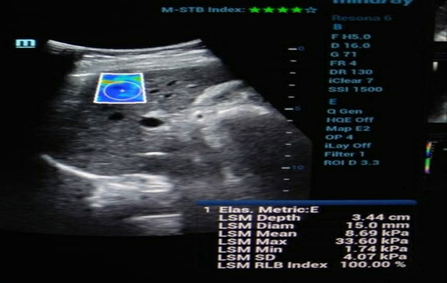
Figure 1. Ultrasound elastography in alcoholic hepatitis.
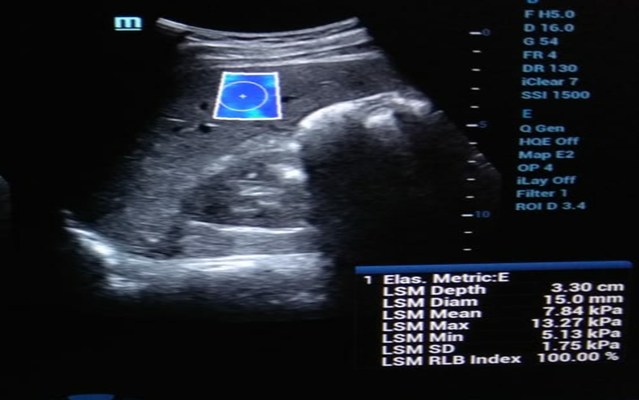
Figure 2. Ultrasound elastography in viral hepatitis.
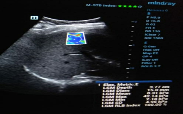
Figure 3. Ultrasound elastography in hepatic steatosis.
DISCUSSION:
Diffuse liver diseases represent a major cause of chronic liver-related morbidity and mortality worldwide. Accurate assessment of hepatic fibrosis is essential because the stage of fibrosis determines prognosis, therapeutic decision-making, surveillance strategies, and the risk of complications including portal hypertension and hepatocellular carcinoma. Although conventional ultrasonography remains the first-line imaging modality for evaluating diffuse liver diseases, its ability to accurately quantify hepatic fibrosis is limited, particularly during the early stages of disease. Ultrasound shear wave elastography has emerged as a valuable non-invasive technique that quantitatively measures liver stiffness, thereby providing an objective assessment of hepatic fibrosis without the limitations associated with liver biopsy. (1–3)
In the present study, hepatic steatosis was the most common diffuse liver disease (32%), followed by alcoholic hepatitis (26%), viral hepatitis (24%), and cirrhosis (12%). This observation is consistent with the increasing global prevalence of metabolic dysfunction-associated steatotic liver disease (MASLD, formerly NAFLD), which has become the leading cause of chronic liver disease worldwide owing to rising rates of obesity, diabetes mellitus, and metabolic syndrome. (4,5)
The present study demonstrated that the mean liver stiffness value was highest in cirrhosis (15.08 kPa). Patients with hepatic steatosis, viral hepatitis, and alcoholic hepatitis exhibited intermediate stiffness values, reflecting varying degrees of hepatic fibrosis. These findings are consistent with the pathological progression of diffuse liver diseases, in which increasing fibrosis results in progressive elevation of liver stiffness measured by shear wave elastography. (6,7)
Using the Castera elastography criteria, 78% of patients demonstrated significant or advanced fibrosis (F2–F4), highlighting the burden of clinically relevant fibrosis among patients with diffuse liver diseases. All patients with clinically diagnosed cirrhosis were correctly classified as F4, indicating excellent concordance between elastography findings and established clinical diagnosis. These observations support the utility of shear wave elastography as a reliable tool for staging hepatic fibrosis in routine clinical practice.
The findings of the present study are in agreement with previous studies evaluating ultrasound elastography. Bavu et al. compared real-time two-dimensional shear wave elastography (2D-SWE) with transient elastography in patients with chronic hepatitis C and reported superior diagnostic accuracy of 2D-SWE for mild and intermediate stages of hepatic fibrosis. (8) Similarly, Ferraioli et al. demonstrated a strong linear correlation between liver stiffness and histological fibrosis stage, with 2D-SWE outperforming transient elastography in identifying significant fibrosis (≥F2). (9) Furthermore, another comparative study reported that 2D-SWE showed higher diagnostic accuracy than both transient elastography and Virtual Touch Quantification (VTQ) for detecting clinically significant and severe fibrosis, emphasizing the robustness of shear wave elastography in chronic liver disease evaluation. (10)
Conventional ultrasonography remains indispensable for identifying morphological abnormalities such as hepatomegaly, altered hepatic echogenicity, coarse echotexture, nodular liver surface, regenerative nodules, splenomegaly, and signs of portal hypertension. However, these sonographic findings generally become apparent only after significant structural changes have occurred. In contrast, ultrasound elastography provides quantitative information regarding tissue stiffness, allowing earlier detection of fibrosis before irreversible morphological changes develop. Therefore, elastography should be considered complementary rather than a replacement for conventional ultrasonography, as the combination of both techniques substantially improves diagnostic confidence. (2,6)
One of the major advantages of ultrasound elastography is its non-invasive nature. Unlike liver biopsy, elastography is painless, rapid, repeatable, and free from the risks of bleeding, infection, sampling error, and interobserver variability. Consequently, it is particularly valuable for serial monitoring of disease progression and assessment of treatment response in patients with chronic liver disease. Since hepatic fibrosis is now recognized as a potentially reversible process following effective treatment of the underlying etiology, repeated non-invasive assessment has become increasingly important in clinical practice. (1–3,7)
The present study has certain limitations. The sample size was relatively small and represented a single tertiary care center. Histopathological confirmation was not available for every patient, limiting direct comparison between elastographic findings and liver biopsy. Additionally, the number of patients with uncommon diffuse liver diseases such as Wilson's disease, amyloidosis, and hemochromatosis was limited, preventing disease-specific statistical analysis. Larger multicenter studies with histopathological correlation are warranted to further validate these findings.
Overall, the findings of the present study demonstrate that ultrasound shear wave elastography is a reliable, reproducible, and non-invasive imaging modality for evaluating diffuse liver diseases and staging hepatic fibrosis. When used in conjunction with conventional ultrasonography, elastography improves diagnostic accuracy, facilitates early identification of significant fibrosis, reduces dependence on invasive liver biopsy, and provides an effective tool for longitudinal monitoring of patients with chronic liver disease.
CONCLUSION:
Ultrasound shear wave elastography is a reliable, non-invasive, and reproducible imaging modality for the evaluation of diffuse liver diseases. In the present study, hepatic steatosis was the most common diffuse liver disease, while cirrhosis demonstrated the highest mean liver stiffness value (15.08 kPa), indicating advanced hepatic fibrosis. The majority of patients were classified as having significant (F2) or severe (F3) fibrosis according to the Castera elastography staging system.
Shear wave elastography provided quantitative assessment of liver stiffness, enabling accurate grading of hepatic fibrosis across various diffuse liver diseases. It proved particularly useful in identifying early stages of fibrosis in patients with hepatic steatosis, allowing timely intervention before progression to irreversible liver damage and cirrhosis.
When used alongside conventional ultrasonography, ultrasound elastography enhances diagnostic accuracy, reduces the need for invasive liver biopsy, and serves as an effective tool for disease staging, treatment monitoring, and long-term follow-up. Therefore, shear wave elastography should be considered an integral component of the routine imaging evaluation of patients with diffuse liver diseases.
REFERENCES:
1. Barr RG, Wilson SR, Rubens D, Garcia-Tsao G, Ferraioli G. Update to the Society of Radiologists in Ultrasound Liver Elastography Consensus Statement. Radiology. 2020;296(2):263-274. doi:10.1148/radiol.2020192437.
2. Ferraioli G, Wong VW, Castera L, Berzigotti A, Sporea I, Dietrich CF, et al. Liver ultrasound elastography: An update to the World Federation for Ultrasound in Medicine and Biology Guidelines and Recommendations. Ultrasound Med Biol. 2018;44(12):2419-2440.
3. Castera L, Friedrich-Rust M, Loomba R. Noninvasive assessment of liver disease in patients with nonalcoholic fatty liver disease. Gastroenterology. 2019;156(5):1264-1281.e4.
4. Rockey DC, Caldwell SH, Goodman ZD, Nelson RC, Smith AD. Liver biopsy. Hepatology. 2009;49(3):1017-1044.
5. Bedossa P, Dargère D, Paradis V. Sampling variability of liver fibrosis in chronic hepatitis C. Hepatology. 2003;38(6):1449-1457.
6. Regev A, Berho M, Jeffers LJ, Milikowski C, Molina EG, Pyrsopoulos NT, et al. Sampling error and intraobserver variation in liver biopsy in patients with chronic hepatitis C. Am J Gastroenterol. 2002;97(10):2614-2618.
7. Dietrich CF, Bamber J, Berzigotti A, Bota S, Cantisani V, Castera L, et al. EFSUMB Guidelines and Recommendations on the Clinical Use of Liver Ultrasound Elastography. Ultraschall Med. 2017;38(4):e16-e47.
8. Bavu E, Gennisson JL, Couade M, Bercoff J, Mallet V, Fink M, et al. Noninvasive in vivo liver fibrosis evaluation using supersonic shear imaging: A clinical study on 113 hepatitis C patients. Radiology. 2011;261(3):895-905.
9. Ferraioli G, Tinelli C, Lissandrin R, Zicchetti M, Dal Bello B, Filice G, et al. Point shear wave elastography method for assessing liver stiffness. Radiology. 2014;273(3):917-924.
10. Cassinotto C, Lapuyade B, Mouries A, Hiriart JB, Vergniol J, Gaye D, et al. Comparison of transient elastography, acoustic radiation force impulse imaging, and shear wave elastography for liver fibrosis assessment. Radiology. 2014;269(1):283-292.
11. European Association for the Study of the Liver (EASL). EASL Clinical Practice Guidelines on non-invasive tests for evaluation of liver disease severity and prognosis. J Hepatol. 2021;75(3):659-689.