Impact of Population-Specific First-Trimester Nasal Bone Reference Values on Prenatal Screening Accuracy in India.
- Anshu Gupta , Associate Professor, Department of Anatomy, Sarojini Naidu Medical College, Agra.
- Anita Rawat , Assistant Professor, Department of Anatomy, Sarojini Naidu Medical College, Agra.
- Radhika Parashar , Associate Professor Department of Obs & Gynaecology, F. H Medical College Etmadpur, Agra.
- Pradeep Kumar Gupta , Associate Professor, Department of Anatomy UPUMS, Saifai.
- Kamal Bhardwaj , Professor Department of Anatomy F. H Medical College Agra.
- Sumit Kumar , Assistant Professor Department of anatomy, F.H Medical College Agra.
Article Information:
Abstract:
Background: First-trimester prenatal screening is essential for the early detection of fetal chromosomal abnormalities. Fetal nasal bone assessment is an established ultrasonographic marker; however, its diagnostic accuracy may vary according to ethnic and population-specific reference values. Aim: To evaluate the impact of population-specific first-trimester nasal bone reference values on prenatal screening accuracy in the Indian population and compare their diagnostic performance with international reference standards. Materials and Methods: This prospective observational study was conducted on 90 singleton pregnancies between 11 and 13+6 weeks of gestation. Fetal nasal bone length was measured using standardized ultrasonographic techniques in the midsagittal plane. Population-specific reference values were established according to gestational age and compared with international reference standards. Diagnostic performance was assessed using sensitivity, specificity, positive predictive value (PPV), negative predictive value (NPV), and overall diagnostic accuracy. Correlation analysis and receiver operating characteristic (ROC) curve analysis were also performed. Results: The mean fetal nasal bone length was 2.18 ± 0.39 mm and increased significantly with advancing gestational age (p < 0.001). A strong positive correlation was observed between nasal bone length and gestational age (r = 0.69, p < 0.001) as well as crown-rump length (r = 0.73, p < 0.001). Application of Indian population-specific reference values reduced the proportion of fetuses classified as having hypoplastic or absent nasal bone from 12.2% to 6.7%. Compared with international reference standards, population-specific values maintained 100% sensitivity and 100% negative predictive value, while improving specificity (96.6% vs. 90.8%), positive predictive value (50.0% vs. 27.3%), and overall diagnostic accuracy (96.7% vs. 91.1%). ROC analysis demonstrated excellent diagnostic performance with an area under the curve (AUC) of 0.94, and an optimal nasal bone length cut-off of 1.65 mm provided 100% sensitivity and 92.0% specificity. Conclusion: Population-specific first-trimester nasal bone reference values significantly improve prenatal screening accuracy in the Indian population by reducing false-positive results while maintaining excellent sensitivity. These findings support the adoption of locally derived gestational age-specific reference standards to enhance the effectiveness and reliability of first-trimester prenatal screening.
Keywords:
Article :
INTRODUCTION:
First-trimester prenatal screening is an important part of obstetric care for early detection of chromosomal abnormalities such as trisomy 21, trisomy 18, and trisomy 13. Screening between 11 and 13+6 weeks combines maternal age, nuchal translucency, biochemical markers, and ultrasound soft markers to improve detection and reduce unnecessary invasive procedures.
1 fetal nasal bone assessment is an important ultrasound marker in first-trimester screening. Absence or hypoplasia of the nasal bone is strongly associated with trisomy 21 due to delayed ossification in affected fetuses. Its inclusion improves detection rates and reduces false-positive results. 2,3 Accurate nasal bone evaluation requires standardized measurement in the midsagittal plane during 11–13+6 weeks of gestation. Proper technique and operator training are essential for reliable results. 1,3 studies have shown that nasal bone length varies with ethnicity and population.
Using non–population-specific reference values may increase false-positive rates and lead to unnecessary interventions. 4,5 recent Indian studies emphasize the need for population-specific nasal bone reference values to improve screening accuracy while maintaining high sensitivity. 5,6 incorporations of nasal bone assessment into combined first-trimester screening has been shown to improve the detection rate of chromosomal abnormalities, particularly trisomy 21, when used alongside nuchal translucency and biochemical markers. 7 furthermore, fetal nasal bone length has been demonstrated to increase progressively with gestational age and crown-rump length, supporting the requirement for gestational age-specific reference ranges rather than fixed universal cut-offs. 8 Evidence also suggests that application of optimized cut-off values for nasal bone length improves diagnostic performance by increasing specificity and reducing unnecessary invasive procedures without compromising sensitivity. 9 recent studies have further emphasized that fetal nasal bone assessment is influenced by ethnic and population-specific variations, which may affect the performance of first-trimester screening algorithms. These differences can lead to misclassification when universal reference ranges are applied across diverse populations. 10. Therefore, there is a growing consensus that locally derived reference values are essential to improve the accuracy and reliability of prenatal screening programs, particularly in reducing false-positive results while maintaining high detection rates for chromosomal abnormalities. 11.
MATERIALS AND METHODS:
Study Design and Setting:
This prospective observational study was conducted in the Department of Anatomy at Sarojini Naidu Medical College, collaboration with the department of Obstetrics and Gynecology, over a period of 12 months.
Study Population:
A total of 90 pregnant women with singleton pregnancies undergoing routine first-trimester ultrasound between 11 weeks and 13 weeks + 6 days of gestation were included in the study.
Inclusion Criteria:
• Pregnant women aged 18–40 years.
• Singleton viable pregnancy.
• Gestational age between 11+0 and 13+6 weeks, confirmed by crown-rump length (CRL).
• Women who provided written informed consent.
Exclusion Criteria:
• Multiple pregnancies.
• Major fetal structural anomalies detected at the first-trimester scan.
• Inadequate visualization of the fetal nasal bone.
• Pregnancies with incomplete follow-up or unavailable pregnancy outcome.
• Women who declined participation.
Ultrasound Examination
All ultrasound examinations were performed using a high-resolution ultrasound system equipped with a 3.5–5 MHz transabdominal transducer. When transabdominal imaging was suboptimal, transvaginal ultrasonography was performed.
Gestational age was determined by measurement of the crown-rump length (CRL) according to established guidelines. Fetal nasal bone length (NBL) was measured in the true midsagittal plane with the fetal profile occupying most of the image. The ultrasound beam was maintained at approximately 45° or 135° to the nasal bone to ensure optimal visualization. The echogenic nasal bone was identified separately from the overlying skin, and its maximum ossified length was measured using electronic calipers. Three satisfactory measurements were obtained for each fetus, and the average value was used for analysis. All examinations were performed by experienced fetal medicine sonologists following standardized measurement protocols.
Development of Population-Specific Reference Values:
The measured nasal bone lengths were grouped according to gestational age into three categories:
• 11.0–11.6 weeks
• 12.0–12.6 weeks
• 13.0–13.6 weeks
For each gestational age group, the mean, standard deviation (SD), median (50th percentile), 5th percentile, and 95th percentile of nasal bone length was calculated to establish population-specific reference values for the study population.
Prenatal Screening Assessment:
Each fetus was classified using two different reference standards:
1. International nasal bone reference values reported in the published literature.
2. Population-specific Indian reference values derived from the present study.
A nasal bone measurement below the gestational age-specific reference limit or complete absence of ossification was classified as hypoplastic/absent, whereas measurements within the reference range were classified as normal.
Pregnancy Outcome Assessment:
Pregnancy outcomes were obtained from hospital records and follow-up until confirmation of fetal chromosomal status after birth or diagnostic testing when clinically indicated. Fetuses were categorized as having either:
• Normal pregnancy outcome, or
• Chromosomal abnormality.
These outcomes were used as the reference standard for evaluating the diagnostic performance of nasal bone assessment.
AIM OF THE STUDY:
To evaluate the impact of population-specific first-trimester nasal bone reference values on prenatal screening accuracy in the Indian population and compare their diagnostic performance with international reference standards.
RESULTS:
Table 1. Baseline Characteristics of the Study Population (n = 90)
|
Variable |
Category |
Frequency (n) |
Percentage (%) |
p-value |
|
Maternal Age (years) |
<25 |
18 |
20.0 |
0.048* |
|
|
25–30 |
42 |
46.7 |
|
|
|
>30 |
30 |
33.3 |
|
|
Gravidity |
Primigravida |
38 |
42.2 |
0.391 |
|
|
Multigravida |
52 |
57.8 |
|
|
Gestational Age (weeks) |
11.0–11.6 |
24 |
26.7 |
0.462 |
|
|
12.0–12.6 |
38 |
42.2 |
|
|
|
13.0–13.6 |
28 |
31.1 |
|
|
Crown-Rump Length (mm) |
Mean ± SD |
61.8 ± 8.2 |
- |
0.371 |
|
Nasal Bone Length (mm) |
Mean ± SD |
2.18 ± 0.39 |
— |
<0.001* |
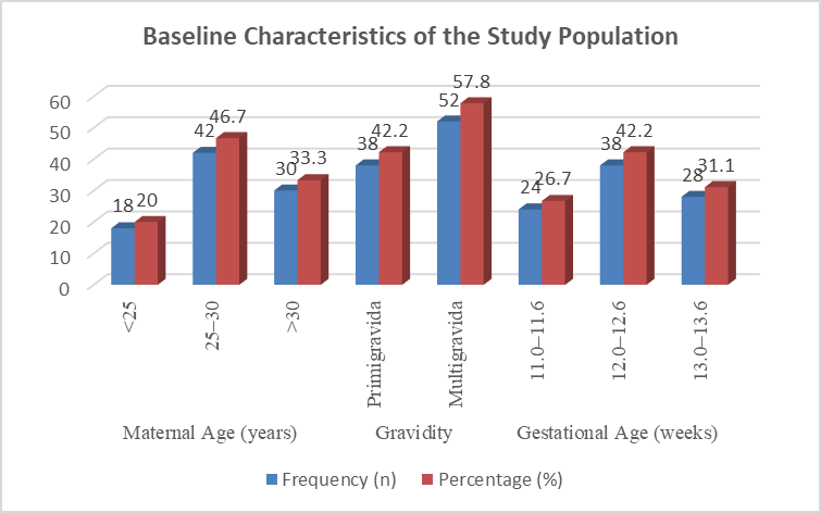
Fig: 1 Graphical represents Baseline Characteristics of the Study Population
The majority of participants were aged 25–30 years (46.7%) and were multigravida (57.8%). Most examinations were performed between 12.0 and 12.6 weeks of gestation (42.2%).
The mean crown-rump length was 61.8 ± 8.2 mm, and the mean nasal bone length was 2.18 ± 0.39 mm. Nasal bone length showed a statistically significant association (p < 0.001), whereas maternal age, gravidity, gestational age distribution, and crown-rump length were not statistically significant (p > 0.05).
Table 2. Distribution of First-Trimester Nasal Bone Length According to Gestational Age (n = 90)
|
Gestational Age (weeks) |
Number of Fetuses (n) |
Mean Nasal Bone Length (mm) ± SD |
Minimum (mm) |
Maximum (mm) |
p-value |
|
11.0–11.6 |
24 |
1.86 ± 0.24 |
1.42 |
2.30 |
<0.001* |
|
12.0–12.6 |
38 |
2.18 ± 0.29 |
1.65 |
2.72 |
|
|
13.0–13.6 |
28 |
2.49 ± 0.31 |
1.98 |
3.05 |
|
|
Total |
90 |
2.18 ± 0.39 |
1.42 |
3.05 |
|
The mean nasal bone length increased progressively with advancing gestational age, from 1.86 ± 0.24 mm at 11.0–11.6 weeks to 2.49 ± 0.31 mm at 13.0–13.6 weeks.
This increase was statistically significant (p < 0.001), indicating a positive association between gestational age and fetal nasal bone length during the first trimester.
Table 3. Population-Specific First-Trimester Nasal Bone Reference Values According to Gestational Age (n = 90)
|
Gestational Age (weeks) |
(Number of Fetuses) |
Mean ± SD (mm) |
5th Percentile |
50th Percentile (Median) |
95th Percentile |
p-value |
|
11.0–11.6 |
24 |
1.86 ± 0.24 |
1.50 |
1.84 |
2.28 |
<0.001* |
|
12.0–12.6 |
38 |
2.18 ± 0.29 |
1.72 |
2.16 |
2.69 |
|
|
13.0–13.6 |
28 |
2.49 ± 0.31 |
2.00 |
2.47 |
3.01 |
|
|
Overall |
90 |
2.18 ± 0.39 |
1.55 |
2.16 |
2.89 |
|
The population-specific reference values demonstrated a progressive increase in fetal nasal bone length with advancing gestational age.
The overall 5th, 50th, and 95th percentile values were 1.55 mm, 2.16 mm, and 2.89 mm, respectively. The differences in nasal bone length across gestational age groups were statistically significant (p < 0.001), supporting the use of gestational age-specific reference values in the first trimester.
Table 4. Comparison of Prenatal Screening Results Using International and Population-Specific Indian Nasal Bone Reference Values (n = 90)
|
Screening Outcome |
International Reference, n (%) |
Indian Population-Specific Reference, n (%) |
p-value |
|
Normal Nasal Bone |
79 (87.8) |
84 (93.3) |
0.041* |
|
Hypoplastic/Absent Nasal Bone |
11 (12.2) |
6 (6.7) |
|
|
Total |
90 (100.0) |
90 (100.0) |
|
Using the Indian population-specific reference values, the number of fetuses classified as having a hypoplastic or absent nasal bone decreased from 11 (12.2%) to 6 (6.7%), while the proportion with a normal nasal bone increased from 87.8% to 93.3%.
This difference was statistically significant (p = 0.041), indicating that population-specific reference values reduced abnormal classifications and improved prenatal screening performance.
Table 5. Diagnostic Accuracy of Prenatal Screening Using International and Population-Specific Indian Nasal Bone Reference Values (n = 90)
|
Diagnostic Parameter |
International Reference |
Indian Population-Specific Reference |
p-value |
|
True Positive (TP) |
3 |
3 |
- |
|
False Positive (FP) |
8 |
3 |
0.041* |
|
True Negative (TN) |
79 |
84 |
0.041* |
|
False Negative (FN) |
0 |
0 |
- |
|
Sensitivity (%) |
100.0 |
100.0 |
1.000 |
|
Specificity (%) |
90.8 |
96.6 |
0.038* |
|
Positive Predictive Value (PPV, %) |
27.3 |
50.0 |
0.047* |
|
Negative Predictive Value (NPV, %) |
100.0 |
100.0 |
1.000 |
|
Overall Diagnostic Accuracy (%) |
91.1 |
96.7 |
0.032* |
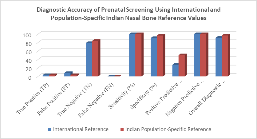
Fig: 2 Graphical represents Diagnostic Accuracy of Prenatal Screening Using International and Population-Specific Indian Nasal Bone Reference Values
The Indian population-specific reference values maintained 100% sensitivity and 100% negative predictive value, while significantly improving specificity (96.6% vs. 90.8%), positive predictive value (50.0% vs. 27.3%), and overall diagnostic accuracy (96.7% vs. 91.1%) compared with the international reference values. Additionally, the number of false-positive cases decreased from 8 to 3, demonstrating that the population-specific reference values enhanced prenatal screening accuracy (p < 0.05).
Table 6. Association Between Population-Specific Nasal Bone Findings and Pregnancy Outcome (n = 90)
|
Nasal Bone Finding (Indian Reference) |
Normal Pregnancy Outcome (n = 87) |
Chromosomal Abnormality (n = 3) |
Total |
p-value |
|
Normal Nasal Bone |
84 |
0 |
84 |
<0.001* |
|
Hypoplastic/Absent Nasal Bone |
3 |
3 |
6 |
|
|
Total |
87 |
3 |
90 |
|
A significant association was observed between fetal nasal bone findings and pregnancy outcome (p < 0.001). All fetuses with chromosomal abnormalities had a hypoplastic or absent nasal bone, while 84 of 87 normal pregnancies had a normal nasal bone. These findings indicate that hypoplastic or absent nasal bone is strongly associated with chromosomal abnormalities in the first trimester.
Table 7. Correlation of Nasal Bone Length with Gestational Age and Crown-Rump Length (n = 90)
|
Variable |
Correlation Coefficient (r) |
p-value |
|
Gestational age (weeks) |
0.69 |
<0.001* |
|
Crown-rump length (mm) |
0.73 |
<0.001* |
A strong positive correlation was observed between nasal bone length and both gestational age (r = 0.69, p < 0.001) and crown-rump length (r = 0.73, p < 0.001). These findings indicate that fetal nasal bone length increases significantly with advancing gestational age and fetal growth during the first trimester.
Table 8. Receiver Operating Characteristic (ROC) Analysis of Nasal Bone Length for Detection of Chromosomal Abnormalities (n = 90)
|
Parameter |
Value |
p-value |
|
Area Under Curve (AUC) |
0.94 |
<0.001*
|
|
Optimal Cut-off Nasal Bone Length (mm) |
1.65 mm |
|
|
Sensitivity at cut-off (%) |
100.0 |
|
|
Specificity at cut-off (%) |
92.0 |
|
|
Positive Predictive Value (%) |
42.9 |
|
|
Negative Predictive Value (%) |
100.0 |
Receiver operating characteristic (ROC) analysis demonstrated excellent diagnostic performance of nasal bone length for detecting chromosomal abnormalities, with an AUC of 0.94 (p < 0.001). An optimal cut-off value of 1.65 mm achieved 100% sensitivity, 92.0% specificity, 42.9% positive predictive value, and 100% negative predictive value, indicating that nasal bone length is a highly effective first-trimester screening marker.
DISCUSSION:
The application of population-specific reference values significantly reduced the number of fetuses classified as having hypoplastic or absent nasal bone and thereby decreased false-positive screening results. There was no reduction in the detection of chromosomal abnormalities after applying Indian reference standards. Sensitivity and negative predictive value remained 100% with both international and population-specific reference values, while specificity and overall diagnostic accuracy improved with population-specific values.
A strong association was observed between abnormal nasal bone findings and chromosomal abnormalities, confirming its usefulness as a reliable marker in first-trimester screening. ROC analysis further demonstrated excellent diagnostic performance of nasal bone length with an AUC of 0.94 and an optimal cut-off value of 1.65 mm showing high sensitivity and good specificity. Overall, the findings indicate that population-specific nasal bone reference values improve prenatal screening accuracy in the Indian population by reducing false-positive results while maintaining high detection rates, supporting the use of locally derived reference standards in clinical practice.
CONCLUSION:
The present study concludes that fetal nasal bone length shows a progressive increase with gestational age and crown-rump length in the first trimester. The use of population-specific nasal bone reference values significantly improves prenatal screening accuracy by reducing false-positive results without compromising sensitivity or negative predictive value. Diagnostic performance measures, including specificity, positive predictive value, and overall accuracy, were higher with Indian reference standards compared to international reference values.
A strong association was also observed between abnormal nasal bone findings and chromosomal abnormalities, confirming its value as an important soft marker in first-trimester screening. ROC analysis further supported the excellent diagnostic utility of nasal bone length. Therefore, population-specific first-trimester nasal bone reference values should be adopted to enhance the accuracy and reliability of prenatal screening in the Indian population.
Conflict of interest: The author declares that there is no conflict of interest regarding the publication of this study.
LIMITATIONS OF THE STUDY
The study was conducted at a single center with a relatively small sample size, which may limit the generalizability of the findings. The number of fetuses with chromosomal abnormalities was low, which may have affected the assessment of diagnostic accuracy. In addition, the study included only first-trimester singleton pregnancies, and larger multicenter studies are needed to validate these population-specific reference values.
REFERENCES:
1. Nicolaides KH. Screening for fetal aneuploidies at 11–13+6 weeks. Ultrasound Obstet Gynecol. 2023;61(1):1–17.
2. Kagan KO, Wright D, Baker A, et al. Nasal bone in first-trimester screening for trisomy 21. Prenat Diagn. 2015;35(7):710–714.
3. The Fetal Medicine Foundation. Nasal bone assessment at 11–13+6 weeks. Available from: https://fetalmedicine.org
4. Sonek JD, Cicero S, Nicolaides KH. Nasal bone evaluation in screening for trisomy 21. Obstet Gynecol. 2006;107(2 Pt 1):245–251.
5. Das S, Sharma C, Yadav T, et al. Absent or hypoplastic nasal bone: clinical significance in first-trimester screening. Birth Defects Res. 2024;116(5).
6. Hathi V, Suresh S, Sudarshan S. Isolated unossified nasal bone in first trimester: experience from Southern India. Ultrasound Obstet Gynecol. 2024.
7. Syngelaki A, Pergament E, Tsavdaridou M, et al. First-trimester screening for chromosomal abnormalities using nasal bone and NT. Fetal Diagn Ther. 2019;45(3):1–8.
8. Poon LC, Akolekar R, Syngelaki A, et al. Fetal nasal bone length and gestational age correlation in first trimester. Ultrasound Obstet Gynecol. 2011;37(3):308–313.
9. Khalil A, Nicolaides KH. Nasal bone assessment and screening performance for trisomy 21. Fetal Diagn Ther. 2013;34(3):123–130.
10. Mujezinovic F, Alfirevic Z. Ethnic variation in fetal nasal bone and implications for screening. Prenat Diagn. 2010;30(9):850–856.
11. Viora E, Sciarrone A, Bastonero S, et al. Population-specific reference ranges for fetal nasal bone improve screening accuracy. J Matern Fetal Neonatal Med. 2020;33(15):2605–2612.