Efficacy of Core Decompression with Bone Marrow Aspirate Concentration and Conservative Management in Treatment of Avascular Necrosis of Femoral Head- A Prospective Study.
- Muruli Manohar Das , Assistant Professor, Department of Orthopaedics, Sri chamundeshwari Medical College Hospital & Research Institute, Channapatna, Ramanagar District, Karnataka, India.
- Nagaraju Honnegowda , Assistant Professor, Department of Orthopaedics, Sri Chamundeshwari Medical College Hospital & Research Institute, Channapatna, Ramanagar District, Karnataka, India.
- Avinash R. , Assistant Professor, Department of Orthopaedics, Sri Chamundeswari Medical College Hospital & Research Institute, Channapatna, Ramanagar District, Karnataka, India.
Article Information:
Abstract:
Background: Avascular Necrosis of the femoral head is a progressive debilitating disease of the hip joint primarily affecting the young adult. This study aims to provide the painless, mobile, functional hip and prevent disease progression by Prevention of early hip sacrificing surgical intervention in patients presenting with stage 2, 3 avascular necrosis of femoral head. Methods: A total of 25 patients (47 hips) with Grade II and III Avascular Necrosis underwent CD+BMAC with conservative management were followed up from 6 weeks, 12 weeks, 24 weeks and every 6 months thereafter. Outcome measures were measured using Harris Hip scores, serial X-rays and MRI. Results: The mean age in our study was 35.28 years, highlighting a higher prevalence of avascular necrosis (AVN) in younger adults, especially those in their 30s and 40s. COVID-19 infection, steroid therapy, and alcohol consumption was identified as significant risk factors for developing AVN, although the most common cause in this study was idiopathic. Post-COVID-19 infections, including those treated with steroids, accounted for about 44% of the cases. The overall mean Harris hip score improved from 57.37 before intervention to 73.52 at 6 weeks, 78.71 at 12 weeks, and 80.19 at 24 weeks. Conclusion: CD+BMAC and conservative management was effective in providing significant pain relief and improving mobility in patients, particularly in the early stages of AVN. This procedure was most effective in early-stage AVN, where it could slow or halt disease progression and delay the need for total hip arthroplasty (THA). Radiographic evidence showed that the treatment could stabilize or partially reverse the necrotic process in 2nd and 3rd Ficat and Arlet-stage cases, although outcomes were less favorable in more advanced stages.
Keywords:
Article :
INTRODUCTION:
Osteonecrosis of the femoral head was first described in 1738 by Munro Konig first described the condition coined the term Osteochondritis Dissecans. Haenish (1925) described first case of idiopathic ischemic necrosis of the femoral head in an adult. Arterial Occlusion (1940) was postulated as the cause of the necrosis. Pietrograndi (1957) described AVN due to steroid therapy.[1]
In approximately 1835, Cruveilhier depicted femoral head morphologic changes secondary to interruption of blood flow.[2] Since 1962, when Mankin described 27 cases of AVN, the number of reported AVN cases has increased steadily.[3] Between 10,000 and 20,000 cases are reported annually in the USA with an estimated prevalence of up to 600,000 10. Overall, the diagnosis of osteonecrosis accounts for 5% to 12% of total hip arthroplasties (THA) performed each year11-12.
Avascular necrosis (Aka -osteochondritis dessicans) -coined by Konig in 1888. In 1925, Haenish describe the TRAUMATIC and IDIOPATHIC cause. Avascular necrosis is death of living elements of involved bone (include cell and marrow) with progressive destruction and alteration of bone architecture as results of compromised vascularity.
In the United States alone, around 10,000 to 20,000 new cases are reported each year.[4] It can occur due to a variety of causes, either traumatic or atraumatic in origin.The main risk factors include alcohol and corticosteroids, which are involved in more than 50% of the cases. Other risk factors encompass radiation exposure, physical injury, and sickle cell disease.[5] However, up to 40% of cases are idiopathic.
Mainly young and active adults between the ages of 20–45 are affected.[6] Several classification systems have been developed to predict the prognosis and to guide the treatment. The FICAT and ARLET classification system is widely used and relies on radiological findings.[7] However, the size, location and extent of the avascular necrosis (AVN) is not taken into consideration.[8] Kerboul et al. quantified the lesion by measuring the necrotic area in the femoral head using anteroposterior and lateral views. The prognosis was better if the angle is less than 200°.
Many treatments have been proposed for AVN of the femoral head. Non-surgical treatments encompass pharmacologic therapies, such as lipid-lowering agents, anticoagulants, vasoactive substances, and bisphosphonates, as well as non-pharmacologic therapies, including extracorporeal shockwave therapy, pulse electromagnetic therapy, and hyperbaric oxygen therapy. Surgical treatment options ranges from core decompression to total hip arthroplasty. Bone marrow aspiration concentrate (BMAC) therapy has been proposed as an adjuvant therapy to core decompression. Several studies have reported improved outcomes when core decompression is combined with BMAC, compared to core decompression alone.[8].
MATERIALS AND METHODS:
This research study was approved by the ethical committee and institutional review board before the commencement of study. The study was conducted on the patients with lateral clavicle fractures admitted under the department of Orthopaedics, at Karnataka institute of Medical sciences, Hubballi. It was conducted between July 2022 to July 2024 for a period of 2 years. It was a prospective type of clinical study
Inclusion Criteria
Patients with Grade 2 & 3 Ficat and Arlet Classification of Avascular necrosis of femoral head.
Exclusion Criteria
Patients with advanced Avascular necrosis of femoral head, Patient on steroid therapy, immunocompromised state, Patient with history of hip surgery, Patients not consenting for the study.
Details of the patients including history, clinical examination and signs and symptoms noted, routine blood investigations and imaging done to assess the grade of avascular necrosis of femoral head.
After obtaining valid, written informed consent for the study, the patient was examined for range of motion of hip joint (Flexion, Extension, Abduction, Adduction, Internal rotation, External rotation) able to sit cross legged, able to squat, patient were recorded with visual analogue score and Harris Hip score to assess the current functional status of the hip at the time of presentation. Routine Investigations were done which includes are subjected to conservative and core decompression with bone marrow aspirate concentration. Harris Hip score is calculated at the time of Presentation and after the procedure.
Relevant investigations such as blood parameters, X-ray Pelvis with bilateral Hip - Anterior posterior, Lateral and Frog leg view, Magnetic Resonance Imaging (MRI) for the diagnosis and staging of the Avascular necrosis of femoral head.
The patients were operated on elective basis after overcoming the avoidable anaesthetic risks. Preoperative medical fitness was taken in all cases. Pre anesthetic examination was carried out a day prior to the surgery. Patients as well as the attenders were explained about the surgery and the risk factors; a written consent for the surgery was taken for all patients. Intravenous antibiotics were given an hour before the surgery. The affected limb was prepared from umbilicus down to knee joint.
Surgical Procedure
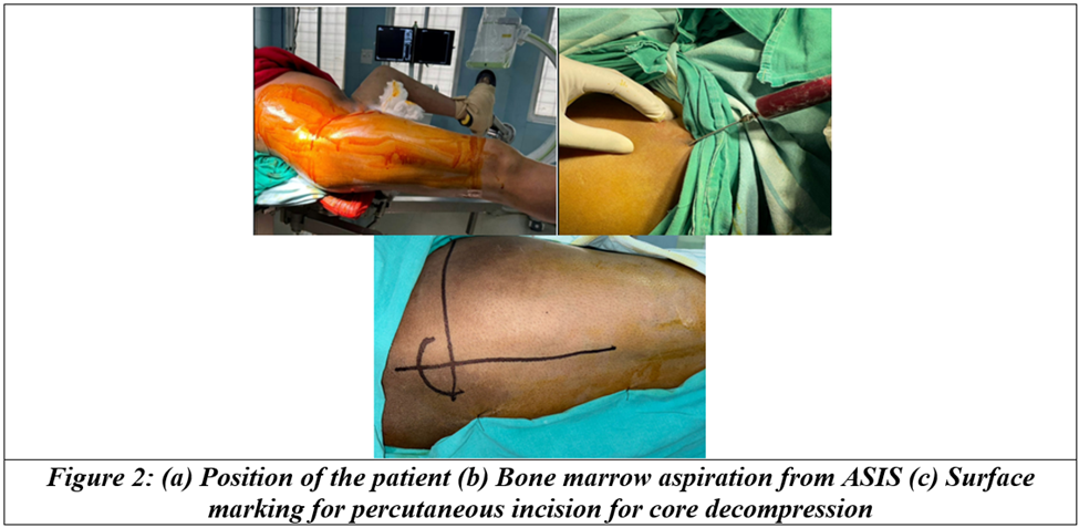
After taking valid written informed consent, patient will be shifted to OT. After inducing under spinal anaesthesia, the patient is positioned supine on a fracture table with both feet secured in traction boots and internally rotated. The lateral aspect of proximal thigh was scrubbed with povidone-Iodine and surgical spirit followed by painting and draping with sterile drapers.
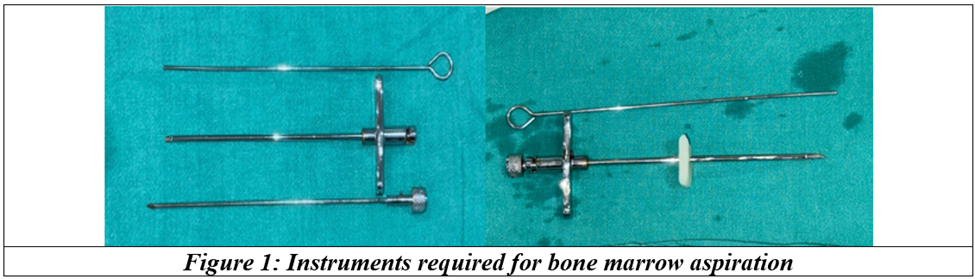
Incision of 2cm long done approximately 2cm above the Anterior Superior Iliac spine and Jamshidi needle was inserted after trochar inserted to the iliac crest, but prior to aspiration, 1 ml of Heparin is loaded into the syringe, to avoid clot formation and coagulation. Approximately 60ml of bone marrow is aspirated. The collected sample is placed in centrifuge and it is then spun at 2000-3000rpm, typically 6ml of sample of bone marrow aspirated concentrate is yielded after processing.
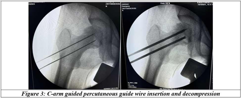
Core decompression is initiated while the blood sample is processing. The C-arm is used to locate the starting point 4mm guide wire is placed over the skin to determine the AP and superoinferior skin markings. A lateral image is also obtained by referencing the 4mm guidewire radiographically, which will provide landmarks for the lateral skin marking. The intersection of these skin markings is the incision location. This should be at the lateral cortex at the location of the lesser tuberosity or proximal to it. A 1-cm incision is then performed at the corresponding entry point over the lateral aspect of the femur, just below the vastus ridge of the trochanter. The starting point is maintained proximal to the level of the lesser trochanter and distal to the vastus ridge.
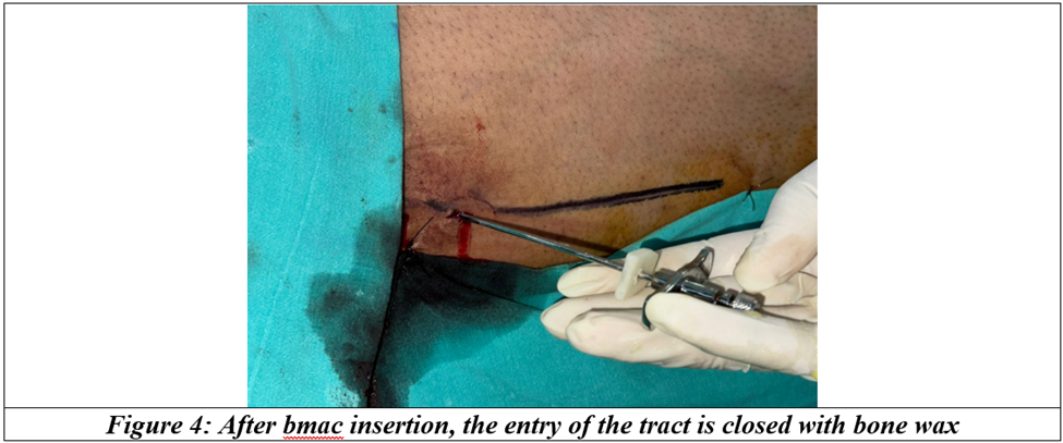
When the ideal starting point has been obtained, the trocar is advanced from lateral to medial under fluoroscopy. This must be performed under the AP and lateral views, the area of necrosis can be entered with the trocar, but the trocar should not be advanced to within 5 mm of subchondral bone to prevent joint violation. Then the reamer is removed. Now the empty core is filled with 6ml of bone marrow aspirate, 3ml each side. Then the core is filled with bone bax to avoid the leakage.
The processed bone marrow aspirate concentrate is filled into the core using Jamshidi needle. The opening was closed with Bone wax. Skin was sutured in layers after hemostasis is achieved. Sterile dressing was done and the patient will be shifted to post op ward.
Post-operative care/ Post-operative management: (Conservative management)
1) Non weight bearing for 2 weeks followed by Weight bearing as tolerated (WBAT) is permitted with the use of crutches for the next 2 weeks.
2) Patient is then given yearly once Inj Zolindronic acid will be given,
3) Along with Tab. Alandronate 70mg once a week will be given.
4) Patients should take oral bisphosphonates as the first medication in the morning and more than 30 minutes before the first food intake, beverage (except water), or any other medication.
5) Patients should receive instruction to stay upright, not to lay down for at least thirty minutes or until the first food intake of the day to reduce esophageal irritation/ulceration. Milk and milk products, coffee, orange juice, and food may decrease the absorption of oral bisphosphonates.
6) Patients should receive supplemental calcium 1000 to 1200 mg/day and vitamin D 800 to 1000 international units/day if dietary intake is inadequate.
7) Tab. Aspirin 75mg once a day at night
8) Tab. Atorvastatin 10mg once a day at night
9) Tab. Vitamin D3 60,000 IU weekly once
10) Analgesics SOS
11) Antacid SOS
12) Non weight bearing hip exercise - side lying hip abduction, side lying clam with resistance, unilate unilateral supine bending, side bridge exercise.
Patient will be followed up at the interval of 1st month, 3months and 6 months. 1st month and 3rd month, patient was clinical followed up with Harris Hip score and Visual analogue score and imaging using plain xray of Pelvis with bilateral hip. At 6th month, patient was clinical followed up with Harris Hip score and Visual analogue score and imaging using MRI
RESULTS:
Age of the patient’s ranges from 22 to 57 years with mean age of around 35 years. Among 25 patients studied, 80% of patients were of 20-40 years of age. It shows increased incidence among the third and fourth decade of life when compare to other population. Among the 25 patients, 2 patients were affected with AVN of left femoral head,1 affected with AVN of Right femoral head and 22 were affected with AVN of bilateral femoral head. Out of 25 patients, 28% of the patients had no prior known risk factors (idiopathic). 12 % of the patients had prior COVID19 infection, 12% of the patients underwent prior steroid therapy, 20% of the patient had COVID19 for which they underwent steroid therapy, which makes 20% were associated with COVID19 infection and steroid therapy, alcohol contributed 16% of risk factor. Collectively, Post COVID19 infections, POST COVID19 infection including Steroid therapies and Steroid therapy alone contributed around 44% of the risk factors.
Out of 21 patients who had Right sided AVN (or with Bilateral AVN who underwent intervention for the right side), 84% of the patients underwent Core decompression with Bone marrow aspiration Concentration with Medications and 08% of the patients underwent conservative approach with medications on the right side. Out of 23 patients who had Left sided AVN (or with Bilateral AVN who underwent intervention for the left side), 92% of the patients underwent Core decompression with Bone marrow aspiration Concentration with Medications and 4% of the patients underwent conservative approach with medications on the right side.
Table 1: Intervention at right side and left side wise distribution of patients
|
Intervention |
Right side |
Left side |
||
|
No of Hips |
% of Hips |
No of Hips |
% of Hips |
|
|
CD + BMAC + C |
21 |
84.00 |
23 |
92.00 |
|
Conservative |
2 |
8.00 |
1 |
4.00 |
Overall Mean Harris Hip before intervention was 57.37. Overall Mean Harris Hip After 6 weeks was 73.52. Overall Mean Harris Hip After 12 weeks was 78.71. Overall Mean Harris Hip After 24 weeks was 80.19.
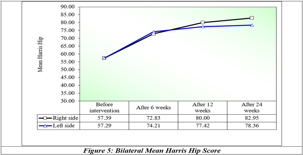
Table 2: Overall before intervention after intervention progression of Ficat Arlet Staging
|
|
Number of Hips |
Percentage |
|
Stage 2 to 3 |
5 out of 29 |
17.24% |
|
Stage 3 to 4 |
5 out of 16 |
31.25% |
DISCUSSION:
The main aim of Core decompression with Bone marrow aspirate concentrate and conservative management of the Hip is to provide painless, stable, mobile hips to patients to carry out activities of daily living. Core decompression with Bone marrow aspirate concentrate and conservative management of the Hip has not only been a gift to patients but also to the economy of the nation as most of the individuals of the Osteonecrosis of the femoral head are rendered crippled and housebound. The mean age group in our study was 35.82 years. Age of the patients ranges from 22 to 57 years. Among 25 patients studied, 80% of patients were of 20-40 years of age. It shows increased incidence among the third and fourth decade of life when compare to other population. Biao Tan et al[9] in his large scale study on the epidemiological analysis of Osteonecrosis of Femoral Head in China had reported the mean age to be 48 years, with majority of the patients being males and aged between 30 to 50 years (peak at 40 years) and 20 to 60 years among females (peak at 50 years). This data is similar to the mean age in our study and reiterates that AVN of femoral head is more common among young Asian population than their western counterparts.
Out of 25 patients, 22 patients were male 3 were female. It comes around 88 % of male predominance and reflects the high prevalence among male population. Harshvardhan et al[10] reported a male to female ratio of 5:1. Male preponderance in our study could be associated with consumption of alcohol, smoking and comparatively higher cases of traumatic. Out of 25 patients, 28% of the patients had no prior known risk factors (idiopathic). 12 % of the patients had prior COVID19 infection, 12% of the patients underwent prior steroid therapy, 20% of the patient had COVID19 for which they underwent steroid therapy, which makes 20% were associated with COVID19 infection and steroid therapy, alcohol contributed 16% of risk factor. Collectively, Post COVID19 infections, POST COVID19 infection including Steroid therapies and Steroid therapy alone contributed around 44% of the risk factors. This data is comparable to the data by Johannson et al[11] who reported Corticosteroid association with AVN in 22% cases and Alcohol consumption in 19% cases.
This is similar to the data by Harsh et al where Steroid abuse (37.3%) was the most commonly associated factor for developing AVN among North Indian population followed by chronic alcohol intake (20.3%) and trauma (15.3%). Steroid intake was associated with Bilateral AVN in 80% cases and Alcohol consumption with bilateral AVN in 60% cases. The exact mechanism of how alcohol causes AVN is not absolutely understood, but various studies have stated various fatty acids are produced and build up in the very small vessels of the bone. The average duration of alcohol use in patients in our study was 13.6 years. Osteonecrosis of the femoral head (ONFH) is a complex and not fully understood condition that primarily affects individuals in their thirties to fifties. If not diagnosed and treated early, ONFH can lead to the collapse of the femoral head. Since ONFH often occurs in younger patients who may not achieve the best outcomes with conventional total hip arthroplasty (THA), joint-preserving techniques are considered crucial in managing this condition. By implementing the most effective joint-preservation procedures, and with early diagnosis and intervention, the prognosis for ONFH patients can be greatly improved. In this study, the patient who is treated with core decompression with bone marrow aspirate concentrate with conservative management including yearly dose of Inj Zolendronic acid and weekly dose of Tab. Alandronate 70mg, Lipid lowering agents like statins, anticoagulants like Aspirin, and Analgesics with Hip strengthening exercies, shows 88% of the patients had avascular necrosis of bilateral femoral head. Among these study participants, 17.24% of hips progressed from stage 2 to 3 and 31.25% of hips progressed from stage 3 to 4, which is statistically significant delay in progression to next stage of AVN of femoral head and preserving the natural hip joint. Core decompression augmented with BMAC was first described by Hernigou and Beaujean in 2002. He presented 189 hips treated with core decompression with a 3-mm trocar and bone marrow grafting. Better results were observed in early stages of AVN. All the groups reported improved Harris Hip Scores after treatment, but 18% of hips required THR at an average of 26 months.
Gangji et al (2005)[12] performed a randomized control study, where 10 patients were given bone marrow stem cells in addition to core decompression (CD), along with 8 control cases where only CD was performed. It was observed that in a period of 24 weeks, level of pain was significantly decreased from 37.8+8.4 mm at baseline to 18.5±6.2 mm at 6 months (p =0.016) compared to no significant decrease in control group even uptill 24 months. The Lequesne index in stem cell group decreased from 7.7±1.5 to 3.0±1.1 at 6 months. The WOMAC score also reduced from 30±5 to 18±7 at 6 months. Subsequently all these changes progressed accordingly in subsequent follow up until 24 months. But in the control group of patients, the similar improvement was not observed neither at 6 months nor subsequently. Zhao et al.[13] compared the results of 51 hips that underwent core decompression and 53 hips that underwent core decompression augmented with bone marrow. Higher Harris Hip Scores were reported in the bone marrow group at the end of follow-up, and significantly fewer patients from the bone marrow group required an additional procedure such as THR or fibular vascularized graft.
Aggarwal S et al[14] study from Jan 2001 to 2017 were retrospectively analysed 432 hips diagnosed from 2001 to 2009. In the alendronate treatment group, at a mean follow-up of 129.6 months (range, 60 to 220 months), 74.5% did not require a surgical procedure, and, in the combination treatment group, at a mean follow-up of 69 months (range, 37 to 105 months), 88.9% did not require a surgical procedure. The clinical failure rate at 3 years was 4% for stage I, 11% for stage II, and 29% for stage III in the alendronate treatment group, and it was 5% for stage I, 9% for stage II, and 32% for stage III in the combination treatment group. Patients in the combination treatment group had a significantly better improvement in VAS score at 6 weeks (from 7.10 to 3.66) compared with patients in the alendronate treatment group (from 7.93 to 7.00).
CONCLUSION:
Avascular necrosis (AVN) of the femoral head is a debilitating condition that primarily affects middle-aged individuals, severely impacting their daily activities. Osteonecrosis of the femoral head (ONFH) is a complex and poorly understood disease. Without early diagnosis and treatment, it can lead to the collapse of the femoral head. Magnetic resonance imaging (MRI) plays a crucial role in detecting the early stages of ONFH, with a sensitivity rate of 90 to 100%. Core decompression combined with bone marrow aspirate concentrate (BMAC) and medical management is currently the most effective joint-preservation procedure. Early diagnosis and intervention significantly improve the prognosis for patients with ONFH.
We conclude that Core decompression + BMAC + conservative management is an effective treatment for AVN of femoral head which delays the disease progression to the advanced stage and provides a pain free, mobile, stable hip with preserving the natural hip joint as there is excellent Harris Hip score outcomes after the intervention. Patient can attain pre disease level of activity without any pain.
REFERENCES:
1. Hamilton TW, Goodman SM, Figgie M. SAS weekly rounds: avascular necrosis. HSS J 2009;5(2):99-113.
2. Lavernia CJ, Sierra RJ, Grieco FR. Osteonecrosis of the femoral head. J Am Acad Orthop Surg 1999;7(4):250-61.
3. Mankin HJ, Brower TD. Bilateral idiopathic aseptic necrosis of the femur in adults: “Chandler’s disease”. Bull Hosp Joint Dis 1962;23:42-57.
4. Barney J, Piuzzi NS, Akhondi H. Femoral head avascular necrosis. 2023 Jul 3. In: StatPearls [Internet]. Treasure Island (FL): StatPearls Publishing 2024.
5. Arbeloa-Gutierrez L, Dean CS, Chahla J, et al. Core Decompression augmented with autologous bone marrow aspiration concentrate for early avascular necrosis of the femoral head. Arthroscopy Techniques 2016;5(3):e615-20.
6. Bakircioglu S, Atilla B. Hip preserving procedures for osteonecrosis of the femoral head after collapse. Journal of Clinical Orthopaedics and Trauma 2021;23:101636.
7. Bakircioglu S, Atilla B. Hip preserving procedures for osteonecrosis of the femoral head after collapse. J Clin Orthop Trauma 2021;23:101636.
8. Arbeloa-Gutierrez L, Dean CS, Chahla J, et al. Core decompression augmented with autologous bone marrow aspiration concentrate for early avascular necrosis of the femoral head. Arthrosc Tech 2016;5(3):e615-20.
9. Tan B, Li W, Zeng P, et al. Epidemiological study based on China osteonecrosis of the femoral head database. Orthop Surg 2021;13(1):153-60.
10. Vardhan H, Tripathy SK, Sen RK, et al. Epidemiological Profile of Femoral Head Osteonecrosis in the North Indian Population. Indian J Orthop 2018;52(2):140-6.
11. Mont MA, Seyler TM, Plate JF, et al. Uncemented Total Hip Arthroplasty in Young Adults with Osteonecrosis of the Femoral Head: A Comparative Study. J Bone Jt Surg 2006;88(suppl_3):104-9.
12. Gangji V. Hauzeur JP Treatment of osteonecrosis of the femoral head with implantation of autologous bone-marrow cells. J Bone Joint Surg Am 2005;87:106.
13. Zhao D, Cui D, Wang B. Treatment of early stage osteonecrosis of the femoral head with autologous implantation of bone marrow-derived and cultured mesenchymal stem cells. Bone 2012;50:325-30.
14. Agarwala S, Banavali SD, Vijayvargiya M. Bisphosphonate Combination therapy in the management of postchemotherapy avascular necrosis of the femoral head in adolescents and young adults: a retrospective study from India. J Glob Oncol 2018;4:1-11.