CLINICAL PROFILE AND RISK FACTORS OF INFERIOR WALL MYOCARDIAL INFARCTION IN A TERTIARY CARE HOSPITAL.
- Susmita Chanda , Ex-Assistant professor, MBBS, MD, Department of General Medicine, JIMS Medical college and hospital, India.
- Manojit Barman , Medical Office, MBBS, MS, FMAS, Department of General Surgery, General Surgery, Alipurduar District Hospital, Alipurduar, India.
Article Information:
Abstract:
Background: Inferior wall myocardial infarction (IWMI) is a common presentation of acute coronary syndrome, often associated with right ventricular involvement and significant morbidity. Identifying clinical profiles, risk factors, and complications is essential for timely management. Objectives: To evaluate the clinical profile, risk factors, pattern, and complications of patients with IWMI admitted to a tertiary care hospital. Methods: The present study was a Prospective observational study. This Study was conducted from November 2019 – May 2021at Department of General Medicine, JIMS Medical college and hospital. Study population 40 patients were taken up for the study. Results: The majority of patients were male (28/40, 70%) with a mean male-to-female ratio of 2.3:1 (P = 0.00034). Most were aged 51–60 years (42.5%), and 62.5% resided in urban areas. Chest pain was the predominant presenting symptom (92.5%), followed by sweating (57.5%) and syncope (47.5%) (P < 0.00001). Elevated jugular venous pressure (60%) and bradycardia (42.5%) were common physical findings. Smoking (47.5%), combined diabetes and hypertension (45%), and alcohol consumption (42.5%) were the most frequent risk factors (P = 0.00804). Isolated IWMI was seen in 50% of patients, while IWMI with right ventricular or posterior wall involvement accounted for 50%. Cardiogenic shock was the most frequent complication (50%), followed by electrical disturbances (42.5%). Right bundle branch block was the most common arrhythmia (22.5%). Conclusion: IWMI predominantly affects middle-aged males, with smoking, diabetes, and hypertension as major risk factors. Early recognition of clinical signs and risk factors, along with prompt management of complications such as cardiogenic shock and arrhythmias, is crucial to improve patient outcomes.
Keywords:
Article :
INTRODUCTION:
Acute myocardial infarction (AMI) is a leading cause of morbidity and mortality worldwide, contributing significantly to the global burden of cardiovascular disease [1]. Inferior wall myocardial infarction (IWMI), typically involving the right coronary artery (RCA), accounts for approximately 30–50% of all AMI cases and presents with distinct clinical and hemodynamic characteristics compared to anterior or lateral infarctions [1,3].
IWMI often involves concomitant right ventricular infarction (RVI), which significantly affects prognosis, predisposing patients to hypotension, arrhythmias, and cardiogenic shock [4–7].
The clinical presentation of IWMI can be variable, often manifesting as chest pain, dyspnea, or epigastric discomfort, and may be accompanied by signs of right ventricular involvement such as elevated jugular venous pressure and hypotension [4,5].
Electrocardiographic changes, including ST-segment elevation in inferior leads and reciprocal changes in anterior leads, play a critical role in early diagnosis [8]. Additionally, anterior ST-segment depression in some patients may reflect posterior or right ventricular involvement, further influencing management and outcomes [8].
Risk factors for IWMI are largely similar to those of other forms of coronary artery disease, including hypertension, diabetes mellitus, smoking, dyslipidemia, and sedentary lifestyle [2]. However, epidemiological studies indicate that the prevalence and impact of these risk factors may differ regionally and over time, highlighting the need for context-specific data [2].
Early recognition of high-risk patients, timely reperfusion therapy, and optimal management of right ventricular involvement have been shown to improve survival and functional outcomes in IWMI [6, 9].
Despite advances in diagnosis and treatment, limited contemporary data exist regarding the clinical profile, risk factors, and outcomes of IWMI in tertiary care settings, particularly in the Indian population. Understanding these patterns is essential for optimizing prevention strategies, risk stratification, and resource allocation in high-burden regions.
Therefore, this study aims to evaluate the clinical characteristics and associated risk factors of inferior wall myocardial infarction among patients admitted to a tertiary care hospital. Aims to study the clinical profile and risk factors of patients presenting with inferior wall myocardial infarction in a tertiary care hospital.
MATERIALS AND METHODS:
Type of study: Prospective observational study.
Place of study: Department of General Medicine, JIMS Medical college and hospital.
Study duration: November 2019 – May 2021.
Sample size: 40 patients were taken up for the study.
Inclusion Criteria:
• Patients aged ≥18 years diagnosed with acute inferior wall myocardial infarction based on clinical features, ECG changes (ST-segment elevation in inferior leads), and/or cardiac biomarkers.
• Patients presenting to the tertiary care hospital within 24 hours of symptom onset.
• Patients willing to provide informed consent to participate in the study.
Exclusion Criteria:
• Patients with concomitant anterior, lateral, or posterior wall myocardial infarction without inferior wall involvement.
• Patients with previous history of myocardial infarction or known structural heart disease.
• Patients with severe comorbid conditions that may confound outcomes, such as advanced renal failure, liver failure, or malignancy.
• Patients who refuse or are unable to give informed consent.
• Patients with incomplete clinical, ECG, or laboratory data.
Study Parameter:
1. Age
2. Sex
3. Body mass index (BMI)
4. Cardiovascular risk factors
5. Presenting symptoms
6. Duration of symptoms before hospital admission
7. Hemodynamic status
8. Electrocardiographic (ECG) findings
9. Cardiac biomarkers
10. Co-morbid conditions
11. In-hospital outcomes and complications
Statistical Analysis:
Data were entered into Excel and analyzed using SPSS and GraphPad Prism. Numerical variables were summarized using means and standard deviations, while categorical variables were described with counts and percentages.
Two-sample t-tests were used to compare independent groups, while paired t-tests accounted for correlations in paired data. Chi-square tests (including Fisher’s exact test for small sample sizes) were used for categorical data comparisons.
P-values ≤ 0.05 were considered statistically significant.
RESULTS:
Table 1: Distribution of Patients Demographic parameters
|
Parameter |
Category |
Frequency |
Percentage |
P value |
|
Gender |
Male |
28 |
70% |
0.00034 |
|
Female |
12 |
30% |
||
|
Total |
40 |
100% |
||
|
Age Group |
21–30 |
1 |
2.50% |
< .00001 |
|
31–40 |
4 |
10% |
||
|
41–50 |
5 |
12.50% |
||
|
51–60 |
17 |
42.50% |
||
|
>60 |
13 |
32.50% |
||
|
Total |
40 |
100% |
||
|
Occupation |
Manual workers |
3 |
7.50% |
0.00262 |
|
Skilled labourers |
6 |
15% |
||
|
Professionals |
14 |
35% |
||
|
Sedentary life |
12 |
30% |
||
|
Housewives |
5 |
12.50% |
||
|
Total |
40 |
100% |
||
|
Residence |
Urban |
25 |
62.50% |
0.0251 |
|
Rural |
15 |
37.50% |
||
|
Total |
40 |
100% |
Table 2: Distribution of Patients Clinical Presentation at Admission
|
Parameter |
Category/Findings |
Frequency |
Percentage |
P value |
|
Symptomatology |
Chest Pain |
37 |
92.50% |
< .00001 |
|
Syncope |
19 |
47.50% |
||
|
Palpitation |
8 |
20% |
||
|
Sweating |
23 |
57.50% |
||
|
Difficulty in Breathing |
4 |
10% |
||
|
Vomiting |
3 |
7.50% |
||
|
Loose Stool |
5 |
12.50% |
||
|
Physical Findings |
Pulse in Normal Range (60–100) |
23 |
57.50% |
< .00001 |
|
Bradycardia (<60) |
17 |
42.50% |
||
|
Hypertension |
13 |
32.50% |
||
|
Normotensive (100–140 / 60–90) |
17 |
42.50% |
||
|
Hypotensive (<100/<60) |
10 |
25% |
||
|
Normal JVP |
16 |
40% |
||
|
Elevated JVP |
24 |
60% |
||
|
Heart Sounds S3/S4 |
2 |
5% |
||
|
Tricuspid Regurgitation Murmur |
11 |
27.50% |
||
|
Respiratory Crepitation |
6 |
15% |
Table 3: Distribution of Patients Complications during Hospital Stay
|
Parameter |
Complication Type / Arrhythmia |
Frequency |
Percentage |
p value |
|
Complications |
Cardiogenic Shock |
20 |
50% |
< .00001 |
|
Electrical Complications |
17 |
42.50% |
||
|
Death |
3 |
7.50% |
||
|
Arrhythmias |
Sinus Bradycardia |
7 |
17.50% |
.17702 |
|
First Degree AV Block |
4 |
10% |
||
|
Second Degree AV Block |
3 |
7.50% |
||
|
Complete Heart Block |
3 |
7.50% |
||
|
Right Bundle Branch Block (RBBB) |
9 |
22.50% |
||
|
Atrial Fibrillation |
3 |
7.50% |
||
|
Ventricular Tachycardia / Fibrillation (VT/VF) |
3 |
7.50% |
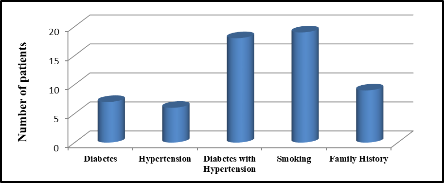
Figure 1: Distribution of Patients According to Risk Factors.
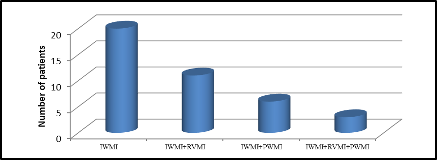
Figure 2: Distribution of Patients According to Pattern of MI
A total of 40 patients with inferior wall myocardial infarction were included in the study. The majority were male (28/40, 70%) with a male-to-female ratio of 2.3:1, which was statistically significant (P = 0.00034). Age distribution showed that most patients were in the 51–60 years age group (17/40, 42.5%), followed by those over 60 years (13/40, 32.5%), while the youngest group (21–30 years) had the lowest representation (1/40, 2.5%) (P < 0.00001) statistically significant. Regarding occupation, professionals comprised the largest proportion (14/40, 35%), followed by individuals with sedentary lifestyles (12/40, 30%), skilled laborers (6/40, 15%), housewives (5/40, 12.5%), and manual workers (3/40, 7.5%) (P = 0.00262) statistically significant. Most patients resided in urban areas (25/40, 62.5%) compared to rural areas (15/40, 37.5%), which was also statistically significant (P = 0.0251). (Table 1)
Among the 40 patients, chest pain was the most common symptom, observed in 37 patients (92.5%), followed by sweating in 23 patients (57.5%) and syncope in 19 patients (47.5%) (P < 0.00001) statistically significant. Other symptoms included palpitations (8/40, 20%), difficulty in breathing (4/40, 10%), loose stools (5/40, 12.5%), and vomiting (3/40, 7.5%). Regarding physical findings, 23 patients (57.5%) had a normal pulse (60–100 bpm), while 17 (42.5%) exhibited bradycardia (<60 bpm) (P < 0.00001) statistically significant. Hypertension was present in 13 patients (32.5%), 17 patients (42.5%) were normotensive, and 10 patients (25%) were hypotensive. Elevated jugular venous pressure (JVP) was observed in 24 patients (60%), while 16 patients (40%) had normal JVP. Heart sounds S3/S4 were noted in 2 patients (5%), tricuspid regurgitation murmur in 11 patients (27.5%), and respiratory crepitations in 6 patients (15%).(Table 2)
During the hospital stay, cardiogenic shock was the most frequent complication, occurring in 20 patients (50%), followed by electrical complications in 17 patients (42.5%), and death in 3 patients (7.5%) (P < 0.00001) statistically significant. Among arrhythmias, right bundle branch block (RBBB) was the most common, seen in 9 patients (22.5%), followed by sinus bradycardia in 7 patients (17.5%). First-degree AV block occurred in 4 patients (10%), second-degree AV block and complete heart block each in 3 patients (7.5%), atrial fibrillation in 3 patients (7.5%), and ventricular tachycardia/fibrillation (VT/VF) in 3 patients (7.5%) (P = 0.17702) statistically not significant. (Table 3)
Among the 40 patients, the most common risk factor was smoking, observed in 19 patients (47.5%), followed by diabetes with hypertension in 18 patients (45%) and alcohol consumption in 17 patients (42.5%) (P = 0.00804) statistically significant. Diabetes alone was present in 7 patients (17.5%), hypertension alone in 6 patients (15%), and a positive family history of coronary artery disease in 9 patients (22.5%). (Figure 1)
Among the 40 patients, isolated inferior wall myocardial infarction (IWMI) was the most common pattern, seen in 20 patients (50%) (P < 0.00001) statistically significant. IWMI with right ventricular MI (IWMI+RVMI) occurred in 11 patients (27.5%), IWMI with posterior wall MI (IWMI+PWMI) in 6 patients (15%), and IWMI involving both right ventricular and posterior wall MI (IWMI+RVMI+PWMI) was observed in 3 patients (7.5%). (Figure 2)
DISCUSSION:
The present study was a Prospective observational study. This Study was conducted from November 2019 – May 2021at Department of General Medicine, JIMS Medical college and hospital. Study population 40 patients were taken up for the study.
Demographics: The majority of patients were male (28/40, 70%), with a male-to-female ratio of 2.3:1. This finding is consistent with previous studies; Azhar et al. reported 89% males, 11% females, and Prabakaran Dorairaj et al. [10] reported 80% males and 20% females. Age-wise, most patients were in the 51–60 years group (42.5%), followed by those above 60 years (32.5%). Jaffe AS et al.[11] observed that 56% of patients were 40–60 years and 30% were above 60 years.
Occupation and Locality: Among occupations, professionals constituted the largest proportion (35%), followed by individuals with a sedentary lifestyle (30%). Grines C et al. reported a majority of IT employees (45%), while Yosikawa T et al. [12] found that 56% of patients had a sedentary lifestyle. Urban residents predominated in this study (62.5%) compared to rural residents (37.5%), similar to findings by Prabakaran Dorairaj [10](urban 73%) and Starling MR (urban 55%).
Clinical Presentation: At the time of admission, chest pain was the most frequent symptom (92.5%), followed by sweating (57.5%) and syncope (47.5%). In contrast, Andreas Yiangou et al.[13] reported syncope in 53% of patients, whereas Starling MR noted chest pain in 70%.
Risk Factors: Smoking was the most prevalent risk factor (47.5%), followed by diabetes with hypertension (45%) and alcohol consumption (42.5%). Armstrong PW et al.[14] reported alcohol consumption as the major risk factor in 62% of patients, and Coma-Canella et al.[15] found diabetes in 75% of patients.
Physical Findings: Elevated jugular venous pressure (JVP) was observed in 60% of patients, while hypertension (32.5%) and tricuspid regurgitation murmur (27.5%) were also notable. Gamallo C et al.[16] reported hypertension in 72% of patients, and Nancy C et al.[17] found tricuspid regurgitation in 80% of patients.
Pattern of Myocardial Infarction: Isolated IWMI was the most common pattern (50%), followed by IWMI with RVMI (27.5%), IWMI with posterior wall MI (15%), and IWMI with RVMI and posterior wall MI (7.5%). Varrier ED et al.[18] reported RVMI+IWMI in 60% of patients, while Nancy C et al. [17]observed RVMI+IWMI+PWMI in 50% of cases.
Complications: Cardiogenic shock occurred in 50% of patients, electrical complications in 42.5%, and death in 7.5%. Armstrong PW et al. [14] reported cardiogenic shock in 65%, while Coma-Canella et al.[15] found electrical complications in 62.5% of patients.
Arrhythmias: The most common arrhythmias observed were right bundle branch block (RBBB) in 22.5% and sinus bradycardia in 17.5%. Siher GPM et al.[19] reported RBBB in 56% of patients, whereas Nancy C et al. [17]observed sinus bradycardia in 52%.
CONCLUSION:
In this study of 40 patients with inferior wall myocardial infarction, the majority were middle-aged males, predominantly residing in urban areas and engaged in professional or sedentary occupations. Chest pain was the most frequent presenting symptom, while elevated jugular venous pressure, bradycardia, and tricuspid regurgitation were notable physical findings. Smoking, combined diabetes and hypertension, and alcohol consumption emerged as the most common risk factors. Isolated IWMI was the most prevalent pattern, though a significant proportion had concomitant right ventricular or posterior wall involvement. Cardiogenic shock was the most frequent complication, and right bundle branch block was the most common arrhythmia observed. These findings highlight the importance of early identification of high-risk patients, prompt management of complications, and targeted preventive strategies in patients with IWMI, especially those with modifiable risk factors such as smoking, hypertension, and diabetes.
REFERENCES:
1. Jameson B, Fauci A, Kasper D, Hauser S, Longo D, Loscalzo J. Harrison’s Principles of Internal Medicine. 20th ed. New York: McGraw-Hill Education; 2018. p. 1872–81.
2. Krishnan A, Asadullah M, Roy A, et al. Change in prevalence of coronary heart disease and its risk between 1991–94 to 2010–12 among rural and urban population of National Capital Region, Delhi. Indian Heart J. 2020;72(5):403–9.
3. Warner MJ, Tivakaran VS. Myocardial infarction, inferior.
4. Albulushi A, Giannopoulos A, Kafkas N, Dragasis S, Pavlides G, Chatzizisis YS. Acute right ventricular myocardial infarction. Expert Rev Cardiovasc Ther. 2018;16(7):455–64.
5. Namana V, Gupta SS, Abbasi AA, Raheja H, Shani J, Hollander G. Right ventricular infarction. Cardiovasc Revasc Med. 2018;19(1):43–50.
6. Zehender M, Kasper W, Kauder E, Schonthaler M, Geibel A, Olschewiski M, et al. Right ventricular infarction as an independent predictor of prognosis after acute inferior myocardial infarction. N Engl J Med. 1993;328:981–8.
7. Asif I, Muddarangappa R, Shah SKD, Vidyasagar S. A study of right ventricular infarction in inferior wall myocardial infarction. J Clin Sci Res. 2013;2:66–71.
8. Mirvis DM. Physiologic bases for anterior ST segment depression in patients with acute inferior wall myocardial infarction. Am Heart J. 1988;116(5 Pt 1):1308–22.
9. Bowers TR, O’Neill WW, Grines C, Pica MC, Safian RD, Goldstein JA. Effect of reperfusion on biventricular function and survival after right ventricular infarction. N Engl J Med. 1998;338(14):933–40.
10. Joshi P, Islam S, Pais P, Reddy S, Dorairaj P, Kazmi K, Pandey MR, Haque S, Mendis S, Rangarajan S, Yusuf S. Risk factors for early myocardial infarction in South Asians compared with individuals in other countries. Jama. 2007 Jan 17;297(3):286-94.
11. Jaffe AS, al-haemodynamic compromise in severe RVMI,1990;82:359-368
12. Yosikawa T, Association between RVMI and pre infarction angina, N Eng Med 1998; 338 :941-947
13. Andreas yiangou, Epsilon waves in RVMI,Texas Heart Journal.2012;39(2)306
14. Armstrong PW, Cardiac Troponin in ACS ,GUSTO II investigators, New England Journal of medicine 1996; 335:1333-1341.
15. Coma-Canella, RV function after MI,2D ECHO study Journal of American College of Cardiology 1983; 51:390-396
16. Gamallo C et al, Haemodynamic criteria of RVMI, circ,1981;64:515-525
17. Nancy C et al, Specific requirement for RVMI management, Journal of American Family Physicians.1999; 60 (6): 1727-1734
18. Varrier ED, Volume loading improves low cardiac output in RVMI-Journal of American College of Cardiology 1983; 2:270-278
19. Siher GPM, atrial arrhythmia and haemodynamic deterioration in RVMI and treatment, Journal of American College of Cardiology 2013; volume 2, issue-3, 270-278.