Histopathological prognostic factors in endometrial carcinoma:A clinicopathological study.
- Utkarsh Joshi , Junior Resident (JR-2) Department: Pathology, Krishna Mohan Medical College & Hospital, Pali Dungra, Sonkh Road, Mathura.
- Deepsheikha Dhand , Assistant Professor Department: Pathology Krishna Mohan Medical College & Hospital, Pali Dungra, Sonkh Road, Mathura.
- Pranshu Sisodia , Senior Resident Department: Pathology Krishna Mohan Medical College & Hospital, Pali Dungra, Sonkh Road, Mathura.
- Tripti Mishra , Senior Resident Department: Pathology, Singareni institute of medical science(GMC), Ramagundam, Telangana.
Article Information:
Abstract:
Background: Endometrial carcinoma is the most common gynecological malignancy in developed countries. Histopathological prognostic factors play a crucial role in predicting disease progression, guiding postoperative management, and determining patient outcomes. Materials and Methods: This hospital-based clinicopathological study included 100 patients with histopathologically confirmed endometrial carcinoma who underwent surgical treatment. Clinical and histopathological data were analyzed, including histological subtype, tumor grade, depth of myometrial invasion, lymphovascular space invasion (LVSI), cervical stromal involvement, lymph node metastasis, adnexal involvement, serosal involvement, and FIGO stage. Statistical analysis was performed using appropriate tests, and a p value <0.05 was considered statistically significant. Results: The mean age of the patients was 58.4 ± 10.8 years, and 76.0% were postmenopausal. Endometrioid carcinoma was the predominant histological subtype (84.0%). Grade 1, Grade 2, and Grade 3 tumors constituted 40.0%, 38.0%, and 22.0% of cases, respectively. Deep myometrial invasion (44.0%), LVSI (30.0%), cervical stromal involvement (18.0%), lymph node metastasis (16.0%), adnexal involvement (10.0%), and serosal involvement (8.0%) were observed. Stage IA was the most common FIGO stage (42.0%). Higher histological grade was significantly associated with deep myometrial invasion, LVSI, cervical stromal involvement, lymph node metastasis, and advanced FIGO stage (all p<0.001). Conclusion: Histological grade, depth of myometrial invasion, LVSI, cervical stromal involvement, lymph node metastasis, and FIGO stage are significant histopathological prognostic factors in endometrial carcinoma. Their comprehensive evaluation is essential for accurate risk stratification, prognostic assessment, and individualized treatment planning.
Keywords:
Article :
INTRODUCTION:
Endometrial carcinoma is the most common malignancy of the female genital tract in developed countries and one of the leading gynecological cancers worldwide[1]. It accounts for approximately 4% of all cancers affecting women, with its incidence steadily increasing because of rising life expectancy, obesity, diabetes mellitus, metabolic syndrome, and other lifestyle-related risk factors.[2] The disease primarily affects postmenopausal women between 55 and 70 years of age, although its incidence in younger women has also increased[3]. In women under 40 years, endometrial carcinoma is uncommon and is usually associated with estrogen-dependent endometrioid tumors or hereditary conditions such as Lynch syndrome, which generally have a favorable prognosis.[4]
Abnormal uterine bleeding is the most common presenting symptom and often leads to diagnosis at an early stage, contributing to favorable survival rates. However, a significant proportion of patients present with advanced disease or develop recurrence and distant metastasis, resulting in poor clinical outcomes.[5] The incidence of endometrial carcinoma is higher in developed countries and urban populations, largely because of the increasing prevalence of obesity, prolonged unopposed estrogen exposure, diabetes mellitus, hypertension, nulliparity, late menopause, and other metabolic and reproductive risk factors.[5]Endometrial carcinoma is a biologically heterogeneous disease with distinct histological and clinical characteristics. According to the World Health Organization (WHO) classification, it is broadly divided into endometrioid and non-endometrioid subtypes, including serous, clear cell, undifferentiated, mixed carcinoma, and carcinosarcoma.[6] Endometrioid carcinoma accounts for nearly 80–90% of cases and generally has a favorable prognosis, whereas non-endometrioid tumors are more aggressive and are associated with higher recurrence rates and reduced survival.[6]
Histopathological evaluation remains fundamental for prognostic assessment and treatment planning. Established prognostic factors include histological subtype, tumor grade, FIGO stage, depth of myometrial invasion, lymphovascular space invasion (LVSI), cervical stromal involvement, lymph node metastasis, adnexal involvement, parametrial extension, and extrauterine spread.[6,7] Deep myometrial invasion, LVSI, poorly differentiated tumors, advanced-stage disease, and non-endometrioid histology are consistently associated with increased risks of recurrence, metastasis, and poor survival. These parameters are therefore incorporated into current risk stratification systems and guide decisions regarding lymph node assessment, adjuvant radiotherapy, chemotherapy, and follow-up.[6,7]Recent advances in molecular pathology have identified four prognostically important molecular subgroups—POLE-mutated, mismatch repair-deficient (MMRd), p53-abnormal, and no specific molecular profile (NSMP)—which have improved risk stratification and individualized treatment.[8] Nevertheless, molecular testing remains unavailable in many low- and middle-income countries, making conventional histopathological examination the cornerstone of diagnosis and prognostic evaluation because of its accessibility, cost-effectiveness, and established clinical utility.[8]Considering the continued importance of histopathological assessment and the potential variation in clinicopathological characteristics across populations, the present study was designed to evaluate histopathological prognostic factors in endometrial carcinoma and determine their association with clinicopathological characteristics.
MATERIALS AND METHODS:
Study Design and Setting
This observational clinicopathological study was conducted in the Department of Pathology in collaboration with the Department of Obstetrics and Gynaecology at Krishna Mohan Medical college and hospital, Mathura. The study included 100 consecutive patients with histopathologically confirmed endometrial carcinoma who underwent surgical treatment during the study period from march 2025 to march 2026. The clinicopathological details of all eligible patients were retrieved from hospital records and pathology archives.
Inclusion Criteria
Patients fulfilling the following criteria were included in the study:
· Histopathologically confirmed diagnosis of endometrial carcinoma.
· Patients who underwent hysterectomy with or without bilateral salpingo-oophorectomy and lymph node dissection.
· Availability of complete clinical details, operative findings, and histopathological records.
· Adequately preserved surgical specimens suitable for detailed histopathological evaluation.
Exclusion Criteria
Patients meeting any of the following criteria were excluded:
· Endometrial hyperplasia without evidence of carcinoma.
· Metastatic tumors involving the endometrium.
· Patients who received neoadjuvant chemotherapy or radiotherapy before surgery.
· Inadequate tissue specimens or poorly preserved histopathological slides.
· Incomplete clinical or pathological records.
Data Collection
Clinical information, including age at diagnosis, presenting complaints, menopausal status, parity, associated comorbidities, radiological findings, operative details, and FIGO stage, was collected from patient’s medical records. Histopathological reports and archived hematoxylin and eosin (H&E) stained slides were reviewed. Whenever necessary, additional sections were prepared from formalin-fixed paraffin-embedded tissue blocks to confirm the histopathological findings.
Histopathological Evaluation
Gross examination findings, including tumor size, location, depth of myometrial invasion, cervical involvement, adnexal involvement, and serosal extension, were recorded. Histopathological evaluation was performed using routine hematoxylin and eosin-stained sections. Tumors were classified according to the latest World Health Organization (WHO) classification of endometrial tumors. Histological grading was performed according to the International Federation of Gynecology and Obstetrics (FIGO) grading system.
The following histopathological prognostic factors were evaluated:
· Histological subtype
· Histological grade
· Tumor size
· Depth of myometrial invasion
· Lymphovascular space invasion (LVSI)
· Cervical stromal involvement
· Lower uterine segment involvement
· Adnexal involvement
· Parametrial extension
· Serosal involvement
· Lymph node metastasis (where lymphadenectomy had been performed)
· Surgical margin status
· FIGO pathological stage
Outcome Measures
The primary outcome was the distribution of histopathological prognostic factors among patients with endometrial carcinoma.The secondary outcome was the association of these prognostic factors with clinicopathological variables, including age, menopausal status, histological subtype, tumor grade, depth of myometrial invasion, lymph node status, and FIGO stage.
Statistical Analysis
The collected data were entered into Microsoft Excel and analyzed using Statistical Package for the Social Sciences (SPSS) software version 25 (IBM Corp., Armonk, NY, USA). Continuous variables were expressed as mean ± standard deviation (SD) or median with interquartile range, as appropriate. Categorical variables were presented as frequencies and percentages. Associations between categorical variables were analyzed using the Chi-square test or Fisher's exact test whenever applicable. Continuous variables were compared using the independent Student's t-test or one-way analysis of variance (ANOVA), depending on the number of comparison groups. Variables showing significant association on univariate analysis were further evaluated using multivariate logistic regression analysis to identify independent prognostic factors. A p value of <0.05 was considered statistically significant.
RESULTS:
A total of 100 patients with histopathologically confirmed endometrial carcinoma were included in the study. The clinicodemographic characteristics of the study population are summarized in Table 1. The majority of patients belonged to the 50–59 years age group (32.0%), followed by the 60–69 years age group (30.0%). The mean age of the study population was 58.4 ± 10.8 years.
Most patients were postmenopausal (76.0%) and multiparous (79.0%). Nearly half of the patients (46.0%) had a body mass index (BMI) of ≥30 kg/m², while 36.0% were overweight (BMI 25–29.9 kg/m²) (Table 1).The histopathological characteristics of endometrial carcinoma are presented in Table 2.
Endometrioid carcinoma was the predominant histological subtype, accounting for 84.0% of cases, whereas serous carcinoma, clear cell carcinoma, mixed carcinoma, and carcinosarcoma constituted 8.0%, 4.0%, 3.0%, and 1.0% of cases, respectively. Regarding tumor differentiation, Grade 1 tumors were observed in 40.0% of patients, followed by Grade 2 (38.0%) and Grade 3 (22.0%). The majority of tumors measured between >2 and 5 cm (58.0%), while 24.0% measured >5 cm and 18.0% measured ≤2 cm (Table 2).
The distribution of major histopathological prognostic factors is shown in Table 3 and Figure 1. Myometrial invasion involving less than 50% of the myometrial thickness was observed in 56.0% of patients, whereas deep myometrial invasion (≥50%) was present in 44.0%. Lymphovascular space invasion (LVSI) was identified in 30.0% of cases. Cervical stromal involvement, adnexal involvement, lymph node metastasis, and serosal involvement were observed in 18.0%, 10.0%, 16.0%, and 8.0% of patients, respectively (Table 3, Figure 1).
The distribution of patients according to the FIGO staging system is presented in Table 4 and Figure 2. Stage IA was the most common stage, accounting for 42.0% of cases, followed by Stage IB (24.0%) and Stage II (14.0%). Advanced-stage disease was less common, with Stage IIIA, IIIB, IIIC, IVA, and IVB accounting for 5.0%, 3.0%, 8.0%, 2.0%, and 2.0% of cases, respectively (Table 4, Figure 2).
The association between histological grade and major prognostic factors is presented in Table 5. Deep myometrial invasion (≥50%), LVSI, cervical stromal involvement, lymph node metastasis, and advanced FIGO stage (Stages III–IV) were significantly more frequent among Grade 3 tumors compared with Grade 1 and Grade 2 tumors.
All these associations were statistically significant (χ² = 18.95–30.11; p < 0.001), indicating that higher histological grade was significantly associated with adverse histopathological prognostic factors (Table 5).
Table 1. Distribution of Patients According to Clinicodemographic Characteristics (n = 100)
|
Variable |
Number (n) |
Percentage (%) |
|
Age Group (years) |
||
|
<40 |
6 |
6.0 |
|
40–49 |
18 |
18.0 |
|
50–59 |
32 |
32.0 |
|
60–69 |
30 |
30.0 |
|
≥70 |
14 |
14.0 |
|
Mean age (years) |
58.4 ± 10.8 |
|
|
Menopausal Status |
||
|
Premenopausal |
24 |
24.0 |
|
Postmenopausal |
76 |
76.0 |
|
Parity |
||
|
Nulliparous |
21 |
21.0 |
|
Multiparous |
79 |
79.0 |
|
BMI (kg/m²) |
||
|
<25 |
18 |
18.0 |
|
25–29.9 |
36 |
36.0 |
|
≥30 |
46 |
46.0 |
Table 2. Histopathological Characteristics (n = 100)
|
Variable |
Number (n) |
Percentage (%) |
|
Histological Type |
||
|
Endometrioid carcinoma |
84 |
84.0 |
|
Serous carcinoma |
8 |
8.0 |
|
Clear cell carcinoma |
4 |
4.0 |
|
Mixed carcinoma |
3 |
3.0 |
|
Carcinosarcoma |
1 |
1.0 |
|
Tumor Grade |
||
|
Grade 1 |
40 |
40.0 |
|
Grade 2 |
38 |
38.0 |
|
Grade 3 |
22 |
22.0 |
|
Tumor Size |
||
|
≤2 cm |
18 |
18.0 |
|
>2–5 cm |
58 |
58.0 |
|
>5 cm |
24 |
24.0 |
Table 3. Histopathological Prognostic Factors ( n = 100)
|
Variable |
Number (n) |
Percentage (%) |
|
Myometrial invasion <50% |
56 |
56.0 |
|
Myometrial invasion ≥50% |
44 |
44.0 |
|
LVSI Present |
30 |
30.0 |
|
LVSI Absent |
70 |
70.0 |
|
Cervical stromal involvement Present |
18 |
18.0 |
|
Absent |
82 |
82.0 |
|
Adnexal involvement Present |
10 |
10.0 |
|
Absent |
90 |
90.0 |
|
Lymph node metastasis Present |
16 |
16.0 |
|
Absent |
84 |
84.0 |
|
Serosal involvement Present |
8 |
8.0 |
|
Absent |
92 |
92.0 |
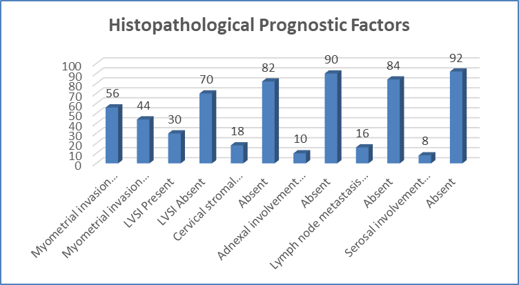
Figure 1 Histopathological Prognostic Factors
Table 4. Distribution According to FIGO Stage ( n = 100)
|
FIGO Stage |
Number (n) |
Percentage (%) |
|
IA |
42 |
42.0 |
|
IB |
24 |
24.0 |
|
II |
14 |
14.0 |
|
IIIA |
5 |
5.0 |
|
IIIB |
3 |
3.0 |
|
IIIC |
8 |
8.0 |
|
IVA |
2 |
2.0 |
|
IVB |
2 |
2.0 |
|
Total |
100 |
100.0 |
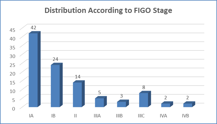
Figure 2 Distribution According to FIGO Stage
Table 5. Association Between Histological Grade and Prognostic Factors
|
Variable |
Grade 1 (n=40) |
Grade 2 (n=38) |
Grade 3 (n=22) |
χ² |
p value |
|
Deep myometrial invasion (≥50%) |
8 (20.0%) |
18 (47.4%) |
18 (81.8%) |
24.86 |
<0.001 |
|
LVSI Present |
4 (10.0%) |
10 (26.3%) |
16 (72.7%) |
30.11 |
<0.001 |
|
Cervical stromal involvement |
2 (5.0%) |
6 (15.8%) |
10 (45.5%) |
19.78 |
<0.001 |
|
Lymph node metastasis |
1 (2.5%) |
5 (13.2%) |
10 (45.5%) |
27.62 |
<0.001 |
|
Advanced FIGO stage (III–IV) |
2 (5.0%) |
8 (21.1%) |
10 (45.5%) |
18.95 |
<0.001 |
DISCUSSION:
The present study demonstrated that endometrial carcinoma predominantly affected older women, with a mean age of 58.4 ± 10.8 years, and 76.0% of patients were postmenopausal. Nearly half of the patients (46.0%) were obese (BMI ≥30 kg/m²), supporting the established association between obesity and endometrial carcinoma. These findings are consistent with Singh et al.[9], who reported that endometrial carcinoma primarily occurs in postmenopausal women and highlighted obesity as a major risk factor through prolonged estrogen exposure and metabolic dysregulation.Endometrioid carcinoma was the predominant histological subtype (84.0%), followed by serous (8.0%), clear cell (4.0%), mixed carcinoma (3.0%), and carcinosarcoma (1.0%).
This distribution closely parallels the observations of Singh et al.[9], who reported that approximately 80–85% of endometrial cancers are of the endometrioid type, whereas serous and clear cell carcinomas are less common but biologically more aggressive. Tumor grading showed that 40.0% of tumors were Grade 1, 38.0% Grade 2, and 22.0% Grade 3. Increasing histological grade was strongly associated with adverse pathological features. Deep myometrial invasion (≥50%) was present in 44.0% of patients overall and increased significantly from 20.0% in Grade 1 to 81.8% in Grade 3 tumors (p<0.001). Similar findings were reported by Singh et al.[9], who identified higher tumor grade and deep myometrial invasion as major predictors of poor prognosis, while the FIGO 2023 Committee further recognizes myometrial invasion as a key determinant of staging and risk stratification.
Lymphovascular space invasion (LVSI) was identified in 30.0% of patients and showed a marked increase with tumor grade, reaching 72.7% in Grade 3 tumors (p<0.001). Comparable findings have been reported by Peters et al.[10], who demonstrated that substantial LVSI independently predicts recurrence and poor survival. Similarly, Barnes et al.[11] reported worse disease-free and overall survival in patients with LVSI, while Raffone et al.[12] confirmed its independent prognostic significance irrespective of molecular subtype. Cervical stromal involvement occurred in 18.0% of patients and increased significantly with tumor grade (p<0.001). These findings agree with McCluggage et al.[13], who reported that cervical stromal invasion is associated with advanced disease and poorer prognosis. Likewise, lymph node metastasis was observed in 16.0% of patients, increasing from 2.5% in Grade 1 to 45.5% in Grade 3 tumors (p<0.001). This is consistent with the landmark study by Creasman et al.[14], which demonstrated that lymph node metastasis is strongly associated with increasing tumor grade and deep myometrial invasion, and with Singh et al.[9], who recognized nodal involvement as a major adverse prognostic factor.
Adnexal and serosal involvement were observed in 10.0% and 8.0% of patients, respectively, reflecting extrauterine disease spread. Most patients were diagnosed at an early stage, with FIGO Stage IA accounting for 42.0% and Stage IB for 24.0%; however, advanced stages increased significantly with tumor grade. Tumor size exceeded 2 cm in 82.0% of cases, a finding that Singh et al.[9] associated with greater myometrial invasion, LVSI, nodal metastasis, and recurrence. Our findings closely correspond with those reported by Singh et al.[9], Peters et al.[10], Barnes et al.[11], Raffone et al.[12], McCluggage et al.[13], Creasman et al.[14], and the FIGO 2023 Committee, confirming that higher histological grade, deep myometrial invasion, LVSI, cervical stromal involvement, lymph node metastasis, and advanced FIGO stage are closely interrelated adverse prognostic factors that should be routinely incorporated into postoperative risk stratification and treatment planning.
CONCLUSION :
The present study demonstrated that histopathological factors, including tumor grade, depth of myometrial invasion, lymphovascular space invasion, cervical stromal involvement, lymph node metastasis, and FIGO stage, are significant prognostic indicators in endometrial carcinoma. Higher histological grade was strongly associated with adverse pathological features and advanced-stage disease. Routine comprehensive histopathological evaluation of these parameters is essential for accurate risk stratification, prognostic assessment, and selection of appropriate adjuvant therapy. Careful assessment of these prognostic factors can facilitate individualized treatment planning and improve the overall management of patients with endometrial carcinoma.
Limitations
The present study was conducted at a single tertiary care center with a relatively small sample size of 100 patients, which may limit the generalizability of the findings. The study primarily evaluated conventional histopathological prognostic factors, and molecular classification and immunohistochemical biomarkers were not included. In addition, long-term follow-up data on recurrence and overall survival were unavailable, precluding assessment of the prognostic impact of these factors on patient outcomes.
REFERENCES:
1. Schindler AE. Progestogen deficiency and endometrial cancer risk. Maturitas. 2009;62(4):334-337.
2. World Cancer Research Fund International. Cancer facts and figures: Endometrial cancer rates. Available from: http://www.wcrf.org/cancer_statistics/cancer_facts/endometrial_cancer_rates.php
3. Siegel R, Ma J, Zou Z. Cancer statistics. CA Cancer J Clin. 2014;64(1):9-29.
4. Garg K, Soslow RA. Endometrial carcinoma in women aged 40 years and younger. Arch Pathol Lab Med. 2014;138(3):335-342.
5. Bohîlțea RE, Ancar V, Cîrstoiu MM. Project for the National Program of Early Diagnosis of Endometrial Cancer. J Med Life. 2015;8(3):305-314.
6. Rosai J. Rosai and Ackerman's Surgical Pathology. 10th ed. Philadelphia: Mosby Elsevier; 2011. p.1492-1500.
7. Florescu MM, Simionescu CE, Stepan AE, Mărgăritescu C, Ciurea RN. Histopathological prognostic factors for endometrial carcinoma. Rom J Morphol Embryol. 2016;57(2 Suppl):719-727.
8. León-Castillo A, Britton H, McConechy MK, McAlpine JN, Nout RA, Kommoss S, et al. Interpretation of the molecular classification of endometrial carcinoma: evidence-based recommendations from the ESGO/ESTRO/ESP guidelines. Int J Gynecol Cancer. 2023;33(7):863-877.
9. Singh N, Hirschowitz L, Zaino R, Alvarado-Cabrero I, Duggan MA, Ali-Fehmi R, et al. Pathologic prognostic factors in endometrial carcinoma (other than tumor type and grade). Int J Gynecol Pathol. 2019;38(Suppl 1):S93-S113.
10. Peters EE, Bosse T, Creutzberg CL, Jürgenliemk-Schulz IM, Jobsen JJ, Mens JWM, et al. Substantial lymph-vascular space invasion (LVSI) is a significant risk factor for recurrence in endometrial cancer: a pooled analysis of the PORTEC 1 and 2 trials. Eur J Cancer. 2015;51(13):1742-1750.
11. Barnes EA, Martell K, Parra-Herran C, Taggar AS, Donovan E, Leung E, et al. Substantial lymphovascular space invasion predicts worse outcomes in early-stage endometrioid endometrial cancer. Brachytherapy. 2021;20(3):527-535.
12. Raffone A, Travaglino A, Saccone G. Lymphovascular space invasion in endometrial carcinoma: prognostic role across molecular subtypes. Diagnostics (Basel). 2023;13.
13. McCluggage WG, Hirschowitz L, Miller K. Recommendations for the pathological reporting of endometrial carcinoma: guidance from the International Society of Gynecological Pathologists. Int J Gynecol Pathol. 2019;38(Suppl 1):S75-S92.
14. Creasman WT, Morrow CP, Bundy BN, Homesley HD, Graham JE, Heller PB, et al. Surgical pathologic spread patterns of endometrial cancer. A Gynecologic Oncology Group study. Cancer. 1987;60(8 Suppl):2035-2041.