A Study of Platelet Indices in Diabetes Mellitus and Their Correlation with Microvascular Complications.
- Purba Biswas , Senior Resident, Department of Pathology, Tripura medical College and Dr BRAM Teaching Hospital.
- Nabaneet Majumder , Professor and Hod, Department of Pathology, Tripura medical College and Dr BRAM Teaching Hospital.
- Swarupa Chakma , Associate Professor, Department of Pathology, Tripura medical College and Dr BRAM Teaching Hospital.
Article Information:
Abstract:
Background: Diabetes mellitus (DM) is a major global health concern associated with significant morbidity and mortality due to its chronic vascular complications. Platelet activation plays a crucial role in the pathogenesis of diabetic microvascular complications. Platelet indices such as Mean Platelet Volume (MPV), Platelet Distribution Width (PDW), Plateletcrit (PCT), and Platelet-Large Cell Ratio (P-LCR) are readily available hematological parameters that may serve as markers of platelet activation and vascular risk. Aim: To evaluate platelet indices in patients with Type 2 Diabetes Mellitus (T2DM) and determine their association with microvascular complications and glycaemic control. Materials and Methods: This hospital-based cross-sectional study was conducted among 150 patients with T2DM. Blood samples were analyzed for fasting blood glucose, post-prandial blood glucose, HbA1c, and platelet indices (MPV, PDW, PCT, and P-LCR). Patients were evaluated for diabetic neuropathy, nephropathy, and retinopathy. Statistical analysis was performed using SPSS version 25. Independent t-test and Pearson correlation analysis were applied, and a p-value <0.05 was considered statistically significant. Results: Microvascular complications were present in 53.33% of patients. Diabetic neuropathy, nephropathy, and retinopathy were observed in 27.33%, 11.33%, and 4.0% of patients, respectively. P-LCR showed a significant association with diabetic neuropathy (p=0.031). Patients with diabetic nephropathy demonstrated significantly higher MPV (p=0.038) and PDW (p=0.041). In diabetic retinopathy, MPV (p=0.006), PDW (p=0.004), and P-LCR (p=0.001) were significantly elevated. HbA1c showed significant positive correlations with MPV (r=0.41, p=0.002), PDW (r=0.37, p=0.006), and P-LCR (r=0.36, p=0.011), while a significant negative correlation was observed with PCT (r=-0.25, p=0.021). Conclusion: Platelet indices, particularly MPV, PDW, and P-LCR, were significantly associated with microvascular complications and poor glycaemic control in patients with T2DM. These indices may serve as simple, inexpensive, and readily available biomarkers for the early detection and monitoring of diabetic microvascular complications. Their routine assessment could aid in risk stratification and timely intervention, thereby reducing diabetes-related morbidity and mortality.
Keywords:
Article :
INTRODUCTION:
Diabetes mellitus (DM) represents a heterogeneous group of metabolic disorders characterized by chronic hyperglycaemia resulting from impaired insulin secretion, defective insulin action, or a combination of both. The disease has emerged as one of the most significant global health challenges due to its rapidly increasing prevalence and its association with substantial morbidity and mortality. DM is a major contributor to end-stage renal disease, blindness, cardiovascular disorders, and other debilitating complications, imposing a considerable burden on healthcare systems worldwide.[1,2] In the South-East Asian region, the diabetic population was estimated at 90 million in 2021 and is expected to rise to 113 million by 2030 and 151 million by 2045. India, often referred to as the “diabetes capital of the world,” has witnessed a steady increase in disease prevalence, rising from 7.1% in 2009 to 8.9% in 2019.[3,4]
The clinical manifestations of diabetes commonly include polyuria, polydipsia, polyphagia, and unexplained weight loss. If inadequately controlled, diabetes can lead to both acute and chronic complications. Acute complications include diabetic ketoacidosis and hyperosmolar hyperglycaemic state, whereas chronic complications are broadly categorized into macrovascular and microvascular disorders. Macrovascular complications encompass coronary artery disease, cerebrovascular accidents, and peripheral vascular disease, while microvascular complications include diabetic retinopathy, nephropathy, and neuropathy.[5–9]
Among these, microvascular complications significantly contribute to long-term disability and reduced quality of life. Persistent hyperglycaemia induces endothelial dysfunction, oxidative stress, and inflammatory changes, which play pivotal roles in the pathogenesis of vascular damage. Furthermore, the presence of microvascular complications has been recognized as an important predictor of future cardiovascular and cerebrovascular events among diabetic individuals.[9,10]
Recent research has increasingly focused on the role of platelets in the development of diabetic vascular complications. Hyperglycaemia promotes non-enzymatic glycation of platelet proteins, resulting in enhanced platelet activation and aggregation. In addition, insulin resistance and insulin deficiency contribute to platelet hyperreactivity by diminishing the inhibitory effects of insulin on platelet function. Consequently, diabetic patients often exhibit a prothrombotic state that may accelerate the progression of vascular complications.[6,7,11–14]
Platelets are essential components of haemostasis, and alterations in platelet morphology and function can be assessed through platelet indices generated by automated haematology analyzers. These indices include Mean Platelet Volume (MPV), Platelet Distribution Width (PDW), Plateletcrit (PCT), and Platelet-Large Cell Ratio (P-LCR). Larger and more reactive platelets possess greater thrombotic potential and are associated with increased production of procoagulant factors.[15]
Several studies have demonstrated elevated platelet indices in individuals with diabetes compared to non-diabetic populations. Increased MPV, in particular, has been associated with diabetes mellitus, metabolic syndrome, coronary artery disease, and cerebrovascular disorders. Similarly, changes in PDW, PCT, and P-LCR have been linked to enhanced platelet activation and vascular complications.[16–18]
Modern automated haematology analyzers provide these platelet parameters rapidly, reliably, and cost-effectively as part of routine complete blood count analysis. Since these indices reflect platelet activation and thrombotic tendency, they may serve as useful biomarkers for identifying diabetic patients at increased risk of vascular complications. Early recognition of such changes could facilitate timely intervention and improved disease management.
In view of the growing burden of diabetes and the need for simple laboratory markers for early detection of complications, the present study was undertaken to evaluate platelet indices in patients with diabetes mellitus and to investigate their association with microvascular complications.
MATERIALS AND METHODS:
The present hospital based cross-sectional study was conducted among diabetic patients in the department of pathology. Patients were selected by convenience sampling technique till desired sample size was reached during the study period.
Sample size: Sample size is denoted by “n” and is calculated by the following formula:
n = 4PQ/L2
where P = Prevalence. Q = 1-P, L = Confidence interval which is 95% in this study
n= 4 x 0.102 x (1- 0.102)/0.05x0.05 = 146.55 which tends to 147. We recruited 150 subjects for the present study.
Inclusion criteria:
• Patients suffering from type 2 diabetes mellitus admitted in the Medicine department who are willing to participate in the study aged >18 years.
Exclusion criteria:
• Abnormal platelet count (<100 and >400x103/microlitre).
• Using drugs affecting platelets functions (aspirin, heparin, warfarin etc.).
• Pregnant females.
• Patients suffering from any connective tissue or auto-immune disorders.
• Patients with malignancy.
Data Collection Procedure:
a. All patients were completely evaluated clinically with special reference to any microvascular complications.
b. Blood sample was taken under all aseptic conditions from ante cubital vein by a clean puncture avoiding any bubbles and froth.
c. About 2 ml of blood sample each was collected in Ethylene Diamine Tetra Acetic acid (EDTA) vial (for complete hemogram and HbA1C) and Fluoride bulb (for Fasting Blood Sugar & Post Prandial Blood Sugar).
d. The testing of blood was done within 2 hours of collection to avoid changing because of ageing of blood cells.
e. Complete hemogram from EDTA vial included Mean Platelet Volume (standard reference range: 8-12 femtolitre), Pateletcrit (0.22-0.24%), Platelet-Large Cell Ratio (15% to 35%) and Platelet Distribution Width (standard reference range: 9-14 femtolitre) and Platelet count (1.5-4 lacs/ cubic millimeter). It was performed using automated blood counter Transasia H 560 from EDTA vial. Mean Platelet Volume (MPV) refers to the average size of platelets in blood whereas Platelet Distribution Width (PDW) measures the variation in size of platelets.
f. Plasma glucose was measured by glucose oxidase method in auto-analyzer Beckman Coulter AU480.
g. HbA1C was recorded by BIO-RAD D-10 High Performance Liquid Chromatography machine. HbA1C is a measure of the average blood sugar level over past 3 months and is an indicator of glycemic control.
h. Additionally, diabetic patients were evaluated for microvascular complications like diabetic retinopathy, neuropathy and nephropathy.
i. The quantitative urine albumin/creatinine ratio (ACR) in the morning spot urine samples was used to diagnose diabetic nephropathy. Sample was collected in a sterile urine pot and should be evaluated within 20 minutes from collection. The semi-automated biochemistry analyzer machine ERBA Mannheim Chem-5 plus v2 was used for analysis. Patients with ACR<30 mg/g was categorized as microalbuminuria negative whereas patients with ACR>30 mg/g was categorized as microalbuminuria positive.
j. Diabetic retinopathy was diagnosed by indirect opthalmoscopy/slit-lamp examination. Before that eyes of the patient had been dilated. Dilatation was done using an eyedrop composed of a combination of Phenylephrine and Tropicamide which were applied in both eyes for 2-3 times at an interval of 15 minutes. After a wait time of 30 to 45 minutes eyes were examined under indirect opthalmoscope/slit-lamp examination. Then according to the findings in opthalmoscopy, based on The International Classification of Diabetic Retinopathy patients were grouped into five clinical categories:
• If no abnormalities detected: No Apparent Retinopathy.
• Microaneurysms only: Mild Non-Proliferative Diabetic Retinopathy (NPDR).
• More than microaneurysms only but less than severe NPDR: Moderate NPDR.
• Any of the following- More than 20 intraretinal hemorrhages in each of 4 retinal quadrants, Definite Venous Beading in 2 or more retinal quadrants, Prominent Intra-retinal Microvascular abnormalities in 1 or more retinal quadrants and no Proliferative Diabetic Retinopathy (PDR): Severe NPDR.
• One or more of retinal neo-vascularisation, vitreous hemorrhage or preretinal hemorrhage: PDR
k. Diabetic neuropathy was diagnosed based on clinical findings. If there is weakness, wasting, impaired vibration, loss of position sense or loss of reflexes, it was categorized as Large Fibre Neuropathy. Whereas presence of superficial pain, electric shock, burning, allodynia, thermal imperception, normal strength and reflexes was categorized as Small Fibre Neuropathy.
• The tools that were used to diagnose diabetic neuropathy at bedside are 128-HZ Tuning Fork and Semmes-Weinstein Monofilament (SWMF).
Data was collected and subjected to statistical analysis.
Statistical analysis: Data so collected was tabulated in an excel sheet. The means and standard deviations of the measurements per group were used for statistical analysis (SPSS 25.00 for windows; SPSS inc, Chicago, USA). Difference between two groups was determined using t test and the level of significance was set at p < 0.05.
RESULTS:
Out of 150 subjects; 69 were males and 81 were females. Maximum subjects were from age group of 51-60 years followed by 60 years while minimum subjects were from 18-30 years followed by 31-40 years. Mean fasting blood glucose (mg/dl) and HbA1c (%) was 152.84 and 8.46 respectively (table 1).
Table 1: Diabetic parameters and platelet indices among the study subjects
|
Variables |
Mean |
SD |
|
Fasting blood glucose (mg/dl) |
152.84 |
8.16 |
|
Post Prandial Blood Sugar (mg/dl) |
211.47 |
30.28 |
|
HbA1c (%) |
8.46 |
1.58 |
|
MPV (fL) |
11.43 |
1.76 |
|
PDW (fL) |
16.24 |
2.18 |
|
PCT (%) |
0.25 |
0.05 |
|
P-LCR |
44.18 |
3.27 |
Microvascular complications were present in 53.33% of the study subjects (graph 1).
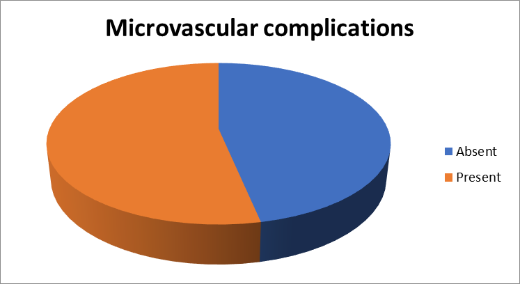
Graph 1: Microvascular complications among the study subjects
Microvascular complications viz. diabetic neuropathy, nephropathy and retinopathy was reported among 27.33%, 11.33% and 4.0% of the subjects respectively. Combination of any 2 microvascular complications was revealed in 10.67% of the subjects (table 2).
Table 2: Distribution of microvascular complications among the study subjects
|
Microvascular complications |
Number |
Percentage |
|
Diabetic Neuropathy |
41 |
27.33 |
|
Diabetic Nephropathy |
17 |
11.33 |
|
Diabetic Retinopathy |
6 |
4.00 |
|
Combination of Any 2 |
16 |
10.67 |
Patients with diabetic neuropathy demonstrated higher MPV, PDW, and P-LCR values and lower PCT values compared to those without neuropathy. However, only P-LCR showed a statistically significant association with diabetic neuropathy (p < 0.05), suggesting increased platelet activation in patients with neuropathic complications (table 3).
Table 3: Comparison of platelet indices according to Diabetic Neuropathy
|
Variables |
Diabetic Neuropathy |
p value |
|||
|
Yes |
No |
||||
|
Mean |
SD |
Mean |
SD |
||
|
MPV (fL) |
12.48 |
1.58 |
11.72 |
1.86 |
0.094 |
|
PDW (fL) |
16.34 |
2.08 |
15.82 |
2.31 |
0.371 |
|
PCT (%) |
0.22 |
0.06 |
0.25 |
0.07 |
0.087 |
|
P-LCR |
45.61 |
3.04 |
42.38 |
3.56 |
0.031* |
*: statistically significant
Patients with diabetic nephropathy exhibited significantly higher MPV and PDW values compared to those without nephropathy (p < 0.05). Although P-LCR was also higher and PCT was lower among nephropathy cases, these differences were not statistically significant. The findings suggest an association between increased platelet activation and the presence of diabetic nephropathy (table 4).
Table 4: Comparison of platelet indices according to Diabetic Nephropathy
|
Variables |
Diabetic Nephropathy |
p value |
|||
|
Yes |
No |
||||
|
Mean |
SD |
Mean |
SD |
||
|
MPV (fL) |
12.94 |
1.63 |
11.38 |
1.79 |
0.038* |
|
PDW (fL) |
16.67 |
2.29 |
15.42 |
2.46 |
0.041* |
|
PCT (%) |
0.23 |
0.05 |
0.25 |
0.06 |
0.128 |
|
P-LCR |
45.12 |
3.18 |
42.89 |
3.61 |
0.164 |
*: statistically significant
Patients with diabetic retinopathy demonstrated significantly elevated MPV, PDW, and P-LCR values compared to those without retinopathy (p < 0.05). Although PCT was slightly lower among retinopathy cases, the difference was not statistically significant. These findings indicate enhanced platelet activation in patients with diabetic retinopathy and support the potential role of platelet indices as markers of microvascular complications in diabetes mellitus (table 5).
Table 5: Comparison of platelet indices according to Diabetic retinopathy
|
Variables |
Diabetic Retinopathy |
p value |
|||
|
Yes |
No |
||||
|
Mean |
SD |
Mean |
SD |
||
|
MPV (fL) |
13.72 |
1.54 |
11.26 |
1.84 |
0.006* |
|
PDW (fL) |
16.89 |
2.36 |
15.24 |
2.18 |
0.004* |
|
PCT (%) |
0.23 |
0.05 |
0.25 |
0.06 |
0.173 |
|
P-LCR |
48.34 |
3.12 |
42.68 |
3.42 |
0.001* |
*: statistically significant
Correlation analysis demonstrated a significant positive relationship between HbA1c levels and MPV, PDW, and P-LCR, indicating that worsening glycaemic control is associated with increased platelet activation. Conversely, PCT showed a weak but significant negative correlation with HbA1c. These findings suggest that platelet indices may reflect the degree of glycaemic control and could serve as useful markers for identifying diabetic patients at increased risk of vascular complications as shown in table 6.
Table 6: Pearson correlation for the association between platelet parameters and glycated haemoglobin (HbA1c) in diabetic patients
|
Variables |
r value |
p value |
|
MPV (fL) and HbA1c |
0.41 |
0.002* |
|
PDW (fL) and HbA1c |
0.37 |
0.006* |
|
PCT (%) and HbA1c |
-0.25 |
0.021* |
|
P-LCR and HbA1c |
0.36 |
0.011* |
*: statistically significant
DISCUSSION:
In the present study, a slight female predominance was observed among patients with Type 2 Diabetes Mellitus (T2DM). The majority of participants belonged to the 51–60 years age group, followed by those aged above 60 years. These findings are comparable with the observations of Khanna P et al. [81], who reported a mean age of 56.96±8.99 years among diabetic cases and a female predominance (57% females and 43% males). Similar demographic characteristics were also documented by Kamilla R. Alhadas et al. [19]. Several epidemiological studies have reported a higher burden of diabetes among women, which has been attributed to factors such as obesity, sedentary lifestyle, hormonal influences, and psychosocial stress. Furthermore, women with diabetes have been shown to experience a disproportionately greater risk of cardiovascular morbidity and mortality compared with men [20].
In the present study, microvascular complications were observed in 53.33% of the study population. Diabetic neuropathy was the most common complication, affecting 27.33% of patients, followed by diabetic nephropathy (11.33%) and diabetic retinopathy (4.0%). A combination of any two microvascular complications was observed in 10.67% of participants. These findings are consistent with those reported by Walinjkar et al. [22], who found microvascular complications in 66 out of 125 diabetic patients, with neuropathy being the most frequent complication. Similarly, Kamilla R. Alhadas et al. [19] reported chronic diabetic complications in 41% of patients, with retinopathy and nephropathy being the predominant manifestations. In the present study, patients with diabetic neuropathy demonstrated higher MPV, PDW, and P-LCR values and lower PCT values compared with those without neuropathy. However, statistically significant association was observed only for P-LCR (p<0.05). The elevated P-LCR in neuropathy patients indicates the presence of larger and more reactive platelets, reflecting enhanced platelet activation. Khanna P et al. [20] similarly reported significantly increased MPV, PDW, and P-LCR values among patients with diabetic neuropathy. Their study demonstrated significant positive correlations between these platelet indices and the presence of neuropathy, suggesting a role of platelet activation in the pathogenesis of diabetic nerve damage.
Patients with diabetic nephropathy in the present study exhibited significantly higher MPV and PDW values compared to those without nephropathy (p<0.05). Although P-LCR was higher and PCT was lower among nephropathy cases, these differences did not reach statistical significance. These findings support the hypothesis that platelet activation contributes to the development of diabetic nephropathy. Unubol et al. [25] reported a significant positive correlation between MPV and microalbuminuria, indicating that larger platelets may play a role in the progression of renal microvascular injury. Similar observations have been reported by several investigators, highlighting MPV as a potential marker for diabetic nephropathy.
Zuberi et al. [16] observed significantly higher MPV values among diabetic patients with vascular complications compared to those without complications. Likewise, Papanas et al., Ates et al. [23], and Tuzcu et al. [24] reported increased MPV levels in patients with diabetic retinopathy. These observations suggest that platelet activation may contribute to retinal microvascular damage through enhanced thrombotic and inflammatory mechanisms. Studies by Buch et al. [26], Dindar et al. [29], Ji et al. [7], and Jindal et al. [28] further demonstrated positive associations between PDW and diabetic microvascular complications, particularly retinopathy and nephropathy. Although Chen et al. [27] did not observe significant differences in PDW among diabetic patients with and without complications, the majority of available evidence supports its role as a marker of platelet activation in diabetes. Prabhat A et al. [30] also reported a significant relationship between elevated MPV and the presence of microvascular complications, reinforcing the findings of the present study.
The present study demonstrated significant positive correlations between HbA1c and MPV (r=0.41, p=0.002), PDW (r=0.37, p=0.006), and P-LCR (r=0.36, p=0.011). Conversely, a significant negative correlation was observed between HbA1c and PCT (r=-0.25, p=0.021). These findings indicate that worsening glycaemic control is associated with increased platelet activation and greater platelet heterogeneity. Similar observations were reported by Khanna P et al. [20], who demonstrated significant associations between HbA1c levels and MPV, PDW, and P-LCR. Kamilla R. Alhadas et al. [19] also reported positive correlations between glycaemic indices and platelet parameters, suggesting that patients with higher fasting blood glucose and HbA1c levels tend to have elevated MPV and PDW values. Yilmaz et al. [32] further demonstrated that MPV correlates positively with the severity of diabetic retinopathy, nephropathy, and HbA1c levels. The underlying mechanisms responsible for increased platelet activity in diabetes include chronic hyperglycaemia, insulin resistance, endothelial dysfunction, increased coagulation activity, and impaired fibrinolysis. These factors collectively promote a prothrombotic state characterized by platelet hyperreactivity, which contributes significantly to the development and progression of diabetic microvascular complications [26]. Overall, the findings of the present study support the growing body of evidence that platelet indices, particularly MPV, PDW, and P-LCR, are closely associated with poor glycaemic control and the presence of diabetic microvascular complications. These indices may therefore serve as simple, inexpensive, and readily available biomarkers for early identification and monitoring of high-risk diabetic patients.
CONCLUSION:
MPV, PDW, and P-LCR were found to be significantly elevated in patients with Type 2 Diabetes Mellitus who had microvascular complications compared to those without such complications. The increased values of these platelet indices suggest enhanced platelet activation and indicate their potential utility as prognostic biomarkers for the early identification of diabetic microvascular complications. As these parameters are readily available through routine hematological investigations, they offer a simple, economical, and non-invasive approach for monitoring disease progression and assessing the risk of complications in diabetic patients.
The incorporation of platelet indices into routine clinical evaluation may facilitate timely recognition of individuals at higher risk, enabling early intervention and implementation of preventive strategies. Such measures could contribute to reducing the burden of diabetes-related morbidity and mortality. Furthermore, longitudinal follow-up studies are warranted to evaluate the relationship between platelet indices and the progression of diabetes, as well as their predictive value for the development and advancement of microvascular complications over time.
REFERENCES:
1. Alberti KGMM, Zimmet PZ. World Health Organization Definition, diagnosis and classification of diabetes mellitus and its complications: Report of a WHO consultation. Part 1. Diagnosis and classification of diabetes mellitus. Geneva. Diabet Med. 2015; 15: 539-53.
2. Keating FK, Sobel BE, Schneider DJ. Effects of increased concentrations of glucose on platelet reactivity in healthy subjects and in patients with and without diabetes. Am J Cardiol. 2003; 92: 1362-65.
3. IDF Diabetes Atlas. International Diabetes Federation. IDF Diabetes Atlas. 10th ed. Brussels, Belgium: International Diabetes Federation; 2021. Available from: http://www.diabetesatlas.org. [Last cited on 2018 Nov 20].
4. IDF Diabetes Atlas. International Diabetes Federation. IDF Diabetes Atlas. 9th ed. Brussels, Belgium: International Diabetes Federation; 2019. Available from: http://www.diabetesatlas.org. [Last cited on 2018 Nov 20].
5. Chawla A, Chawla R, Jaggi S: Microvasular and macrovascular complications in diabetes mellitus. Distinct or continuum?. Indian J Endocrinol Metab. 2016; 20: 546-51.
6. Radha RK, Selvam D: MPV in uncontrolled & controlled diabetics- its role as an indicator of vascular complication. J Clin Diagn Res. 2016; 10: EC22-6.
7. Ji S, Zhang J, Fan X, Wang X, Ning X, Zhang B, et al. The relationship between mean platelet volume and diabetic retinopathy: A systematic review and meta-analysis. Diabetol Metab Syndr 2019; 11: 25.
8. Goyal R, Jialal I: Diabetes Mellitus Type 2. StatPearls [Internet], Treasure Island (FL); 2022.
9. Gosmanov AR, Gosmanova EO, Kitabchi AE: Hyperglycemic crises: diabetic ketoacidosis and hyperglycemic hyperosmolar state. Endotext. MDText.com, Inc, South Dartmouth (MA); 2021.
10. Sociedade Brasileira de Diabetes. Diretrizes da Sociedade Brasileira de Diabetes. 2013-2014. São Paulo: AC Farmacêutica.
11. Balasubramaniam K, Viswanathan GN, Marshall SM, Zaman AG. Increased atherothrombotic burden in patients with diabetes mellitus and acute coronary syndrome: a review of antiplatelet therapy. Cardiol Res Pract [Internet]. 2012; 2012.
12. Papanas N, Symeonidis G, Maltezos E, Mavridis G, Karavageli E, Vosnakidis TH, Lakasas G. Mean platelet volume in patients with type 2 diabetes mellitus. Platelets. 2004; 15(8): 475-8.
13. Hekimsoy Z, Payzin B, Ornek T, Kandoğan G. Mean platelet volume in type 2 diabetic patients. J Diabetes Complications. 2004; 18(3): 173-6.
14. Wendland AE, Farias MG, Manfroi WC. Volume plaquetário médio e doença cardiovascular. J Bras Patol Med Lab [Internet]. 2009; 45(5): 371-8.
15. Elsherbiny IA, Shoukry A, Tahlawi MAE. Mean platelet volume and its relation to insulin resistance in non‑diabetic patients with slow coronary flow. J Cardiol 2012; 59: 176‑81.
16. Zuberi BF, Akhtar N, Afsar S. Comparison of mean platelet volume in patients with diabetes mellitus, impaired fasting glucose and non‑diabetic subjects. Singapore Med J 2008; 49: 114‑6.
17. Khandekar MM, Khurana AS, Deshmukh SD, Kakrani AL, Katdare AD, Inamdar AK. Platelet volume indices in patients with coronary artery disease and acute myocardial infarction: An Indian scenario. J Clin Pathol 2006; 59: 146‑9.
18. Ulutas KT, Dokuyucu R, Sefil F, Yengil E, Sumbul AT, Rizaoglu H, et al. Evaluation of mean platelet volume in patients with type 2 diabetes mellitus and blood glucose regulation: a marker for atherosclerosis?. International journal of clinical and experimental medicine. 2014;7(4):955.
19. Alhadas KR, Santos SN, Freitas MM, Viana SM, Ribeiro LC, Costa MB. Are platelet indices useful in the evaluation of type 2 diabetic patients?. Jornal Brasileiro de Patologia e Medicina Laboratorial. 2016; 52: 96-102.
20. Khanna P, Salwan SK, Sharma A. Correlation of Platelet Indices in Patients with Type 2 Diabetes Mellitus and Associated Microvascular Complications: A Hospital-Based, Prospective, Case-Control Study. Cureus. 2024; 16(3).
21. Martyn CN, Matthews DM, Popp-Snijders C, Tucker J, Ewing DJ, Clarke BF. Effects of Sorbinil treatment on erythrocytes and platelets of persons with diabetes. Diabetes Care 1986; 9: 36-9.
22. Walinjkar RS, Khadse S, Kumar S, Bawankule S, Acharya S. Platelet indices as predictor of microvascular complications in type 2 diabetes. Indian J Endocr Metab 2019; 23: 206-10.
23. Ateş O, Kiki İ, Bilen H, Keleş M, Koçer İ, Kulaçoğlu DN, et al. Association of mean platelet volume with the degree of retinopathy in patients with diabetes mellitus. Eur J Gen Med [Internet]. 2009; 6(2): 99-102.
24. Ayhan Tuzcu E, Arıca S, Ilhan N, Daglioglu M, Coskun M, Ilhan O, et al. Relationship between mean platelet volume and retinopathy in patients with type 2 diabetes mellitus. Graefe's Archive for Clinical and Experimental Ophthalmology. 2014; 252(2): 237-40.
25. Ünübol M, Ayhan M, Güney E. The relationship between mean platelet volume with microalbuminuria and glycemic control in patients with type II diabetes mellitus. Platelets. 2012; 23(6): 475-80.
26. Buch A, Kaur S, Nair R, Jain A. Platelet volume indices as predictive biomarkers for diabetic complications in Type 2 diabetic patients. J Lab Physicians. 2017; 9(2):84-8.
27. Chen X, Fang L, Lin H, Shen P, Zhang T, LI H, et al. The Relationship between Type 2 Diabetes and Platelet Indicators. Iran J Public Health. 2017; 46(9):1211-6.
28. Jindal S, Gupta S, Gupta R, Kakkar A, Singh HV, Gupta K, et al. Platelet indices in diabetes mellitus: indicators of diabetic microvascular complications. Hematol. 2011; 16(2):86-9.
29. Dindar S, Cinemre H, Sengul E, Annakkaya AN. Mean platelet volume is associated with glycaemic control and retinopathy in patients with type 2 diabetes mellitus. West Indian Med J 2013; 62: 519-23.
30. Agrawal P, Singh S, Singh AK, Pursnani N, Keshari CK, Gautam A, et al. Platelet indices as a predictor of microvascular complications of type 2 diabetes mellitus. J Diabetol 2023; 14: 74-7.
31. Subramanian S, Green SR, Vaithy A. A prospective observational study of relation between platelet indices and microvascular complica tions in type 2 diabetic patients in a tertiary care hospital. Int J Recent Sci Res. 2020; 11(03): 37915–37919.
32. Yilmaz T, Yilmaz A. Relationship between Altered Platelet Morphological Parameters and Retinopathy in Patients with Type 2 Diabetes Mellitus. J Ophthalmol. 2016; 2016: 1-5.