Evaluation of Modified Mallampati Grading and Skin-to-Epiglottis Distance as Independent Predictors of Difficult Laryngoscopy in Patients with Oral and Oropharyngeal Malignancies.
- Khushpreet Kaur , MD Resident, Department of Anaesthesiology & Critical Care, Sri Guru Ram Das Institute of Medical Sciences and Research (SGRDIMSAR) Amritsar 143501, Punjab, India.
- Shubhdeep Kaur , MD, Associate Professor, Department of Anaesthesiology & Critical Care Sri Guru Ram Das Institute of Medical Sciences and Research (SGRDIMSAR) Amritsar 143501, Punjab, India.
- Anita Kumari , MD, Professor Department of Anaesthesiology & Critical Care Sri Guru Ram Das Institute of Medical Sciences and Research (SGRDIMSAR) Amritsar 143501, Punjab, India.
- Ruchi Gupta , MD, DNB Professor & Head Department of Anaesthesiology & Critical Care Sri Guru Ram Das Institute of Medical Sciences and Research (SGRDIMSAR) Amritsar 143501, Punjab, India.
Article Information:
Abstract:
Background: Airway assessment in patients with oral and oropharyngeal malignancies is challenging because tumour-related anatomical changes increase the risk of difficult laryngoscopy. Clinical and ultrasonographic parameters may improve preoperative airway prediction. Methods: This cross-sectional observational study included 85 patients undergoing surgery for oral and oropharyngeal malignancies. Preoperative airway assessment was performed using Modified Mallampati Grading (MMG) and ultrasonographic Skin-to-Epiglottis Distance (SED). Difficult laryngoscopy was defined as a Modified Cormack–Lehane grade > IIb. Diagnostic performance was assessed using sensitivity, specificity, predictive values, odds ratio, and receiver operating characteristic analysis. Results: Difficult laryngoscopy occurred in 71.8% of patients. MMG Grade III/IV was significantly associated with difficult laryngoscopy (OR 10.8, p < 0.001). An SED >2.5 cm predicted difficult laryngoscopy with good sensitivity and specificity, supporting its diagnostic utility. Conclusion: Both MMG and SED are useful independent predictors of difficult laryngoscopy. Ultrasonographic airway assessment provides an objective and valuable adjunct to routine clinical evaluation.
Keywords:
Article :
INTRODUCTION:
Airway management is a central pillar of anaesthesia practice, playing a critical role in ensuring patient safety during the perioperative period as well as in emergency and critical care settings. Maintaining a patent airway is non-negotiable, as any lapse can rapidly lead to life-threatening complications such as hypoxemia, cardiac arrest, neurological damage, or death. Despite ongoing advancements in airway tools and techniques, the problem of difficult airway management persists and continues to be a significant cause of anaesthesia-related morbidity and mortality worldwide. ¹
A difficult airway refers to a clinical situation in which an experienced anaesthesiologist faces difficulty in achieving effective mask ventilation, tracheal intubation, or both. Recognising such situations in beforehand is essential, as unexpected airway difficulty can rapidly escalate into a critical, life-threatening event. This concern is particularly significant in patients with oral and oropharyngeal malignancies. In these individuals, tumour infiltration can alter and distort normal airway anatomy, while factors such as mucosal oedema, post-radiotherapy fibrosis, and trismus further compromise mouth opening and cervical mobility. These alterations hinder proper alignment of the airway axes during laryngoscopy, thereby increasing the chances of difficult laryngoscopy and intubation. Therefore, timely identification and meticulous preoperative planning are crucial to ensure safe airway management in this vulnerable patient population. ²
The Modified Mallampati grading, first described by Seshagiri Mallampati et al., is a simple bedside method based on the visibility of oropharyngeal structures and has been widely used to anticipate difficult laryngoscopy, with higher classes (III–IV) indicating increased difficulty. However, subsequent work by Michael Wilson et al. demonstrated that although Mallampati grading is helpful as an initial screening tool, its predictive accuracy is limited when used in isolation and improves significantly when combined with other clinical airway assessment parameters3,4
The Skin-to-Epiglottis Distance, evaluated using ultrasonography, has emerged as an objective measure of anterior neck soft tissue thickness and is increasingly used to predict difficult laryngoscopy. Jing Wu et al. demonstrated that higher SED values were significantly associated with difficult laryngoscopy, with good diagnostic accuracy and a proposed cut-off around 2.7 cm. Similarly, Salvatore Falcetta et al. reported that increased SED correlated strongly with airway difficulty, suggesting threshold values of approximately 2.5–2.75 cm. However, despite its promising predictive performance, SED is best used as an adjunct rather than a standalone parameter, as combining it with clinical predictors enhances overall airway assessment accuracy.4,5
This study evaluates MMG and SED as independent predictors of difficult laryngoscopy in patients with oral and oropharyngeal malignancies.
MATERIALS AND METHODS:
This cross-sectional observational study was conducted in the Department of Anaesthesiology at Sri Guru Ram Das Institute of Medical Sciences and Research, Amritsar, from July 2024 to December 2025. A total of 85 adult patients undergoing elective surgery for oral and oropharyngeal malignancies were included after obtaining institutional ethical approval and informed consent from patients.
Inclusion criteria encompassed patients aged 18–80 years scheduled for elective oral and oropharyngeal cancer surgery who were willing to participate, while exclusion criteria included previous head and neck surgery, pre-existing tracheostomy, planned fiberoptic intubation, and BMI > 30 kg/m².
All patients underwent a detailed pre-anaesthetic check-up one day prior to surgery, with special attention to airway assessment along with other routine investigations.
Modified Mallampati Grading (MMG) was assessed in the sitting position with the head in neutral alignment; patients opened their mouth maximally and protruded the tongue fully without phonation, and visibility was graded as Class I (soft palate, fauces, uvula, and pillars visible), Class II (soft palate, fauces, and uvula visible, pillars not visible), Class III (soft palate and base of uvula visible), or Class IV (only hard palate visible), with Classes III and IV considered predictors of difficult laryngoscopy. 7
Skin-to-Epiglottis Distance (SED) was measured using ultrasonography with a Wipro GE ultrasound machine and a high-frequency linear probe in the transverse plane at the thyrohyoid membrane level in the midline of the anterior neck; the hyoid bone was identified as a hyper echoic structure with posterior acoustic shadowing, followed by caudal probe movement to visualise the epiglottis as a curved hypo echoic structure with a bright anterior interface, with the skin-to-epiglottis distance measured in centimetres and values > 2.5 cm deemed predictive of difficult laryngoscopy.
After pre-anaesthetic check-up review, patients were shifted to the operating room where standard monitoring was instituted, including heart rate, non-invasive blood pressure, peripheral oxygen saturation (SpO₂), electrocardiography (ECG), and end-tidal carbon dioxide (EtCO₂). Following confirmation of NPO status, patients received premedication with glycopyrrolate (0.01 mg/kg) and either butorphanol (0.02 mg/kg) or fentanyl (2 µg/kg) intravenously; anticipating difficult airways, they were pre oxygenated with 100% oxygen for 5 minutes, anaesthesia was induced with propofol (2–2.5 mg/kg IV), and after confirming adequate mask ventilation, neuromuscular blockade was achieved with succinylcholine (1–1.5 mg/kg IV). All patients were planned for nasotracheal intubation under direct laryngoscopy; nasal passages were prepared with oxymetazoline drops and 2% lignocaine gel applied to the maxillofacial endotracheal tube for atraumatic insertion.
Laryngoscopy was performed, and the glottic view on the first attempt was graded using the Modified Cormack–Lehane classification, with grade > II considered difficult laryngoscopy. Modified Cormack–Lehane (CL) Grading:
• Grade I: Full view of the glottis
• Grade Ila: Partial view of the glottis
• Grade II: Only the arytenoids or posterior part of the vocal cords visible
• Grade IIIa: Only the epiglottis visible, but it can be lifted
• Grade IIIb: Only the epiglottis visible and cannot be lifted
• Grade IV: Neither the glottis nor the epiglottis visible
In cases of difficulty, the blade was changed from Macintosh to McCoy or a video-laryngoscope to achieve successful intubation. The number of attempts and any change in technique were recorded.
Intraoperative vitals were monitored continuously, recorded every 5 minutes and documented every 30 minutes. At the end of surgery, patients with primary closure were extubated on table after meeting standard criteria, while those requiring ventilatory support were shifted to the intensive care unit.
Statistical Analysis
Data analysis was performed using SPSS version 26. Categorical variables were presented as frequencies and percentages, their associations were evaluated with the Chi-square test, odds ratios (OR) along with 95% confidence intervals were calculated, sensitivity and specificity were determined from ROC analysis, AUC and a p-value below 0.05 was deemed statistically significant.
RESULTS:
A total of 85 patients were included in the study, with a marked male predominance (74 males [87.06%], 11 females [12.94%]). Most patients were overweight, with 65 (76.47%) having a BMI of 25–29.9 kg/m², while 20 (23.53%) had normal BMI.
Based on laryngoscopic findings, 61 patients (71.8%) were classified as having difficult laryngoscopy, whereas 24 patients (28.2%) had easy laryngoscopy. The distribution of Modified Cormack–Lehane grades was Grade Ila – 24 (28.2%), Grade II – 29 (34.1%), Grade IIIa – 26 (30.6%), and Grade IIIb – 6 (7.1%), indicating a predominance of higher grades.
Macintosh blade was used in 70 patients (82.4%), while a video laryngoscope was utilised in 15 patients (17.6%), primarily in anticipated or encountered difficult airway situations. In cases of difficulty, modification of technique, including change of blade or use of videolaryngoscopy, was performed to facilitate intubation.
Successful intubation was achieved on the first attempt in 72 patients (84.7%), while 10 patients (11.8%) required a second attempt. A third attempt was needed in 2 patients (2.4%), and only 1 patient (1.2%) required four attempts, indicating a high overall success rate despite the high incidence of difficult laryngoscopy. (Table 1).
Table 1: Demographic Profile of Study Population (n = 85)
|
Parameter |
Category |
Frequency (n) |
Percentage (%) |
Mean ± SD |
|
Age (years) |
30–39 |
8 |
9.4 |
56.8 ± 10.7 |
|
40–49 |
19 |
22.3 |
||
|
50–59 |
28 |
32.94 |
||
|
60–69 |
19 |
22.35 |
||
|
70–79 |
11 |
12.94 |
||
|
Gender |
Male |
74 |
87.06 |
|
|
Female |
11 |
12.94 |
|
|
|
BMI (kg/m²) |
18.5–24.9 |
20 |
23.53 |
26.6 ± 2.1 |
|
25–29.9 |
65 |
76.47 |
Modified Mallampati Grading
Modified Mallampati Grade III–IV was considered predictive of difficult laryngoscopy. Based on this criterion, 64 out of 85 patients (75.3%) were predicted to have difficult laryngoscopy, out of these predicted difficult (Grade III–IV), 54 patients (84.4%) were actually difficult whereas 10 patients (15.6%) were easy. 21 patients (24.7%) were predicted to have easy laryngoscopy out of these predicted easy (Grade I–II), 14 patients (66.7%) were actually easy, while 7 patients (33.3%) were difficult, representing false-negative predictions.
Difficult laryngoscopy was observed more frequently in patients with higher Mallampati grades, with 54 out of 64 patients (84.4%) in Grade III–IV compared to 7 out of 21 patients (33.3%) in Grade I–II. This difference was statistically significant, as reflected by an odds ratio of 10.80 with a corresponding p-value of <0.001. The magnitude of the odds ratio indicates that patients with Mallampati Grade III–IV had nearly elevenfold higher odds of difficult laryngoscopy, highlighting its strong predictive value. (Table 2)
Table 2: Diagnostic Accuracy of Modified Mallampati Grade in Predicting Difficult Laryngoscopy
|
Modified Mallampati Grade |
Actual Easy Laryngoscopy |
Actual Difficult Laryngoscopy |
|
Predicted easy laryngoscopy (I, II) |
14 |
7 |
|
Predicted difficult laryngoscopy (III, IV) |
10 |
54 |
|
Odds Ratio (95% CI) |
10.8(3.4863 to 33.4568) |
|
|
p value |
0.000† |
|
Data presented as n = number of patients, p<0.001 highly significant †
Skin to Epiglottis Distance
Skin-to-epiglottis distance was evaluated using a cut-off value of >2.5 cm to predict difficult laryngoscopy. Based on this threshold, 43 out of 85 patients (50.6%) were predicted to have difficult laryngoscopy; among these predicted difficult cases (SED >2.5 cm), 38 patients (88.4%) were actually difficult, while 5 patients (11.6%) were easy. A total of 42 patients (49.4%) were predicted to have easy laryngoscopy; among these predicted easy cases (SED ≤2.5 cm), 19 patients (45.2%) were actually easy, whereas 23 patients (54.8%) were difficult, representing false-negative predictions.
Difficult laryngoscopy was more commonly observed in patients with higher SED values, with 38 out of 43 patients (88.4%) in the >2.5 cm group compared to 23 out of 42 patients (54.8%) in the ≤2.5 cm group. This difference was statistically significant, as indicated by an odds ratio of 6.28 with a corresponding p-value of 0.001. The magnitude of the odds ratio suggests that patients with SED >2.5 cm had more than sixfold higher odds of experiencing difficult laryngoscopy, indicating good predictive ability. (Table 3)
Table 3: Diagnostic Accuracy of Skin to Epiglottis Distance (in cms) in Predicting Difficult Laryngoscopy
|
Skin-to-Epiglottis Distance |
Easy Laryngoscopy (n=24) |
Difficult Laryngoscopy (n=61) |
|
Predicted easy laryngoscopy (< 2.5 cm) |
19 |
23 |
|
Predicted difficult laryngoscopy (> 2.5 cm) |
5 |
38 |
|
Odds Ratio (95% CI) |
6.27(2.0630 to 19.1065) |
|
|
p value |
0.001* |
|
Data presented as n = number of patients, p<0.05 significant
Receiver operating characteristic analysis showed good diagnostic performance, with an area under the curve of 0.798 (95% CI: 0.698–0.878). The optimal cut-off derived from ROC analysis was >2.32 cm, which provided a sensitivity of 68.85% and specificity of 83.33%. The positive predictive value was 91.3%, while the negative predictive value was 51.3%, indicating that SED is particularly effective in identifying true difficult airways. (Table 4),(Figure1)
Table 4 : Diagnostic Performance of Skin-to-Epiglottis Distance (in cms) for Predicting Difficult Laryngoscopy
|
Parameter |
Value |
95% Confidence Interval |
|
Area Under the ROC Curve (AUC) |
0.798 |
0.698 – 0.878 |
|
Optimal Cut-off (Youden Index) |
> 2.32 cm |
— |
|
Sensitivity (%) |
68.85% |
55.7 – 80.1 |
|
Specificity (%) |
83.33% |
62.6 – 95.3 |
|
Positive Likelihood Ratio (+LR) |
4.13 |
1.66 – 10.27 |
|
Negative Likelihood Ratio (−LR) |
0.37 |
0.25 – 0.57 |
|
Positive Predictive Value (PPV) (%) |
91.3% |
80.9 – 96.3 |
|
Negative Predictive Value (NPV) (%) |
51.3% |
41.0 – 61.4 |
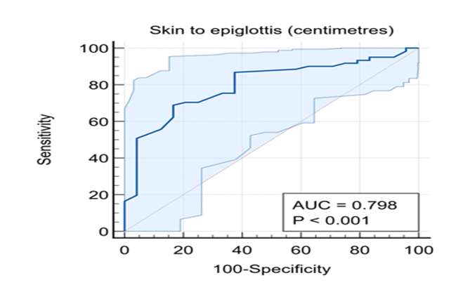
Figure 1: ROC Curve
DISCUSSION:
Airway management is a fundamental responsibility of the anaesthesiologist, and failure to anticipate a difficult airway can lead to serious perioperative complications. This challenge is particularly pronounced in patients with oral and oropharyngeal malignancies, where tumour-related distortion, fibrosis, and restricted. The present study was conducted to evaluate the predictive value of Modified Mallampati Grading (MMG) and ultrasonographic Skin-to-Epiglottis Distance (SED) in patients with oral and oropharyngeal malignancies. A total of 85 patients undergoing elective surgery under general anaesthesia were included. All patients underwent preoperative airway assessment followed by standardised induction with propofol and neuromuscular blockade after adequate pre oxygenation. Nasotracheal intubation under direct laryngoscopy was performed in all cases, and the glottic view was graded using the Modified Cormack–Lehane classification, with grade > II considered as difficult laryngoscopy.8 The intraoperative findings were then correlated with preoperative clinical and ultrasonographic parameters.
A markedly high incidence of difficult laryngoscopy (71.8%) was observed in the present study, which is greater than that reported in general surgical populations. In comparison, Varghese Zachariah et al.,9reported an incidence of around 15% among patients undergoing elective surgeries under general anaesthesia, while Senapathi et al.,10 documented a rate of 28.1% in their study population. This higher incidence in the present study is likely due to the unique nature of the study group. In the present study, Modified Mallampati Grading demonstrated a strong association with difficult laryngoscopy. A total of 64 patients were predicted to have difficult laryngoscopy, of whom 54 were actually difficult and 10 were easy, indicating false positives. Among the 21 patients predicted to have easy laryngoscopy, 14 were truly easy while 7 were actually difficult, representing false negatives. Thus, difficult laryngoscopy was encountered in 84.4% of patients predicted to be difficult compared to 33.3% of those predicted to be easy. This difference was statistically significant, as reflected by an odds ratio of 10.80 with a corresponding p-value of <0.001.
These findings are comparable to previous studies, although variations exist in diagnostic accuracy. Greeshma et al., 11 reported very high sensitivity (95%) and specificity (99.6%) for MMG, while Varghese Zachariah et al.,9 found moderate sensitivity (75.8%) and specificity (78.06%)¹⁰. Another study also showed only a weak to moderate relationship between Mallampati grade and laryngoscopic view, suggesting that exact prediction is not always reliable. 12 The differences seen in the present study may be due to the type of patients included. In general surgical patients, airway anatomy is usually normal, so prediction is more accurate. However, in patients with malignancies, changes such as tumour growth, fibrosis, and reduced mouth opening can affect the airway, leading to higher false-negative (33.3%) and false-positive (15.6%) results, and reducing the reliability of Modified Mallampati grading when used alone. Skin-to-epiglottis distance also showed significant predictive value in the present study. A total of 43 patients were predicted to have difficult laryngoscopy, of whom 38 were actually difficult and 5 were easy. Among the 42 patients predicted to have easy laryngoscopy, 19 were truly easy while 23 were actually difficult. Thus, difficult laryngoscopy was observed in 88.4% of patients predicted difficult compared to 54.8% of those predicted easy, with a statistically significant association (p = 0.001) and an odds ratio of 6.28. Receiver operating characteristic analysis demonstrated good diagnostic performance, with an AUC of 0.798, sensitivity of 68.85%, specificity of 83.33%, positive predictive value of 91.3%, and negative predictive value of 51.3%.
These findings are consistent with previous studies evaluating ultrasonographic predictors of airway difficulty. Senapathi et al.,10 reported a cut-off value of approximately 2.6 cm with sensitivity of 69.4% and specificity of 93.5%. Falcetta et al., 6 also demonstrated that SED around 2.5–2.75 cm had good predictive performance, with sensitivity in the range of 80–85% and specificity around 70–75%¹⁴. Pinto et al.,13 reported similar findings, with sensitivity of approximately 75–80% and specificity of 70–80%, supporting the reliability of SED as a predictor. Fulkerson et al.,14 further emphasised that anterior neck soft tissue measurements, including SED, show high specificity and are particularly useful in identifying true difficult airways.
Despite similar cut-off values across studies, the diagnostic performance varies due to differences in study populations. In general populations, increased SED is often related to adipose tissue distribution, whereas in the present study, it is more likely due to tumour-related changes such as infiltration, oedema, and fibrosis, which directly affect airway anatomy. This explains the higher proportion of patients exceeding the SED threshold and the stronger association with difficult laryngoscopy observed in this study. Overall, both MMG and SED were found to be significant predictors of difficult laryngoscopy. MMG demonstrated a higher odds ratio, indicating stronger association, but was limited by false predictions. In contrast, SED provided a more objective and reproducible assessment with better specificity and positive predictive value. The markedly high incidence of difficult laryngoscopy in this study highlights the importance of population-specific airway assessment and suggests that a combination of clinical and ultrasonographic parameters may provide a more reliable approach in patients with oral and oropharyngeal malignancies.
CONCLUSION:
The present study demonstrated a high incidence of difficult laryngoscopy (71.8%) in patients with oral and oropharyngeal malignancies. Modified Mallampati Grading showed a strong association with difficult laryngoscopy, with an odds ratio of 10.80 (p < 0.001). Skin-to-epiglottis distance also demonstrated significant predictive value, with an odds ratio of 6.28 (p = 0.001), along with a sensitivity of 68.85% and specificity of 83.33%, indicating good diagnostic performance. Although both parameters were useful, their predictive accuracy varied in this high-risk population. Incorporating ultrasonographic assessment alongside clinical evaluation may enhance preoperative prediction and improve airway management preparedness.
REFERENCES:
1. Apfelbaum JL, Hagberg CA, Connis RT, Abdelmalak BB, Agarkar M, Dutton RP, et al. 2022 American Society of Anesthesiologists practice guidelines for management of the difficult airway. Anesthesiology. 2022;136(1):31–81. doi:10.1097/ALN.0000000000004002
2. Nikhar SA, Saraf R, Badhe AS. Airway management in patients with oral cancer: a retrospective analysis. Turk J Anaesthesiol Reanim. 2017;45(2):108–111. doi:10.5152/TJAR.2017.93747
3. Mallampati SR, Gatt SP, Gugino LD, Desai SP, Waraksa B, Freiberger D, et al. A clinical sign to predict difficult tracheal intubation. Can Anaesth Soc J. 1985;32(4):429–434. doi:10.1007/BF03011357
4. Wilson ME, Spiegelhalter D, Robertson JA, Lesser P. Predicting difficult intubation. Br J Anaesth. 1988;61(2):211–216. doi:10.1093/bja/61.2.211
5. Wu J, Dong J, Ding Y, Zheng J. Role of anterior neck soft tissue measurements by ultrasound in predicting difficult laryngoscopy. Med Sci Monit. 2014;20:2343–2350. doi:10.12659/MSM.891036
6. Falcetta S, Cavallo S, Gabbanelli V, Pelaia P, Sorbello M. Evaluation of anterior neck soft tissue thickness by ultrasound as a predictor of difficult direct laryngoscopy. Anaesthesia. 2018;73(5):605–610. doi:10.1111/anae.14201
7. Samsoon GLT, Young JRB. Difficult tracheal intubation: a retrospective study. Anaesthesia. 1987;42(5):487–490. doi:10.1111/j.1365-2044.1987.tb04039.x
8. Cormack RS, Lehane J. Difficult tracheal intubation in obstetrics. Anaesthesia. 1984;39(11):1105–1111. doi:10.1111/j.1365-2044.1984.tb08932.x
9. Varghese A, Mohamed T. Validation of bedside airway assessment tests in predicting difficult laryngoscopy. J Anaesthesiol Clin Pharmacol. 2010;26(3):326–330.
10. Senapathi TGA, Widnyana IMG, Aribawa IGNM, Wiryana M. Ultrasonographic airway assessment as a predictor of difficult laryngoscopy. Anesth Pain Med. 2017;7(2):e13410. doi:10.5812/aapm.13410
11. Greeshma M, Radhakrishnan M, Kumar N. Evaluation of Modified Mallampati grading in predicting difficult airway. Indian J Anaesth. 2018;62(9):683–688. doi:10.4103/ija.IJA_130_18
12. Nørskov AK, Rosenstock CV, Wetterslev J, Lundstrøm LH. Diagnostic accuracy of airway tests for predicting difficult laryngoscopy. Br J Anaesth. 2015;115(5):824–831. doi:10.1093/bja/aev265
13. Pinto J, Cordeiro L, Pereira C, Gama R, Fernandes HL, Assunção J. Predicting difficult laryngoscopy using skin-to-epiglottis distance. J Crit Care. 2016;33:26–31. doi:10.1016/j.jcrc.2016.01.029
14. Fulkerson JS, Moore HM, Anderson TS, Lowe RF. Ultrasonography in the preoperative difficult airway assessment. J Clin Anesth. 2015;27(6):483–489. doi:10.1016/j.jclinane.2015.05.012.