A STUDY OF MEAN PLATELET VOLUME IN PATIENTS WITH ACUTE CORONARY SYNDROME IN A TERTIARY CARE HOSPITAL.
- Ajay Peter Topno , Assistant Professor, Dept. of Medicine, Nagaon Medical College.
- Sermili Rini Singnarpi , Assistant Professor, Dept. of Dermatology, Lakhimpur Medical College.
- Jacquiline D Shira , Assistant Professor, Dept. of Medicine, Nagaon Medical College.
Article Information:
Abstract:
Background: Platelets have been implicated in the pathogenesis of cardiovascular disorders including atherosclerosis and its complications such as Acute Coronary Syndrome (ACS) and sudden cardiac death. Platelet indices, especially the mean platelet volume correlate with functional status of platelets and are an emerging risk marker for atherothrombosis. Aim: To study the Mean platelet volume in the spectrum of Acute Coronary Syndrome and to assess their usefulness in predicting acute coronary events. Materials and Method: An observational cross-sectional hospital based study was carried out on 100 cases diagnosed with Acute Coronary Syndromes (ACS) and 100 Non-ACS patients from June 2021 to May 2022 considering the inclusion and exclusion criteria. The Mean Platelet Volume (MPV) was compared between ACS and Non-ACS patients as well as among the spectrum of ACS. Observations: Mean Platelet Volume was significantly higher in ACS patients in comparison to Non-ACS group (p<0.001). In patients with ACS, the mean value of MPV was 12.32 fL and in the Non-ACS group it was 10.9 fL. Conclusion: Mean Platelet Volume, one of the platelet indices can be used as simple and cost effective predictive parameters to predict ACS. Their use in a risk stratification system to predict ACS and in response to intervention are worthy of consideration.
Keywords:
Article :
Introduction:
Cardiovascular diseases accounts for approximately 12 million deaths annually and is the commonest cause of death globally. Acute coronary syndrome (ACS) includes the full spectrum of clinical manifestations from unstable angina (UA) to non-ST elevation myocardial infarction (MI) and ST-segment elevation MI. Platelets are heterogeneous blood elements with diverse sizes and densities. Platelet activation is a hallmark of acute coronary syndrome. It has been seen that mean platelet volume (MPV), which indicates platelet size is a marker of platelet function and is positively associated with indicators of platelet activity.
An increased MPV, an indicator of larger and more reactive platelets, has been associated with myocardial damage in ACS and has been found to be predictive of an unfavorable outcome among survivors of AMI , . Platelet activation leads to the formation of free arachidonic acid, which can be transformed into prostaglandins, such as thromboxane A2, one of the most potent vasoconstriction and platelet-aggregating substances, or into leukotrienes, which can amplify the acute inflammatory response . Platelet function and size correlate because larger platelets, produced from activated megakaryocyte in the bone marrow, are likely to be more reactive than normal platelets. There are more secretory granules and mitochondria in the larger platelets and are therefore known to be more active than small platelets.
Materials and Methods:
This observational cross-sectional study was conducted in the Dept. of Medicine, Lskhimpur Medical College and Hospital, Lakhimpur from 1st June 2021 to 31st May 2022 on 200 patients presenting in Outdoor and Emergency room with chest pain. The patients were classified as having STEMI, NSTEMI, Unstable angina and Non-ACS based on the ECG and Cardiac Biomarker reports. Patients who are unwilling for investigations or from whom a valid consent could not be obtained, who were less than 18 years of age, who were receiving anti-platelet drugs, anticoagulant therapy, who had bleeding disorders or other blood dyscrasias, who were diagnosed with cancer or severe liver and renal disease were excluded from the study. Written informed consent was taken from all the patients. The socio-demographic data, clinical history and details about the risk factors of CAD were collected using a semi-structured questionnaire and interview method. 5ml of venous blood samples were drawn at the time of admission before initiation of treatment from the median cubital vein into EDTA vials. It was analysed using an automated analyser SYSMEX XN 550 for Mean Platelet Volume.
Statistical Analysis:
Values were expressed as mean ± standard deviation. The means of continuous variables were compared with independent samples t-test (between two groups) and the one way analysis of variance (ANOVA) test (among the three study groups). Chi-square tests were used to compare categorical variables, such as status of smoking, diabetes, gender, hypercholesterolemia and hypertension. All of the tests were two-tailed; p<0.05 was considered statistically significant. SPSS 20.0 was used for data analysis.
OBSERVATIONS:
A total of 200 subjects were included in the study of which 100 were diagnosed to have ACS and the rest 100 were non-ACS patients. Out of the 100 patients with ACS, 37 had STEMI, 35 had NSTEMI and 28 had Unstable angina. The mean age of ACS and Non-ACS patients were 60.55 ± 11.29 and 60.37 ± 10.53 years respectively.
Table 1: General Characteristics of the Patients
|
Patient characteristics |
ACS |
STEMI |
NSTEMI |
UA |
Non-ACS |
p value |
|
Sex (male/female) |
74/26 |
26/11 |
28/7 |
20/8 |
69/31 |
0.434 |
|
Smoker |
40 |
20 |
13 |
7 |
38 |
0.115 |
|
Diabetes Mellitus |
42 |
15 |
18 |
9 |
39 |
0.451 |
|
Hypertension |
63 |
10 |
17 |
10 |
36 |
0.000* |
|
Hypercholesterolemia |
34 |
16 |
8 |
10 |
11 |
0.000* |
The baseline characteristics viz., gender, hypertension, diabetes mellitus, smoking, were not significantly different among ACS and non-ACS patients except for Hypercholesterolemia and Hypertension which were significantly higher among ACS than Non-ACS patients.
Table 2: Difference in MPV in ACS and Non-ACS
|
Group |
Number |
Mean |
Std. Deviation |
P value |
|
ACS |
100 |
12.32 |
1.230 |
<0.001 |
|
NON-ACS |
100 |
10.9 |
1.0939 |
Table 2: Comparison of MPV between groups
|
Group |
MPV |
P value |
|
STEMI |
12.96 ± 1.010 |
0.002 |
|
NSTEMI |
12.06 ± 1.172 |
|
|
STEMI |
12.96 ± 1.010 |
0.000 |
|
UNSTABLE ANGINA |
11.84 ± 1.216 |
|
|
NSTEMI |
12.06 ± 1.172 |
0.219 |
|
UNSTABLE ANGINA |
11.84 ± 1.216 |
|
|
ACUTE MI (STEMI+NSTEMI) |
12.63 ± 1.215 |
0.002 |
|
UNSTABLE ANGINA |
11.84 ± 1.216 |
The primary aim was to see the difference of MPV values between ACS and Non-ACS. There was significant (P<0.05) difference in MPV values between case group and control group. The obtained mean value of MPV of 12.32±1.230 in case group was greater than the mean value of MPV of 10.9±1.0939 in control group and it is statistically significant.
One secondary aim was to look for a significant difference in MPV values between patients with STEMI, NSTEMI and Unstable Angina. The obtained mean value of MPV in STEMI is 12.96 ± 1.010 fl , NSTEMI is 12.06 ± 1.172 fl and Unstable Angina is 11.84 ± 1.216 fl and their difference is statistically significant (P<0.05).
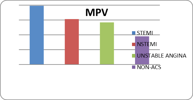
Graph 1: Comparison of mean MPV values among the study groups.
Discussion:
The mean MPV of the ACS group was significantly higher than the Non-ACS group.
Lippi et al in their study conducted in 2304 patients reported that patients with ACS had significantly higher MPV values than patients without ACS. Ranjith et al in their study conducted in India found similar results showing mean MPV values in ACS and Non-ACS groups as 10.97±0.58 fl and 9.12±0.63 fl respectively.
We also found a significant difference in MPV amongst the ACS spectrum i.e. STEMI and NSTEMI, STEMI and Unstable Angina. Although there was no significant difference in MPV between NSTEMI and Unstable Angina groups. Rifat et al found that the difference in MPV was statistically significant in STEMI and NSTEMI patients, which was 8.7±1 and 7.9±0.7 fl (p<0.01) respectively, which was in concordance with this study. The results of a study conducted by Sermin et al were also not in discordance with the results obtained in this study.
When putting the STEMI and NSTEMI patients into one group as Acute Myocardial Infarction (AMI) and then comparing with the Unstable Angina, it was seen that the difference in MPV in the two groups was statistically different. This finding is supported by a study conducted by Chu et al , who found that MPV values were higher in patients with acute myocardial infarction (AMI) as compared with patients with unstable angina (UA) (11.06±0.79 fl vs 10.66±0.87 fl, p=0.027). Although Dehghani et al and Biradar et al did not find any significant difference in MPVs between AMI and UA.
Conclusion:
Mean Platelet Volume in patients with ACS was significantly increased when compared to Non-ACS patients. Mean Platelet Volume can be utilised as simple and cost effective predictive parameters with other laboratory tests (particularly in remote centres) to predict impending cardiovascular events especially acute coronary events.
LIMITATIONS:
As the study was cross sectional, no follow up details are available and the prognostic utility of the values cannot be elucidated.
References:
1. Shah SN, Anand MP, Billimoria AR, Kamath SA, Karnad DR et al. API textbook of Medicine. Eighth edition. New Delhi. 2008:507-526.
2. Fauci AS, Kasper DL, Longo DL, Braunwald E, Hauser S et al. Harrison’s Principles of Internal Medicine. Seventeenth edition. New Delhi. Mc Graw Hill Medical. 2008(2):1514-1530.
3. Boos CJ, Lip GY. Platelet activation and cardiovascular Outcomes in acute coronary syndromes. J Thromb Haemost 2006; 4: 2542–3.
4. Tsiara S, Elisaf M, Jagroop IA, Mikhailidis DP. Platelets as predictors of vascular risk: is there a practical index of platelet Activity. ClinAppl Thromb Hemost 2003; 9:177–90.
5. Ross R. Atherosclerosis: an inflammatory disease. N Engl J Med1999; 340:115-26
6. Dalby K S, Milner P C, Martin J F: Bleeding time and platelet volume in acute myocardial infarction. A 2 year follows up study. ThrombosHaemost1988; 59: 353-6.
7. Lippi G, Filippozzi L, Salvagno GL, Montagnana M, Franchini M, Guidi GC, et al. Increased mean platelet volume in patients with acute coronary syndromes. Arch Pathol Lab Med 2009;133:1441-3.
8. Ranjith MP, Divya R, Mehta VK, Krishnan MG, KamalRaj R, Kavishwar A. Significance of platelet volume indices and platelet count in ischaemic heart disease. J Clin Pathol 2009;62:830-3.
9. Ulusoy RE, Yokuşoğlu M, Kırılmaz A, Nevruz O, Baysan O, Kılıçaslan F, et al. Mean platelet volume in ST elevation and non-ST elevation myocardial infarction. Gulhane Med J. 2011;53(2):114-8.
10. Yekeler S, Akay K, Borlu F. Comparison of MPV and PLT Values in Patients with and without Diagnosis of Acute Coronary Syndrome. J Am Coll Cardiol. 2013;62(18_S2):C116.
11. Chu H, Chen WL, Huang CC, Chang HY, Kuo HY, Gau CM, Chang YC, Shen YS. Diagnostic performance of mean platelet volume for patients with acute coronary syndrome visiting an emergency department with acute chest pain: the Chinese scenario. Emerg Med J. 2011 Jul;28(7):569-74.
12. Dehghani MR, Taghipour-Sani L, Rezaei Y, Rostami R. Diagnostic importance of admission platelet volume indices in patients with acute chest pain suggesting acute coronary syndrome. Indian Heart J. 2014 Nov-Dec;66(6):622-8.
13. Biradar, S., Kashinakunti, S., & R., M. (2017). Platelet volume indices in acute coronary syndrome - a case control study. International Journal of Advances in Medicine, 3(2), 349-352.