Concomitant Ipsilateral Femoral Shaft and Ankle Fractures: A Case Series and Proposal of a Novel Classification System.
- Juvin Joseph , Assistant Professor, Department of Orthopedics, Christian Medical College, Ludhiana, Punjab, India.
- Abhishek Samuel , Associate Professor, Department of Orthopedics, Christian Medical College, Ludhiana, Punjab, India
Article Information:
Abstract:
Background: The concomitant occurrence of a ipsilateral diaphyseal femoral shaft fracture and an ankle fracture is uncommon, and the incidence of this combined injury pattern has not been clearly defined in literature. The aims of this case series were to describe the clinical presentation, radiological characteristics, surgical management, and outcomes of patients with concomitant diaphyseal femoral shaft and ankle fractures; to highlight practical considerations regarding fixation sequence and implant selection; and to provide a focused review of relevant literature. Methods: A narrative review was conducted using selected peer-reviewed articles focusing on femur fracture and related ipsilateral lower limb injuries, including epidemiological studies, case reports. Results: Skeletal injury patterns consisted of diaphyseal femur fractures (AO/OTA 32) paired with malleolar ankle fractures (AO/OTA 44). Surgical stabilization was standardized across the cohort, utilizing intramedullary interlocking (IMIL) nailing for all femur fractures and open reduction and internal fixation (ORIF) plating for all ankle fractures. Surgical staging varied; simultaneous single-sitting stabilization was achieved in 33.3% (n=1) of cases, while the remaining 66.7% (n=2) underwent staged ankle fixation at a median of 4 days post-injury (range: 2–6 days). The mean total time from injury to definitive surgical fixation was 27.3 ± 23.1 hours. The mean timeline to achieve full weight-bearing (FWB) status was 7.3 ± 2.3 weeks. Conclusion: This case series highlights the rare and challenging injury pattern of concomitant diaphyseal femoral shaft and ipsilateral ankle fractures. We propose a novel classification system that incorporates injury definition, soft-tissue modifiers, surgical sequencing, implant selection, and postoperative rehabilitation protocols for these complex segmental lower-limb injuries. This classification provides a practical framework for clinical decision-making and may facilitate standardized management and future research in this uncommon injury pattern.
Keywords:
Article :
INTRODUCTION:
Femoral diaphyseal fractures are commonly caused by high-energy trauma in younger patients and low-energy falls in older adults. They are frequently associated with multisystem trauma and ipsilateral musculoskeletal injuries.[1,2] Commonly reported associated injuries include ipsilateral femoral neck fractures, tibial shaft fractures, and knee ligamentous or bony injuries.[3-8]
The concomitant occurrence of a diaphyseal femoral shaft fracture and an ankle fracture is uncommon, and the incidence of this combined injury pattern has not been clearly defined in literature. Most available evidence concerning multilevel ipsilateral lower-limb fractures consists of case reports or small clinical series describing fractures involving the femoral neck, shaft, distal femur, patella, or ankle region.[9-13]
The Winquist and Hansen classification remains a commonly used system for describing the degree of comminution in femoral shaft fractures, whereas the AO/OTA classification provides a standardized framework for reporting femoral shaft and malleolar fracture patterns.[14,15] Because of the rarity of this pattern, limited guidance exists regarding optimal surgical timing, fixation sequence, postoperative rehabilitation, and expected functional outcome.
The aims of this case series were to describe the clinical presentation, radiological characteristics, surgical management, and outcome of patients with concomitant diaphyseal femoral shaft and ankle fractures; to highlight practical considerations regarding fixation sequence and implant selection; and to provide a focused review of relevant literature.
MATERIALS AND METHODS:
A single tertiary center retrospective observational review was conducted of three patients presenting with concomitant diaphyseal femoral shaft fracture and ankle fracture who were treated surgically at Christian Medical College and Hospital, Ludhiana, Punjab, India, between March 2024 and December 2025.
Inclusion Criteria
· Patients with a diaphyseal femoral shaft fracture associated with an ipsilateral ankle fracture
· Both open and closed injuries
· Managed operatively.
Exclusion Criteria
· A non-salvageable limb, non-operative management
· Femoral shaft fractures associated primarily with foot fractures rather than ankle fractures.
Diagnosis was based on plain radiographs of the pelvis with both hips, full-length anteroposterior and lateral radiographs of the femur, and anteroposterior, lateral, and mortise views of the ankle. Computed tomography was obtained when additional characterization of the ankle fracture or associated injury was required.
Clinical records and radiographs were reviewed for demographic details, mechanism of injury, fracture classification, open injury status, fixation method, surgical sequence, postoperative complications, radiological union, and functional recovery.
Data Collected
· Age and gender
· Mechanism of injury
· Fracture patten
· Time of presentation after injury
· Time to surgery after injury
· Sequence of fracture fixation
· Time from Injury to Presentation
· Type of fixation
· Complications
Written informed consent was obtained from all patients.
Case 1
A 28-year-old man with no significant medical history presented to the emergency department after a high-energy motor vehicle accident. He had pain and swelling in the right thigh and ankle, inability to bear weight on the right lower limb, and a post-traumatic soft-tissue defect over the posteromedial aspect of the middle third of the right leg. Examination revealed marked swelling and localized tenderness over the right thigh and ankle.
Radiographs demonstrated a diaphyseal femoral shaft fracture classified as AO/OTA 32A1.b and a bimalleolar ankle fracture classified as AO/OTA 44A2.3.[15] Computed tomography of the ankle was performed for preoperative planning. Nail length and diameter were measured preoperatively to guide implant selection.
The patient underwent staged fixation. The femoral fracture was treated first with closed reduction and intramedullary interlocking nailing. The ankle fracture was subsequently treated with open reduction and internal fixation using a one-third tubular plate and cannulated cancellous screws.
The soft-tissue defect was managed with serial vacuum-assisted closure dressings followed by split-thickness skin grafting. The postoperative course was uneventful. Immediate postoperative radiographs showed satisfactory length, alignment, and rotation.
Walker-assisted mobilization with limited weight-bearing was initiated. Radiographs at 6 weeks and 3 months showed progressive fracture healing. At 1 year, complete union of both femur and ankle fractures was confirmed radiologically, and the patient had returned to pre-injury activity without persistent pain or functional limitation.
Case 1 Images
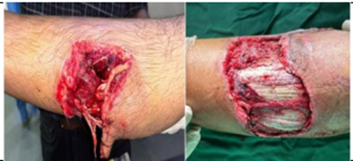
Figure 1: Pre op clinical images.
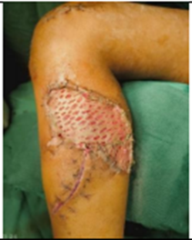
Figure 2: Post op clinical images
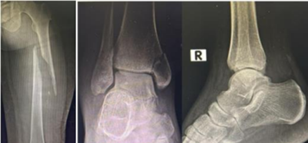
Figure 3: Preoperative radiograph depicting diaphyseal femur and ankle fracture of case 1
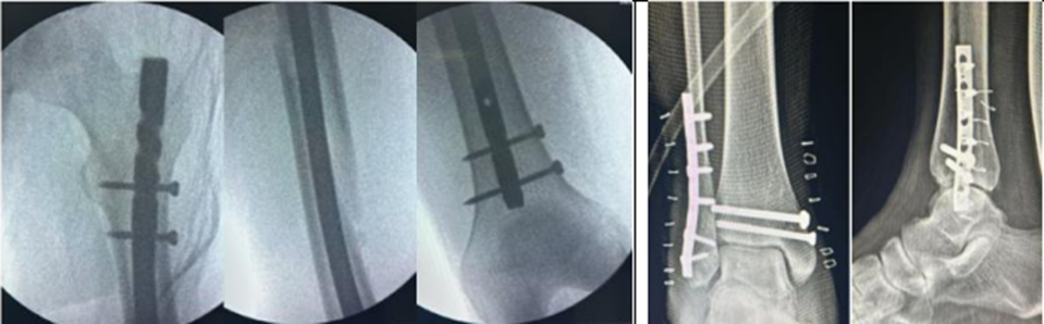
Figure 4: Post operative radiograph depicting diaphyseal femur and ankle fracture
Case 2
A 45-year-old man with no significant medical history presented after a high-energy motor vehicle accident with pain and swelling of the right thigh and ankle and inability to bear weight on the right lower limb. Clinical examination revealed swelling and localized tenderness over the right thigh and ankle. Radiographs showed a diaphyseal femoral shaft fracture classified as AO/OTA 32A2 and a bimalleolar ankle fracture classified as AO/OTA 44B2.[15] Computed tomography of the ankle was performed to define the fracture configuration and aid preoperative planning. Nail length and diameter were measured before surgery. The femoral fracture was treated with closed reduction and intramedullary interlocking nailing. The ankle fracture was treated with open reduction and internal fixation using a reconstruction plate and cannulated cancellous screws. Immediate postoperative radiographs demonstrated satisfactory length, alignment, and rotation. Walker-assisted mobilization with limited weight-bearing was advised. Radiographs at 6 weeks and 3 months demonstrated progressive fracture healing. At 1 year, both fractures had united radiologically, and the patient had resumed pre-injury activity without persistent pain or functional restriction.
Case 2 Images
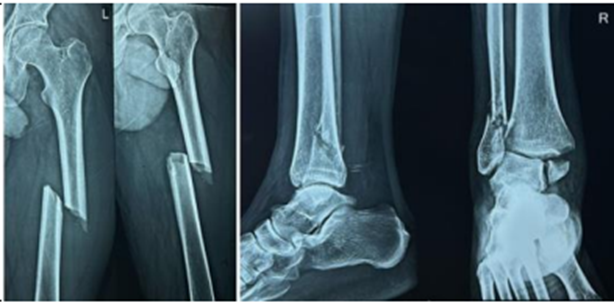
Figure 5: Preoperative radiograph depicting diaphyseal femur and ankle fracture of case 2
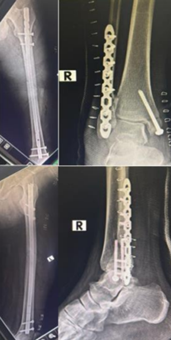
Figure 6: Post operative radiograph depicting diaphyseal femur and ankle fracture of case 2
Case 3
A 62-year-old man with hypertension presented after a high-energy motor vehicle accident with pain and swelling of the right thigh and ankle, inability to bear weight, and an open deep lacerated wound over the lateral aspect of the heel. He also had an associated head injury. After initial resuscitation and stabilization, a secondary survey was conducted. Examination revealed heel-pad avulsion with wound contamination, minimal soft-tissue loss, and localized tenderness over the right thigh and ankle.
Radiographs demonstrated a segmental diaphyseal femoral shaft fracture classified as AO/OTA 32C2, a bimalleolar ankle fracture classified as AO/OTA 44B1, and an extra-articular calcaneal fracture classified as AO/OTA 82A2.[15] The open calcaneal injury was classified as Gustilo-Anderson grade IIIB.[16] Computed tomography of the head revealed bitemporal subdural hematoma with left frontal extradural hematoma. Computed tomography was also used to further assess the ankle fracture for operative planning.
The patient underwent staged management. Initial wound debridement and heel-pad fixation with Kirschner wires were performed on the day of admission. After stabilization, the femoral fracture was treated with closed reduction and retrograde intramedullary nailing, and the ankle fracture was treated with open reduction and internal fixation using an anatomical plate. Serial wound debridement and vacuum-assisted closure were performed for the heel wound. Implant removal was required 2 months after surgery because of surgical-site infection after radiological evidence of union at the ankle and calcaneum. Early knee and ankle mobilization with restricted weight-bearing was followed. The ankle and calcaneal fractures united radiologically; however, the segmental femoral shaft fracture progressed to hypertrophic non-union.
Case 3 Images
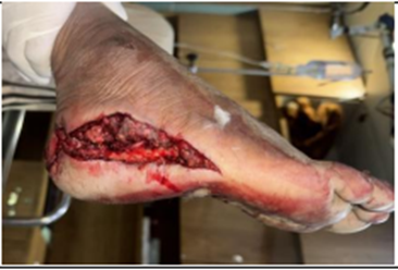
Figure 7: Pre op clinical images of case 3
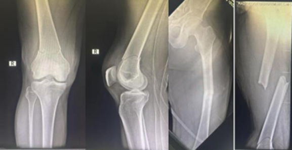
Figure 8: Preoperative radiograph depicting diaphyseal femur and ankle fracture of case 3
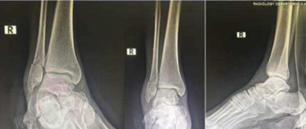
Figure 9: Preoperative radiograph depicting diaphyseal femur and ankle fracture of case 3
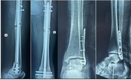
Figure 10: Post operative radiograph depicting diaphyseal femur and ankle fracture of case 3
RESULTS:
Surgical and rehabilitation outcomes in this case series (n = 3), all patients were male with a mean age of 45 ± 17 years. The primary mechanism of injury was road traffic accidents (RTA, 100%). Skeletal injury patterns consisted of diaphyseal femur fractures (AO/OTA 32) paired with malleolar ankle fractures (AO/OTA 44). Surgical stabilization was standardized across the cohort, utilizing intramedullary interlocking (IMIL) nailing for all femur fractures and open reduction and internal fixation (ORIF) plating for all ankle fractures. Surgical staging varied; simultaneous single-sitting stabilization was achieved in 33.3% (n=1) of cases, while the remaining 66.7% (n=2) underwent staged ankle fixation at a median of 4 days post-injury (range: 2–6 days). The mean total time from injury to definitive surgical fixation was 27.3 ± 23.1 hours. The mean timeline to achieve full weight-bearing (FWB) status was 7.3 ± 2.3 weeks. Delayed functional recovery (10 weeks to FWB) and adverse events—specifically femur non-union and an ankle surgical site infection-were isolated to a single patient (n=1, 33.3%) who presented with a complex, multi-fragmentary 32C2 femur fracture pattern.
Using the proposed classification system, two cases were categorized as Type I and one case as Type III.
All patients underwent surgical management, either as a single-stage procedure or through a staged approach based on injury complexity. Simple femoral fractures were treated with antegrade intramedullary nailing, while complex femoral fractures mainly distal segmental fractures were managed using retrograde femoral nailing.
At a mean follow-up of three months:
· Two patients had no complications
· One patients developed surgical site infection at ankle and non union at femur.
Statistical dashboard: Concomittant FEMUR & ankle fractures (n=3 Cases)
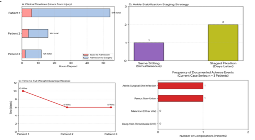
Graph 1: Analysis of patient case data.
DISCUSSION:
Injury Configuration and Clinical Findings
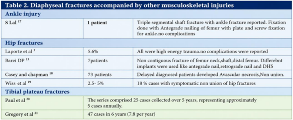
This case series highlights a rare injury pattern involving simultaneous diaphyseal femoral shaft and ankle fractures. Previous literature has documented the association between femoral shaft fractures and ipsilateral ankle fractures; however, this injury pattern is rare, and consequently, the available literature remains limited.[17] The combination may be overlooked in the initial assessment because attention is often directed toward the more obvious femoral shaft fracture and associated systemic injuries. Careful examination of the entire limb and dedicated ankle radiographs are therefore essential in high-energy trauma. In all cases, the mechanism of injury was road traffic accidents, which generate substantial axial, rotational, and bending forces capable of producing fractures at multiple levels of the lower limb. The coexistence of femoral and ankle fractures likely reflects the transmission of energy through the entire extremity during impact, resulting in combined skeletal injuries.
Clinical Implications and Management
In our series, fixation strategy was individualized according to fracture morphology, soft-tissue condition, and patient stability. Femoral stabilization was generally prioritized to restore limb length and facilitate mobilization, while ankle fixation was performed during the same sitting or as part of a staged protocol when soft-tissue management was required. Open injuries and contaminated wounds required staged debridement, temporary or definitive fixation as appropriate, and soft-tissue reconstruction.
The principal complication occurred in the patient with associated heel-pad avulsion and open calcaneal injury, emphasizing the importance of soft-tissue status in determining outcome. Although ankle and calcaneal union was achieved, the segmental femoral fracture progressed to hypertrophic non-union. This suggests that high-energy segmental femoral fractures with associated open injuries may require close surveillance for delayed union or non-union.
STRENGTHS AND LIMITATIONS
Strengths
· First institutional series focusing on ipsilateral femur and ankle fractures
· Highlights orthopaedic surgical decision-making
· Provides clinical learning points and proposed classification
Limitations
· Small sample size
· Retrospective design.
· Single-centre study.
· Limited long-term functional outcome data.
CONCLUSION:
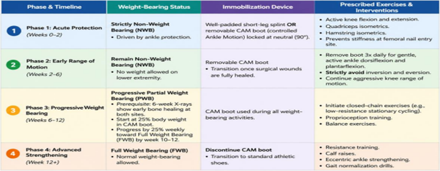
Concomitant diaphyseal femoral shaft and ankle fractures are rare injuries usually resulting from high-energy trauma. A systematic trauma evaluation, dedicated imaging of the entire injured limb, early recognition of associated ankle and soft-tissue injuries, and individualized surgical sequencing are essential for optimal outcomes. Intramedullary nailing remains suitable for most femoral shaft fractures, while implant selection should be modified in patients with pre-existing deformity, previous surgery, or complex fracture morphology. Larger multicentre studies are required to define the incidence, optimal fixation sequence, rehabilitation protocols, and long-term outcomes of this uncommon injury pattern. We have proposed a classification system for such injuries (Table 3.1 & 3.2) along with definitive surgical sequencing (table 4), implant choice (Table5) and post op rehabilitation protocol (table 6).
Table 1: Juvin & Samuel Proposed Classification for Concomittant Diaphyseal femur and Ankle Fracture
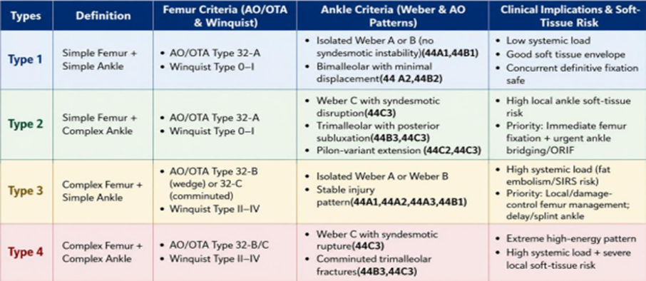
Table 2: Soft-Tissue Modifiers ("S" Modifier)
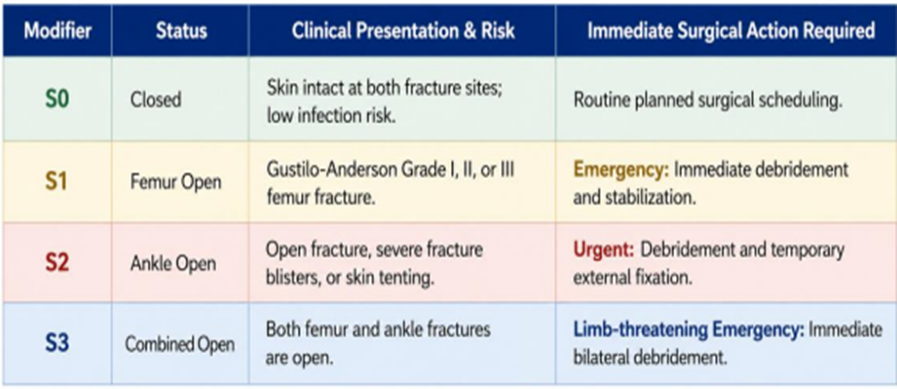
Table 3: Definitive Surgical Sequencing
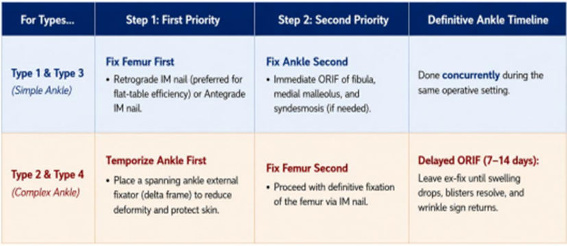
Table 4: Specific Implant Choices for Complex Ankle Patterns
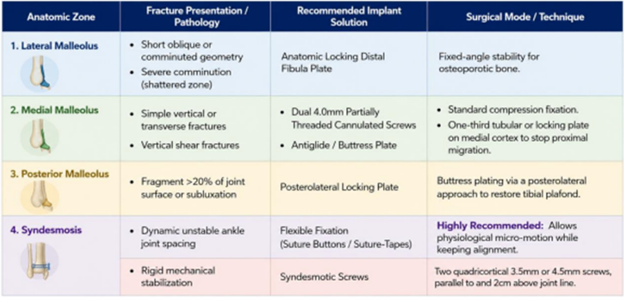
Table 5: Postoperative Rehabilitation Protocol.
REFERENCES:
1. Salminen ST, Pihlajamaki HK, Avikainen VJ, et al. Population-based epidemiologic and morphologic study of femoral shaft fractures. Clin Orthop Relat Res 2000;(372):241-9.
2. Regel G, Lobenhoffer P, Grotz M, et al. Treatment results of patients with multiple trauma: an analysis of 3406 cases treated between 1972 and 1991 at a German level I trauma center. J Trauma 1995;38:70-8.
3. Laporte C, Benazet JP, Scemama P, et al. Ipsilateral hip and femoral shaft fractures: components of therapeutic choice. Rev Chir Orthop Reparatrice Appar Mot 1999;85:24-32.
4. Watson JT, Moed BR. Ipsilateral femoral neck and shaft fractures: complications and their treatment. Clin Orthop Relat Res 2002;(399):78-86.
5. Barquet A, Fernandez A, Leon H. Simultaneous ipsilateral trochanteric and femoral shaft fracture. Acta Orthop Scand 1985;56:36-9.
6. Walling AK, Seradge H, Spiegel PG. Injuries to the knee ligaments with fractures of the femur. J Bone Joint Surg Am 1982;64-A:1324-7.
7. Ostrum RF, Tornetta P, Watson JT, et al. Ipsilateral proximal femur and shaft fractures treated with hip screws and a reamed retrograde intramedullary nail. Clin Orthop Relat Res 2014;472:2751-8.
8. Tsarouhas A, Hantes ME, Karachalios T, et al. Reconstruction nailing for ipsilateral femoral neck and shaft fractures. Strategies Trauma Limb Reconstr 2011;6:69-75.
9. Douša P, Bartoníček J, Luňáček L, et al. Ipsilateral fractures of the femoral neck, shaft and distal end: long-term outcome of five cases. Int Orthop 2011;35(7):1083-8.
10. Bartoníček J, Stehlik J, Douša P. Ipsilateral fractures of the hip, femoral shaft, distal femur and patella. Hip Int 2000;10(3):174-7.
11. Griffin M, Dick AG, Umarji S. Outcomes after trifocal femoral fractures. Case Rep Surg 2014;2014:528061.
12. Kim BS, Cho JW, Yeo DH, et al. Surgical treatment of ipsilateral multi-level femoral fracture treated using antegrade intramedullary nail. J Trauma Inj 2018;31(2):96-102.
13. Barei DP, Schildhauer TA, Nork SE. Noncontiguous fractures of the femoral neck, femoral shaft, and distal femur. J Trauma 2003;55(1):80-6.
14. Winquist RA, Hansen ST. Comminuted fractures of the femoral shaft treated by intramedullary nailing. Orthop Clin North Am 1980;11(3):633-48.
15. Meinberg EG, Agel J, Roberts CS, et al. Fracture and dislocation classification compendium-2018. J Orthop Trauma 2018;32(Suppl 1):S1-170.
16. Gustilo RB, Anderson JT. Prevention of infection in the treatment of one thousand and twenty-five open fractures of long bones: retrospective and prospective analyses. J Bone Joint Surg Am 1976;58(4):453-8.
17. Lal S, Jha AK, Agrawal P, et al. Triple-segmental shaft femur fracture: a case report. J Orthop Case Rep 2025;15(9):149-53.
18. Casey MJ, Chapman MW. Ipsilateral concomitant fractures of the hip and femoral shaft. J Bone Joint Surg Am 1979;61-A:503-9.
19. Wiss DA, Sima W, Brien WW. Ipsilateral fractures of the femoral neck and shaft. J Orthop Trauma 1992;6:159-66.
20. Paul GR, Sawka MW, Whitelaw GP. Fractures of the ipsilateral femur and tibia: Emphasis on intra-articular and soft tissue injury. J Orthop Trauma 1990;4:309-14.
21. Gregory P, DiCicco J, Karpin K, et al. Ipsilateral fractures of the femur and tibia: treatment with retrograde femoral nailing and undreamed tibial nailing. J Orthop Trauma 1996;10:309-16.