ROLE OF HIGH-RESOLUTION COMPUTED TOMOGRAPHY [HRCT] OF TEMPORAL BONE IN EVALUATION OF MIDDLE EAR DISEASES AND ITS CORRELATION WITH INTRA OPERATIVE FINDINGS.
- Dipti Parmar , R3, Radiodiagnosis, Parul Sevashram Hospital.
- Dharmesh Baria , Associate Professor, MD Radiodiagnosis, Parul Sevashram Hospital.
- Anil Rathva , Professor and Head, MD Radiodiagnosis, Parul Sevashram Hospital.
- Shreya Panchani , R3, Radiodiagnosis, Parul Sevashram Hospital.
Article Information:
Abstract:
Background: High-resolution computed tomography (HRCT) is widely used for preoperative assessment of middle ear disease, but its diagnostic accuracy varies across temporal bone structures. Objective: To evaluate the diagnostic performance of HRCT temporal bone findings by correlating them with intraoperative findings in patients undergoing surgery for middle ear disease. Methods: This cross-sectional observational study included 50 patients with clinically suspected middle ear disease who underwent preoperative HRCT followed by surgical intervention. HRCT findings were assessed for disease extent and involvement of the scutum, ossicular chain, individual ossicles, aditus, antrum, mastoid air cells, tegmen tympani, sinus tympani, facial canal, sigmoid sinus plate, posterior external auditory canal wall, and lateral semicircular canal. Intraoperative findings were considered the reference standard. Sensitivity, specificity, predictive values, observed agreement, and Cohen’s kappa were calculated. Results: Mastoid air-cell involvement was present in all patients, while aditus and antrum involvement were each detected in 94%. HRCT showed high sensitivity for mastoid involvement (100%), tegmen erosion (100%), incus erosion (94.9%), aditus and antrum disease (94.0% each), and ossicular-chain erosion (93.8%). Specificity was 100% for scutum erosion, sinus tympani involvement, posterior external auditory canal wall erosion, and lateral semicircular canal erosion. Agreement was strongest for posterior canal-wall, sigmoid sinus plate, and lateral semicircular canal erosion. Facial canal and tegmen abnormalities were frequently overestimated. Conclusion: HRCT is a valuable preoperative anatomical roadmap for middle ear disease, particularly for defining disease extent and detecting major bony erosions. Facial canal and tegmen findings should be interpreted cautiously because of lower specificity.
Keywords:
Article :
INTRODUCTION:
Chronic middle ear disease is a significant cause of chronic otorrhoea, hearing loss and may lead to potentially serious complications of the temporal bone. Inflammatory middle ear disease is a relatively broad spectrum of disease, but in particular, cholesteatoma can cause locally destructive effects and lead to erosion of the ossicular chain, scutum, facial nerve canal, tegmen tympani, sigmoid sinus plate and labyrinth. The disease may also spread through the aditus into the mastoid antrum and air-cell system and the extent of the disease may not be fully assessed by clinical examination alone [1,2].
Prior to surgical exploration proper evaluation of the extent of the disease is necessary. While otoscopy and audiological tests are useful in showing abnormalities of the TM and functional hearing loss, they are not effective in reliably demonstrating the presence of hidden recesses of the middle ear, the mastoid compartment, or subtle erosions of critical bony structures. Diseases can be present outside the direct line of clinical sight in the sinus tympani, facial recess, epitympanum, aditus ad antrum and mastoid air cells. Preoperative imaging has thus emerged as an essential part of surgical planning, especially if there is likely to be extensive disease or complications [2,3].
High-resolution computed tomography (HRCT) of the temporal bone is uniquely suited for this purpose, because it gives the thin-section visualization of the complicated bony structure of the middle and mastoid ear. The characteristic CT findings of cholesteatoma and other destructive processes of the middle ear are nondependent soft-tissue opacification, scutal blunting, ossicular erosion, aditus widening, mastoid involvement, and erosion of adjacent bony boundaries [3,4]. Multiplanar reformations further enhance anatomical localization and yield a view of surgically important structures such as the tegmen, facial nerve canal, sigmoid sinus plate, external auditory canal wall, and lateral semicircular canal [4,5].
The use of HRCT is not confined to the extent of proving the presence of middle ear soft tissue. Instead, it serves as an anatomical map to guide surgeons through the disease distribution, to identify areas of structural erosion, to predict the technically challenging zones and to detect potential complications that may guide the surgical approach [2,6]. Awareness of the ossicular-chain disruption may help with the reconstruction of the hearing system, and the presence of a labyrinthine fistula, erosion of the tegmen, involvement of the sigmoid plate, or abnormalities of the facial canal can signal an increased risk of complications during surgery. Therefore, the role of describing the extent and staging of disease by CT has been proposed to enhance the communication between radiologists and otologists as well as to guide surgical decisions [6,7].
However, there are some limitations that HRCT has identified. The soft-tissue attenuation of inflammatory mucosa, granulation tissue, fluid and cholesteatoma can be similar, which may make it difficult to confidently distinguish the tissues from each other in conventional CT. Partial-volume averaging can occur in very small erosions, and thin normal bony coverings can be mistaken for dehiscent (especially in the tympanic facial nerve canal). Therefore, the diagnostic efficacy of HRCT may be quite variable in different anatomical areas [5,8]. There have been a number of previous radiological–surgical correlation studies, with good results for disease extent, scutal erosion, ossicular, sinus and labyrinthine erosion, but less consistent results for facial canal dehiscence, tegmen and individual ossicular defect [6,9].
The clinical significance of these differences is that imaging may be very sensitive for the detection of disease and yet give a false positive result at certain structures. On the other hand, erosions of small size may not be diagnosed, but are nonetheless clinically significant. It is therefore crucial to compare directly with intraoperative observation to assess the real predictive value of HRCT and to select the anatomical structures that can be best diagnosed from HRCT. Correlation is particularly helpful in institutions where chronic ear disease is advanced and/or has extensive involvement, and in institutions where CT findings often affect the nature of surgery.
The current study aimed to assess the value of preoperative HRCT temporal bone in the surgical management of patients with clinical suspicion of middle ear diseases. The HRCT results were compared to the intraoperative observations for the presence and extent of disease as well as for the ossicular chain and individual ossicles, for the aditus, mastoid antrum and air cells, for the tegmen tympani and sinus tympani, for the facial nerve canal, the sigmoid sinus plate, the posterior external auditory canal wall, and the involvement of the lateral semicircular canal. The other objective of the study was to calculate the sensitivity, specificity, predictive value, observed agreement and intermethod agreement of HRCT in each surgically relevant finding. This study aimed to shed light on the use of HRCT as a preoperative road map map and to inform the surgical planning process in middle ear disease, by establishing its strengths and limitations in particular temporal bone structures.
MATERIALS AND METHODS:
Study design and setting
This cross-sectional observational study was conducted in the Department of Radio-diagnosis, Parul Sevashram Hospital, Vadodara, Gujarat, India. The study was carried out over a period of one year, from December 2024 to December 2025, after obtaining approval from the Institutional Ethics Committee. The study evaluated the role of high-resolution computed tomography (HRCT) of the temporal bone in patients with clinically suspected middle ear disease and correlated preoperative HRCT findings with intraoperative findings.
Study population
Patients clinically suspected to have middle ear disease and referred for HRCT temporal bone evaluation were screened for eligibility. Patients were included if they subsequently underwent surgical intervention for middle ear pathology and provided written informed consent.
Patients were excluded if they had a previous operative history involving the temporal bone, history of temporal bone trauma, did not undergo surgical intervention, or were unwilling to participate in the study. A convenience sampling method was used. Based on institutional case flow, the expected sample size was approximately 45–50 patients over one year. A total of 50 eligible patients were included in the final analysis.
Clinical assessment
Detailed clinical history and examination findings were recorded for all participants before imaging. Demographic data included age and sex. Clinical variables included presenting symptoms such as ear discharge, hearing loss, otalgia, tinnitus, headache, vertigo, fever, and facial weakness. Laterality of the affected ear was also documented.
HRCT temporal bone acquisition protocol
HRCT temporal bone imaging was performed using an Alexion 16-slice CT scanner (Toshiba). Helical acquisition was used with a pitch of 0.8–1.2 and rotation time of 0.75 seconds. The scan field of view was adjusted to the head region. Imaging was performed using 120 kV and 150 mAs.
Patients were positioned supine with appropriate head positioning to minimize radiation exposure to the ocular lens. The scan was planned from the arcuate eminence superiorly to the mastoid tip inferiorly on the lateral scout topogram. Axial source images were obtained with 0° gantry tilt, with the scan plane parallel to the inferior orbitomeatal line.
Image reconstruction was performed using a high-resolution bone algorithm. Reconstructed images were obtained with 0.5-mm slice thickness and 0.75-mm reconstruction interval. Multiplanar reformations were generated in axial, coronal, and sagittal planes.
HRCT image analysis
All HRCT images were reviewed on an OSIRIX MD workstation by an experienced radiologist. Imaging findings were recorded in a structured format before correlation with operative findings.
The following HRCT parameters were assessed:
· presence and extent of middle ear soft tissue lesion;
· scutum erosion;
· ossicular-chain erosion;
· malleus erosion;
· incus erosion;
· stapes erosion;
· aditus involvement;
· mastoid antrum involvement;
· mastoid air-cell involvement;
· tegmen tympani erosion;
· sinus tympani involvement;
· bony facial canal dehiscence or erosion;
· sigmoid sinus plate erosion;
· posterior external auditory canal wall erosion;
· lateral semicircular canal erosion;
· disease extension and associated complications.
Intraoperative correlation
Intraoperative findings documented during surgical exploration were used as the reference standard. HRCT findings were compared with corresponding surgical findings for each anatomical site and pathological feature. Each finding was classified as true positive, false positive, false negative, or true negative according to the agreement between HRCT and intraoperative assessment.
Outcome measures
The primary outcome was radiological-surgical correlation between HRCT temporal bone findings and intraoperative findings in middle ear disease.
Secondary outcomes included the diagnostic performance of HRCT for individual temporal bone findings, including sensitivity, specificity, positive predictive value, negative predictive value, observed agreement, and Cohen’s kappa agreement.
Statistical analysis
Data were entered into Microsoft Excel and analyzed using SPSS version 25.0. Categorical variables were summarized as frequency and percentage in the format n (%).
For each HRCT finding, intraoperative findings were considered the reference standard. Diagnostic indices were calculated using the following formulae:
Sensitivity = TP / (TP + FN)
Specificity = TN / (TN + FP)
Positive predictive value = TP / (TP + FP)
Negative predictive value = TN / (TN + FN)
Observed agreement = (TP + TN) / total cases
Cohen’s kappa statistic was used to assess agreement between HRCT and intraoperative findings. McNemar’s chi-square test was used to assess paired disagreement between HRCT and intraoperative findings wherever applicable. A p-value <0.05 was considered statistically significant.
Specificity and negative predictive value were not estimated for structures where no intraoperative-negative cases were available.
RESULTS:
A total of 50 patients with middle ear disease underwent preoperative HRCT temporal bone assessment followed by intraoperative evaluation. The study cohort included 31 (62.0%) males and 19 (38.0%) females. The largest age group was 10–20 years, comprising 13 (26.0%) patients, followed by 21–30, 31–40, and 51–60 years, each with 9 (18.0%) patients. Right-sided involvement was observed in 29 (58.0%) patients and left-sided involvement in 21 (42.0%) patients.
Ear discharge was the most common presenting symptom, reported by 42 (84.0%) patients, followed by hearing loss in 38 (76.0%) and otalgia in 24 (48.0%). Tinnitus, headache, vertigo, fever, and facial weakness were less frequent clinical features. Baseline clinical details are summarized in Table 1.
Table 1. Baseline demographic and clinical characteristics of the study population (N = 50).
|
Characteristic |
n |
% |
|
Age group |
|
|
|
10–20 years |
13 |
26.0 |
|
21–30 years |
9 |
18.0 |
|
31–40 years |
9 |
18.0 |
|
41–50 years |
5 |
10.0 |
|
51–60 years |
9 |
18.0 |
|
>60 years |
5 |
10.0 |
|
Sex |
|
|
|
Male |
31 |
62.0 |
|
Female |
19 |
38.0 |
|
Presenting symptom |
|
|
|
Ear discharge (otorrhoea) |
42 |
84.0 |
|
Hearing loss |
38 |
76.0 |
|
Ear pain (otalgia) |
24 |
48.0 |
|
Tinnitus |
15 |
30.0 |
|
Headache |
10 |
20.0 |
|
Vertigo |
8 |
16.0 |
|
Fever |
6 |
12.0 |
|
Facial weakness |
2 |
4.0 |
|
Affected ear |
|
|
|
Right |
29 |
58.0 |
|
Left |
21 |
42.0 |
Symptoms were not mutually exclusive; therefore, symptom percentages do not sum to 100%.
On HRCT temporal bone, mastoid air-cell involvement was the most frequent finding, present in 50 (100.0%) patients.
Aditus and antrum involvement were each identified in 47 (94.0%) patients, followed by scutum erosion in 44 (88.0%) and ossicular-chain erosion in 42 (84.0%).
Among individual ossicles, incus erosion was the most frequently detected abnormality on HRCT, followed by stapes and malleus involvement. The spectrum of HRCT-positive findings is shown in Figure 1, and the corresponding intraoperative findings are presented in Table 2.
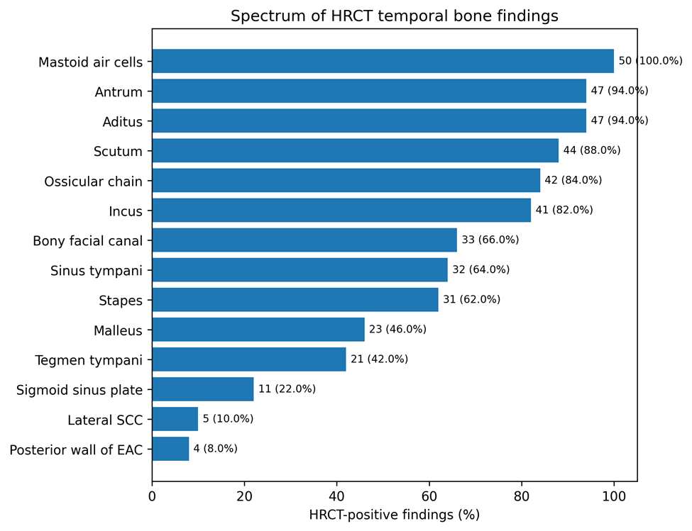
Figure 1. Spectrum of HRCT temporal bone findings in middle ear disease. Values represent HRCT-positive findings among 50 patients.
Table 2. HRCT temporal bone findings and intraoperative correlation.
|
Structure |
HRCT positive n (%) |
Intraoperative positive n (%) |
TP/FP/FN/TN |
Observed agreement n (%) |
|
Scutum |
44 (88.0%) |
49 (98.0%) |
44/0/5/1 |
45 (90.0%) |
|
Ossicular chain |
42 (84.0%) |
32 (64.0%) |
30/12/2/6 |
36 (72.0%) |
|
Malleus |
23 (46.0%) |
19 (38.0%) |
17/6/2/25 |
42 (84.0%) |
|
Incus |
41 (82.0%) |
39 (78.0%) |
37/4/2/7 |
44 (88.0%) |
|
Stapes |
31 (62.0%) |
23 (46.0%) |
20/11/3/16 |
36 (72.0%) |
|
Aditus |
47 (94.0%) |
50 (100.0%) |
47/0/3/0 |
47 (94.0%) |
|
Antrum |
47 (94.0%) |
50 (100.0%) |
47/0/3/0 |
47 (94.0%) |
|
Mastoid air cells |
50 (100.0%) |
50 (100.0%) |
50/0/0/0 |
50 (100.0%) |
|
Tegmen tympani |
21 (42.0%) |
7 (14.0%) |
7/14/0/29 |
36 (72.0%) |
|
Sinus tympani |
32 (64.0%) |
37 (74.0%) |
32/0/5/13 |
45 (90.0%) |
|
Bony facial canal |
33 (66.0%) |
13 (26.0%) |
12/21/1/16 |
28 (56.0%) |
|
Sigmoid sinus plate |
11 (22.0%) |
12 (24.0%) |
10/1/2/37 |
47 (94.0%) |
|
Posterior wall of EAC |
4 (8.0%) |
5 (10.0%) |
4/0/1/45 |
49 (98.0%) |
|
Lateral SCC |
5 (10.0%) |
7 (14.0%) |
5/0/2/43 |
48 (96.0%) |
TP, true positive; FP, false positive; FN, false negative; TN, true negative. Intraoperative findings were used as the reference standard.
HRCT showed high sensitivity for most surgically assessed temporal bone findings. Sensitivity was 50/50 (100.0%) for mastoid air-cell involvement and 7/7 (100.0%) for tegmen tympani erosion, while sensitivity for scutum erosion, ossicular-chain erosion, malleus erosion, incus erosion, stapes erosion, aditus involvement, antrum involvement, sinus tympani disease, and bony facial canal dehiscence ranged from 86.5% to 94.9%. Lower sensitivity was observed for posterior wall of external auditory canal erosion and lateral semicircular canal erosion, although both remained highly specific. Detailed diagnostic indices are provided in Table 3.
Specificity was 100.0% for scutum erosion, sinus tympani disease, posterior external auditory canal wall erosion, and lateral semicircular canal erosion. The strongest agreement with intraoperative findings was observed for posterior wall of external auditory canal erosion, sigmoid sinus plate erosion, and lateral semicircular canal erosion, whereas lower agreement was observed for bony facial canal and tegmen tympani assessment.
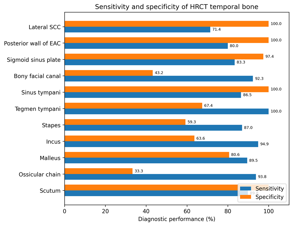
Figure 2. Sensitivity and specificity of HRCT temporal bone for surgically verified findings. Structures without intraoperative-negative cases were not included in the specificity display.
Table 3. Diagnostic performance and agreement of HRCT temporal bone using intraoperative findings as reference standard.
|
Structure |
Sensitivity n/N (%) |
Specificity n/N (%) |
PPV n/N (%) |
NPV n/N (%) |
Cohen’s κ |
McNemar χ² |
p value |
|
Scutum |
44/49 (89.8%) |
1/1 (100.0%) |
44/44 (100.0%) |
1/6 (16.7%) |
0.26 |
3.20 |
0.074 |
|
Ossicular chain |
30/32 (93.8%) |
6/18 (33.3%) |
30/42 (71.4%) |
6/8 (75.0%) |
0.31 |
5.79 |
0.016 |
|
Malleus |
17/19 (89.5%) |
25/31 (80.6%) |
17/23 (73.9%) |
25/27 (92.6%) |
0.67 |
1.12 |
0.289 |
|
Incus |
37/39 (94.9%) |
7/11 (63.6%) |
37/41 (90.2%) |
7/9 (77.8%) |
0.63 |
0.17 |
0.683 |
|
Stapes |
20/23 (87.0%) |
16/27 (59.3%) |
20/31 (64.5%) |
16/19 (84.2%) |
0.45 |
3.50 |
0.061 |
|
Aditus |
47/50 (94.0%) |
NE |
47/47 (100.0%) |
NE |
0.00 |
1.33 |
0.248 |
|
Antrum |
47/50 (94.0%) |
NE |
47/47 (100.0%) |
NE |
0.00 |
1.33 |
0.248 |
|
Mastoid air cells |
50/50 (100.0%) |
NE |
50/50 (100.0%) |
NE |
NE |
0.00 |
1.000 |
|
Tegmen tympani |
7/7 (100.0%) |
29/43 (67.4%) |
7/21 (33.3%) |
29/29 (100.0%) |
0.37 |
12.07 |
<0.001 |
|
Sinus tympani |
32/37 (86.5%) |
13/13 (100.0%) |
32/32 (100.0%) |
13/18 (72.2%) |
0.77 |
3.20 |
0.074 |
|
Bony facial canal |
12/13 (92.3%) |
16/37 (43.2%) |
12/33 (36.4%) |
16/17 (94.1%) |
0.24 |
16.41 |
<0.001 |
|
Sigmoid sinus plate |
10/12 (83.3%) |
37/38 (97.4%) |
10/11 (90.9%) |
37/39 (94.9%) |
0.83 |
0.00 |
1.000 |
|
Posterior wall of EAC |
4/5 (80.0%) |
45/45 (100.0%) |
4/4 (100.0%) |
45/46 (97.8%) |
0.88 |
0.00 |
1.000 |
|
Lateral SCC |
5/7 (71.4%) |
43/43 (100.0%) |
5/5 (100.0%) |
43/45 (95.6%) |
0.81 |
0.50 |
0.480 |
PPV, positive predictive value; NPV, negative predictive value; EAC, external auditory canal; SCC, semicircular canal; NE, not estimable. Specificity and NPV were not calculated where no intraoperative-negative cases were available.
Overall, HRCT temporal bone demonstrated strong preoperative utility in defining the extent of middle ear disease and identifying surgically relevant bony erosions. It was particularly useful for detecting scutum erosion, ossicular involvement, aditus/antrum disease, mastoid air-cell involvement, sinus tympani disease, sigmoid sinus plate erosion, posterior external auditory canal wall erosion, and lateral semicircular canal erosion.
DISCUSSION:
The present study demonstrates that high-resolution computed tomography (HRCT) is valuable for preoperative assessment of surgically treated middle ear disease, although its diagnostic performance varies among temporal bone structures. HRCT showed high sensitivity for mastoid air-cell, aditus, antrum, scutum, ossicular, sinus tympani, and facial canal involvement. Specificity was particularly high for scutum erosion, sinus tympani disease, sigmoid sinus plate erosion, posterior external auditory canal wall erosion, and lateral semicircular canal erosion. By contrast, lower specificity and agreement were observed for composite ossicular-chain erosion, tegmen tympani erosion, and bony facial canal abnormalities. These findings support the use of HRCT as a preoperative anatomical roadmap while emphasizing that suspected defects in thin bony structures require cautious interpretation.
The extensive distribution of disease in our cohort was reflected by universal mastoid air-cell involvement at surgery, aditus and antrum involvement in all patients, scutum erosion in 98%, and ossicular-chain erosion in 64%. Gomaa et al. studied 56 patients with unsafe chronic suppurative otitis media and similarly documented advanced destructive disease, including incus erosion in 88.2%, scutal or lateral attic-wall erosion in 64.3%, mastoid sclerosis in 60.7%, and facial canal erosion in 21.4% [10]. Lateral semicircular canal involvement was present in 9% of their patients, compared with 14% intraoperatively in our study. These observations indicate that patients selected for surgery frequently have disease extending beyond the clinically accessible middle ear, thereby reinforcing the need for cross-sectional imaging to identify occult mastoid and bony complications.
Dashottar et al. evaluated 50 patients with suspected middle ear cholesteatoma and found mesotympanic involvement in 96%, mastoid involvement in 80%, widening of the aditus in 48%, and incus erosion in 70% [11]. HRCT achieved sensitivities and specificities ranging from 82% to 100% for scutum, tegmen, and incus erosion, whereas sensitivity decreased to 68.4% for stapes erosion and 50% for facial canal erosion. In our study, mastoid disease was more prevalent, and HRCT showed 94% sensitivity for both aditus and antrum involvement and 94.9% sensitivity for incus erosion. The higher sensitivity for stapes and facial canal abnormalities in our cohort may reflect the use of 0.5-mm reconstructions and multiplanar reformations, differences in disease severity, or a lower radiological threshold for diagnosing erosion. The reduced specificity for facial canal disease in our study suggests that improved sensitivity may have been achieved at the cost of more false-positive interpretations.
Scutum erosion was identified with 89.8% sensitivity, 100% specificity, and 90% observed agreement. No radiologically positive case was disproved at surgery, indicating that definite scutal erosion on HRCT is a highly reliable sign. Kataria et al., in a prospective analysis of 70 surgically treated patients, reported 100% sensitivity and 98.1% specificity for scutum erosion [12]. They also recorded 92.3% sensitivity and 100% specificity for ossicular erosion, 100% sensitivity and 98.5% specificity for lateral semicircular canal erosion, and 100% sensitivity and 97.0% specificity for sinus plate erosion. Our results similarly showed high specificity for scutum, lateral semicircular canal, and sigmoid sinus plate erosion. However, specificity for composite ossicular-chain erosion was substantially lower in our cohort. This discrepancy may be related to differences in disease spectrum, definitions of subtle erosion, and the effect of grouping abnormalities of three ossicles into a single composite outcome.
Assessment of the ossicular chain produced a more complex pattern. Composite ossicular-chain erosion showed 93.8% sensitivity but only 33.3% specificity, with 12 false-positive findings and a kappa value of 0.31. Analysis of individual ossicles was more informative. Malleus erosion demonstrated 89.5% sensitivity, 80.6% specificity, and a kappa value of 0.67; incus erosion showed 94.9% sensitivity, 63.6% specificity, and a kappa value of 0.63; and stapes erosion showed 87.0% sensitivity, 59.3% specificity, and a kappa value of 0.45. Singh and Bhardwaj reported sensitivity and specificity of 78.5% and 78.1% for malleus erosion, 73.1% and 57.8% for incus erosion, and 52.0% and 57.1% for stapes erosion in 60 patients with safe or limited squamous disease [13]. Their study demonstrated progressively poorer detection from the malleus to the stapes, whereas our cohort showed higher sensitivity for all three ossicles. The difference may reflect more extensive erosive disease in our surgically selected population, which would make defects more conspicuous. Nevertheless, both studies identify the stapes as a particularly difficult structure to evaluate because of its small size, orientation, and frequent obscuration by surrounding soft tissue.
Stefanescu et al. also demonstrated structure-dependent ossicular performance, reporting 85.7% sensitivity, 96.0% specificity, and a kappa value of 0.82 for malleus erosion [14]. For incus erosion, sensitivity was 80.6%, specificity 86.6%, and kappa 0.62, whereas stapes assessment yielded 57.1% sensitivity, 84.3% specificity, and kappa 0.44. Their evaluation of lateral semicircular canal fistula showed 75% sensitivity and 97.6% specificity. Our findings followed the same general pattern of stronger agreement for the malleus and incus than for the stapes. In addition, our lateral semicircular canal results—71.4% sensitivity, 100% specificity, and kappa 0.81—closely resembled their high-specificity, moderate-sensitivity profile. Thus, identification of a lateral semicircular canal defect on HRCT should be regarded as a credible preoperative warning, although a negative examination cannot completely exclude a small fistula.
Ram et al. reported comparatively high diagnostic performance in a 60-patient radiological–surgical correlation study [15]. Sensitivity, specificity, and accuracy were 100%, 90.9%, and 96.6% for malleus erosion; 92.3%, 97.0%, and 95.0% for incus erosion; and 95.6%, 93.3%, and 95.0% for stapes erosion, respectively. They also found 84.6% sensitivity and 93.6% specificity for facial canal dehiscence, 100% sensitivity and specificity for tegmen erosion, 60% sensitivity and 100% specificity for labyrinthine fistula, and 75% sensitivity and 100% specificity for sinus plate erosion. Although our sensitivities for individual ossicles were broadly comparable, specificity was lower for the incus and stapes. More importantly, our study showed marked overestimation of facial canal and tegmen abnormalities. Differences in scanner characteristics, reconstruction techniques, reader thresholds, operative documentation, and disease prevalence may account for these variations between studies.
Sinus tympani involvement showed 86.5% sensitivity, 100% specificity, and substantial agreement with surgery in our cohort. This is clinically important because the sinus tympani is a concealed recess in which residual disease may remain difficult to visualize during conventional microscopic surgery. The absence of false-positive findings indicates that radiological involvement of this recess should be explicitly communicated to the surgeon. However, five surgically positive cases were not detected, indicating that a normal HRCT appearance cannot exclude limited sinus tympani disease. Similarly, the sigmoid sinus plate and posterior external auditory canal wall showed high agreement, with kappa values of 0.83 and 0.88, respectively. These findings support the reliability of HRCT for identifying well-defined cortical defects in surgically relevant boundaries.
The bony facial canal was the least reliable structure in the present study. Despite a sensitivity of 92.3%, specificity was only 43.2%, positive predictive value was 36.4%, observed agreement was 56%, and kappa was 0.24. Twenty-one radiological diagnoses of facial canal dehiscence or erosion were not confirmed during surgery. This systematic overestimation was also reflected by a statistically significant McNemar test. The facial canal may have an extremely thin bony covering, particularly along its tympanic segment, and partial-volume averaging, congenital microdehiscence, oblique orientation, and adjacent inflammatory tissue may simulate a cortical defect. Consequently, suspected facial canal dehiscence should be regarded as an important operative caution rather than a definitive diagnosis.
Tegmen tympani assessment showed a similar imbalance. HRCT detected all seven surgically confirmed erosions, producing 100% sensitivity and 100% negative predictive value, but 14 false-positive findings reduced specificity to 67.4%, positive predictive value to 33.3%, and kappa to 0.37. Radiological overcalling may result from marked thinning, focal irregularity, or partial-volume effects being interpreted as complete cortical interruption. Future reporting may benefit from distinguishing an intact tegmen, a thinned or equivocal tegmen, and a definite bony defect rather than classifying all suspicious abnormalities as erosion.
Kapoor et al. assessed 54 patients with cholesteatoma and reported overall HRCT accuracy above 90% for most evaluated disease features, with lower accuracy principally affecting erosion of the malleus, incus, and stapes [16]. Their clinical cohort included posterosuperior retraction pockets in 51.9% of patients, moderate hearing impairment in 44.4% of right ears, and high radiological–operative agreement for several measures of disease extension and complications. The authors concluded that HRCT was most effective for mapping cholesteatoma extent and detecting major complications, whereas precise evaluation of individual ossicular defects was less consistent. Our findings support this interpretation but further demonstrate that diagnostic uncertainty also applies to the tegmen tympani and facial nerve canal. The inclusion of predictive values, kappa statistics, and paired-disagreement testing in our analysis helps differentiate findings that are dependable when positive from those that are primarily useful as precautionary warnings.
The present study has several strengths. All patients underwent both HRCT and surgical exploration, allowing direct site-specific comparison with an operative reference standard. Thin-section bone-algorithm images and multiplanar reformations were used, and a broad range of clinically relevant structures was assessed systematically. Diagnostic evaluation included sensitivity, specificity, predictive values, observed agreement, Cohen’s kappa, and McNemar testing. This approach is useful because sensitivity and specificity alone may not adequately represent performance when the prevalence of a finding is very high or very low.
Several limitations should also be acknowledged. This was a single-center study with convenience sampling and a relatively small sample of 50 patients. The surgically selected cohort had a high prevalence of advanced disease, which may have increased sensitivity while limiting the precision or calculation of specificity for frequently involved structures. The broad classification of middle ear disease was not stratified according to cholesteatoma subtype, disease stage, histopathological diagnosis, or surgical procedure. Image interpretation by a single radiologist prevented assessment of interobserver variability. Intraoperative findings, although the most appropriate practical reference standard, may also be observer dependent, particularly for subtle facial canal and tegmen defects. Finally, conventional HRCT cannot reliably differentiate cholesteatoma from granulation tissue, effusion, or inflamed mucosa on the basis of attenuation alone.
CONCLUSION:
HRCT provides substantial preoperative value by defining the distribution of middle ear and mastoid disease and identifying surgically important bony abnormalities. Definite scutum, sinus tympani, sigmoid sinus plate, posterior external auditory canal wall, and lateral semicircular canal abnormalities showed high specificity and can be interpreted with considerable confidence. HRCT was also highly sensitive for aditus, antrum, mastoid, and ossicular involvement. In contrast, facial canal and tegmen abnormalities were frequently overestimated and should be reported with appropriate qualification. HRCT should therefore be used as a detailed anatomical roadmap that complements, rather than replaces, careful surgical exploration.
REFERENCES:
1. Jackler RK, Dillon WP, Schindler RA. Computed tomography in suppurative ear disease: a correlation of surgical and radiographic findings. Laryngoscope. 1984;94(6):746–752. doi:10.1288/00005537-198406000-00004
2. Yates PD, Flood LM, Banerjee A, Clifford K. CT scanning of middle ear cholesteatoma: what does the surgeon want to know? Br J Radiol. 2002;75(898):847–852. doi:10.1259/bjr.75.898.750847
3. Gaurano JL, Joharjy IA. Middle ear cholesteatoma: characteristic CT findings in 64 patients. Ann Saudi Med. 2004;24(6):442–447. doi:10.5144/0256-4947.2004.442
4. Baráth K, Huber AM, Stämpfli P, Varga Z, Kollias S. Neuroradiology of cholesteatomas. AJNR Am J Neuroradiol. 2011;32(2):221–229. doi:10.3174/ajnr.A2052
5. Corrales CE, Blevins NH. Imaging for evaluation of cholesteatoma: current concepts and future directions. Curr Opin Otolaryngol Head Neck Surg. 2013;21(5):461–467. doi:10.1097/MOO.0b013e328364b473
6. Ng JH, Zhang EZ, Soon SR, Tan VY, Tan TY, Mok PK, Yuen HW. Pre-operative high resolution computed tomography scans for cholesteatoma: has anything changed? Am J Otolaryngol. 2014 Jul-Aug;35(4):508-13. doi: 10.1016/j.amjoto.2014.02.015.
7. Razek AA, Ghonim MR, Ashraf B. Computed Tomography Staging of Middle Ear Cholesteatoma. Pol J Radiol. 2015 Jun 25;80:328-33. doi: 10.12659/PJR.894155.
8. Gulati M, Gupta S, Prakash A, Garg A, Dixit R. HRCT imaging of acquired cholesteatoma: a pictorial review. Insights Imaging. 2019;10(1):92. doi:10.1186/s13244-019-0782-y
9. Manik S, Dabholkar Y, Bhalekar S, Velankar H, Chordia N, Saberwal A. Sensitivity and specificity of high-resolution computed tomography of the temporal bone in diagnosing cholesteatoma and its correlation with intraoperative findings. Indian J Otolaryngol Head Neck Surg. 2021;73(1):25–29. doi:10.1007/s12070-020-01892-z
10. Gomaa MA, Abdel Karim ARA, Abdel Ghany HS, Elhiny AA, Sadek AA. Evaluation of temporal bone cholesteatoma and the correlation between high resolution computed tomography and surgical finding. Clin Med Insights Ear Nose Throat. 2013;6:21–28. doi:10.4137/CMENT.S10681
11. Dashottar S, Bucha A, Sinha S, Nema D. Preoperative temporal bone HRCT and intra-operative findings in middle ear cholesteatoma: a comparative study. Int J Otorhinolaryngol Head Neck Surg. 2019;5(1):77–82. doi:10.18203/issn.2454-5929.ijohns20185070
12. Kataria T, Sehra R, Grover M, Sharma S, Verma N, Sharma MP. Correlation of preoperative high-resolution computed tomography temporal bone findings with intra-operative findings in various ear pathologies. Indian J Otolaryngol Head Neck Surg. 2022;74(Suppl 2):1526–1534. doi:10.1007/s12070-020-01950-6
13. Singh J, Bhardwaj B. How efficacious is HRCT temporal bone in determining the ossicular erosion in cases of safe and limited squamous type CSOM? Indian J Otolaryngol Head Neck Surg. 2019;71(Suppl 2):1179–1182. doi:10.1007/s12070-018-1250-6
14. Stefanescu EH, Balica NC, Motoi SB, Grigorita L, Georgescu M, Iovanescu G. High-resolution computed tomography in middle ear cholesteatoma: how much do we need it? Medicina (Kaunas). 2023;59(10):1712. doi:10.3390/medicina59101712
15. Ram B, Gujral GS, Sarkar K, Sharma P. Comparative study of pre-operative high-resolution computed tomography in middle ear cholesteatoma with per-operative findings. Int J Res Med Sci. 2023;11(5):1568–1574. doi:10.18203/2320-6012.ijrms20231317
16. Kapoor AA, Kapoor A, Nimkar NU, Soni HD, Ojha VS, Biswas R. High-Resolution Computed Tomography and Intraoperative Correlation in Cholesteatoma: Enhancing Preoperative Evaluation and Surgical Management. Cureus. 2023 Aug 29;15(8):e44333. doi: 10.7759/cureus.44333.