Methods of managing anterior perforations in Safe type chronic otitis media.
- Balbir Kaur , (MBBS) Sri Guru Ram Das Institute of Medical Sciences and Research, Amritsar.
- Arvinder Singh Sood , (MBBS, MS ENT) Sri Guru Ram Das Institute of Medical Sciences and Research, Amritsar.
- Pooja Pal , (MBBS, MS ENT) Sri Guru Ram Das Institute of Medical Sciences and Research, Amritsar.
Article Information:
Abstract:
Background: Anterior tympanic membrane perforations in tubotympanic chronic otitis media (COM) pose surgical challenges due to poor vascularity and limited graft support and poor visualization. Multiple methods of repair have been proposed like single layer with anterior tucking and Double-layer temporalis fascia grafting, to improve outcomes in terms of graft uptake and hearing gain. Objective: To compare graft uptake and hearing gain between single-layer and double-layer temporalis fascia grafts in Type I tympanoplasty for anterior perforations. Methods: This interventional study was conducted on 60 patients with tubotympanic COM and anterior perforations. Patients were randomly divided into two groups of 30 each: Group A (single-layer fascia) and Group B (double-layer fascia). Outcomes assessed included graft uptake at 3 weeks, 6 weeks, and 3 months, and hearing gain using air-bone gap (ABG) closure at 3 months. Results: At 3 months, graft uptake was 90% in Group A and 96.7% in Group B (p = 0.301). Mean ABG closure was 9.67 ± 4.14 dB in Group A and 10.83 ± 3.96 dB in Group B (p = 0.269). Although the difference was not statistically significant but double-layer grafting showed higher uptake rates. Conclusion: Double-layer temporalis fascia grafting provides better graft uptake compared to single-layer technique in anterior perforations, though hearing outcomes are comparable. It may be preferred in high-risk anterior perforations.
Keywords:
Article :
INTRODUCTION:
Chronic otitis media (COM) is a significant cause of preventable hearing loss worldwide, particularly in developing countries. ¹ The primary cause of tubotympanic COM is chronic Eustachian tube dysfunction, which leads to mucosal oedema, middle ear infection, and negative middle ear pressure. ² The fibrous layer of the tympanic membrane degenerates due to repeated inflammation, resulting in permanent perforation. ² The tympanic membrane plays a crucial role in protecting the middle ear and transmitting sound energy into mechanical vibrations. It consists of three layers: squamous epithelial, fibrous, and mucosal. ³ When perforation occurs, this mechanism is disrupted, leading to conductive hearing loss. The size and location of the perforation influence the degree of hearing loss as well as the potential for recovery.
Type I tympanoplasty is the standard surgical procedure for repairing tympanic membrane perforations when the ossicular chain is intact. ⁴ However, anterior perforations remain a surgical challenge due to poor vascularity, limited anterior support, and increased risk of graft medialization. The anterior canal wall overhang further complicates visualization and graft placement.⁵ Temporalis fascia is the most commonly used graft material because of its ease of harvest, biocompatibility, and favourable acoustic properties.6,7 However, single-layer grafting may be insufficient in anterior perforations due to lack of support.8
To overcome these limitations, techniques such as anterior tucking, cartilage reinforcement, and double-layer fascial grafting have been introduced. Modifications of underlay grafting for subtotal perforations of tympanic membrane was done by anterior tucking of the graft for effective surgical outcomes. In anterior tucking graft is secured at the anterior margin to prevent medialization and improve uptake.9 Double-layer grafting aims to improve graft stability and vascular integration, thereby enhancing anatomical success rates.10 The present study was undertaken to compare the results of different methods of single-layer versus double-layer temporalis fascia grafting in terms of graft uptake and hearing improvement in anterior tympanic membrane perforations.
MATERIALS AND METHODS:
This was an Interventional comparative study and total of 60 patients were taken with division into two groups of 30 each Group A and Group B. In group A single layer fascia was used and in group B double layer fascial grafting was done using temporalis fascia. Patients with age group of 18-45 years were included in the study with conductive hearing loss in safe chronic otitis media.
Exclusion criteria: The patients having unsafe pathology with posterior perforations and sensorineural/mixed hearing loss were excluded from the study. Also the revision cases were not included.
All patients underwent underlay Type I tympanoplasty under general anaesthesia. Post auricular wilde’s incision was given and temporalis fascia was harvested. Group A underwent single-layer temporalis fascia grafting and in Group B double-layer fascial grafting technique was used. In this technique the second harvested smaller fascial graft was inserted anteriorly between the large graft medially and the anterior remnants of the tympanic membrane laterally.
The outcomes were measured in terms of graft uptake at 3 weeks, 6 weeks, and 3 months with the help of otoscope and hearing gain was measured by mean Air bone gap (ABG) at 3 months on pure tone audiometry (PTA).
The Statistical Analysis was done by using appropriate statistical tools and software and valid conclusion was drawn. IBM SPSS version 20 was used in the statistical analysis. All the collected data was recorded on a structured data collection form.
RESULTS:
A total of 60 patients with tubotympanic chronic otitis media with anterior tympanic membrane perforations were included in the study. Patients were randomly divided into two groups of 30 each. Group A and Group B with single-layer temporalis fascia grafting and double-layer temporalis fascia grafting respectively.
Demographic Profile
The majority of patients were in the 36–45 years age group (41.6%), followed by 26–35 years (36.7%) and 15–25 years (21.7%). The mean age in Group A was 34.80 ± 9.19 years and in Group B was 31.77 ± 9.66 years. The difference between the two groups was not statistically significant (p = 0.218).
TABLE 1 STUDY PARTICIPANTS ACCORDING TO AGE GROUPS IN GROUP A AND GROUP B
|
Age Group (Years) |
Group A(n=30) |
Group B(n=30) |
Total (%) (n=60) |
|
15 – 25 |
7 (23.3) |
6 (20.0) |
13(21.7) |
|
26 – 35 |
10 (33.4) |
12 (40.0) |
22(36.7) |
|
36 – 45 |
13 (43.3) |
12 (40.0) |
25(41.6) |
|
Total |
30 |
30 |
60 (100) |
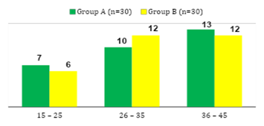
FIGURE 1: DISTRIBUTION OF STUDY PARTICIPANTS ACCORDING TO AGE GROUPS IN GROUP A AND GROUP B
Gender Distribution
Out of 60 patients, 38 (63.3%) were females and 22 (36.7%) were males. In Group A, 21 (70%) were females and 9 (30%) were males. In Group B, 17 (56.7%) were females and 13 (43.3%) were males.
TABLE 2: GENDER DISTRIBUTION BETWEEN GROUP A AND GROUP B
|
Sex |
Group A n=30 (%) |
Group B n=30 (%) |
Total (%) n=60 |
|
Male |
9 (30.0) |
13 (43.3) |
22 (36.7) |
|
Female |
21 (70.0) |
17 (56.7) |
38 (63.3) |
|
Total |
30 (100) |
30 (100) |
60 (100) |
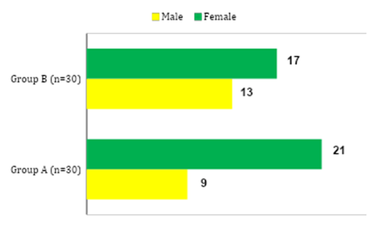
FIGURE 2: GENDER DISTRIBUTION BETWEEN GROUP A AND GROUP B
Perforation Characteristics
Subtotal and multi-quadrant perforations were the most common (40%), followed by combined anterior and posterior perforations (28.3%), anteroinferior perforations (23.3%), and anterior-only perforations (8.4%). p = 0.812 which was not statistically significant.
TABLE 3: COMPARISON OF QUADRANTS INVOLVED BETWEEN GROUP A AND GROUP B
|
Quadrant Involved |
Group A n=30 (%) |
Group B n=30 (%) |
Total n=60 (%) |
χ2 (DOF) |
p-value |
|
Anteroinferior (AI) |
6 (20.0) |
8 (26.7) |
14 (23.3) |
0.957 (3) |
0.812 |
|
Anterior + Posterior (AI+PI) |
10 (33.3) |
7 (23.3) |
17 (28.3) |
|
|
|
AS + AI Anterior only |
3 (10.0) |
2 (6.7) |
5 (8.4) |
|
|
|
Subtotal / Large (AI+PI+AS+PS) |
11(36.7) |
13 (43.3) |
24 (40.0) |
|
|
|
Total |
30 (100) |
30 (100) |
60 (100) |
|
|
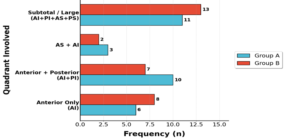
FIGURE 3: COMPARISON OF QUADRANTS INVOLVED BETWEEN GROUP A AND GROUP B
Graft Uptake
At 3 weeks, graft uptake was successful in 26 patients (86.7%) in Group A and 27 patients (90%) in Group B, with an overall success rate of 88.3%. The difference was not statistically significant (p = 1.000). At 6 weeks follow up, the graft uptake was observed in 27 patients (90%) in Group A and 29 patients (96.7%) in Group B, with an overall success rate of 93.3%. The final results at 3 months showed the graft uptake was successful in 27 patients (90%) in Group A and 29 patients (96.7%) in Group B, with an overall success rate of 93.3%. p value was 0.301which was statistically non- significant.
TABLE 4: COMPARISON OF EARLY GRAFT UPTAKE STATUS AT 3 WEEKS BETWEEN GROUP A AND GROUP B
|
3-Week Uptake Status |
Group A n=30 (%) |
Group B n=30 (%) |
Total n=60 (%) |
χ2 (DOF) |
p-value |
|
Success (Y) (Success rate) |
26 (86.7) |
27 (90.0) |
53 (88.3) |
0.000 (1) |
1.000 |
|
Failure (N) (failure rate) |
4 (13.3) |
3 (10.0) |
7 (11.7) |
|
|
|
Total |
30 |
30 |
60 |
|
|
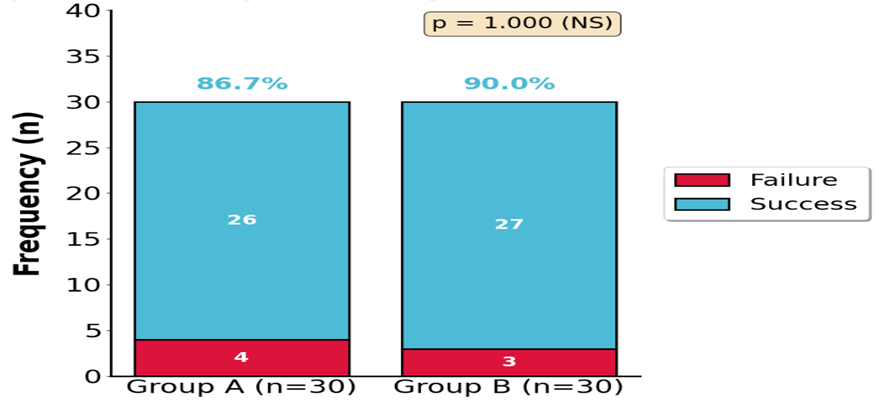
FIGURE 4: COMPARISON OF EARLY GRAFT UPTAKE STATUS AT 3 WEEKS BETWEEN GROUP A AND GROUP B
TABLE 5: COMPARISON OF FINAL GRAFT UPTAKE SUCCESS RATE AT 3 MONTHS BETWEEN GROUP A AND GROUP B
|
Outcome (at 3 Months) |
Group A n=30 (%) |
Group B n=30 (%) |
Total (%) |
(χ²) (DOF) |
p-value |
|
Success (Y) (success rate%) |
27 (90.0) |
29 (96.7) |
56 (93.3) |
1.071 (1) |
0.301 |
|
Failure (N) (failure rate%) |
3 (10) |
1 (3.3) |
4 (66.7) |
|
|
|
Total |
30 |
30 |
60 |
|
|
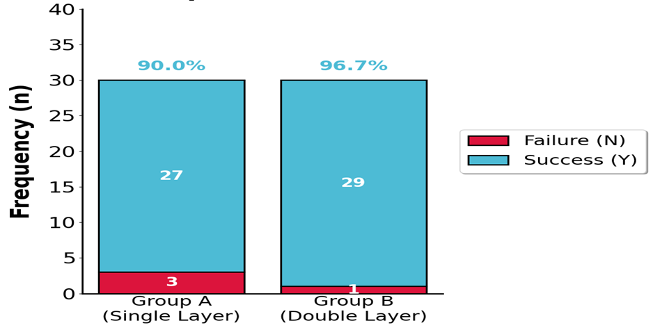
FIGURE 5: FINAL GRAFT UPTAKE SUCCESS RATE AT 3 MONTHS.
Audiological Outcomes
The mean pre-operative Air Bone Gap of Group A was29.17+9.19 dB, which was comparable to Group B with mean ABG of 29.0+9.03dB. At 3 months postoperatively, the mean air-bone gap was 19.50 ± 4.14 dB in Group A and 18.17 ± 3.96 dB in Group B. The difference was statistically non-significant (p = 0.460).
TABLE 6: BASELINE AUDIOLOGICAL PROFILE: COMPARISON OF PRE-OPERATIVE AIR BONE GAP ABG (dB) BETWEEN GROUP A AND GROUP B
|
Parameter |
Group A n=30 |
Group B n=30 |
t-value (DOF) |
p-value |
|
Mean Pre-op ABG |
29.17 ± 9.19 dB |
29.00 ± 9.03 dB |
0.078 (58) |
0.938 |
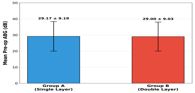
FIGURE 6: COMPARISON OF MEAN PRE-OP ABG.
TABLE 7: POST-OPERATIVE ABG (DB) AT 3 MONTHS — COMPARISON BETWEEN GROUP A AND GROUP B
|
Parameter |
Group A n=30 |
Group B n=30 |
t-value (DOF) |
p-value |
|
Mean Post-op ABG |
19.50 ± 4.14 dB |
18.17 ± 3.96 Db |
0.744 (58) |
0.460 |
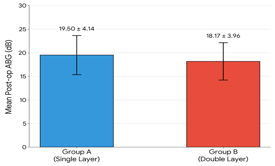
FIGURE 7: POST-OPERATIVE ABG (dB) AT 3 MONTHS — COMPARISON BETWEEN GROUP A AND GROUP B
Operative Time
The mean operative time was 54.90 ± 2.58 minutes in Group A and 55.93 ± 3.39 minutes in Group B. The difference was not statistically significant (p = 0.187).
DISCUSSION:
Anterior tympanic membrane perforations are associated with lower success rates due to poor vascularity, limited anterior support, and increased risk of graft medialization.11 These anatomical factors significantly influence graft stability and surgical outcomes in tympanoplasty.
In the study, the majority of patients were in the 36–45 years age group (41.6%), followed by 26–35 years (36.7%), with no statistically significant difference between the two groups (p = 0.218). This finding was consistent with the natural history of chronic otitis media, which predominantly affects young and middle-aged adults.12 A female predominance (63.3%) was observed in the present study; however, gender has not been shown to significantly influence tympanoplasty outcomes.13
Most patients had subtotal and multi-quadrant perforations (40%), with comparable distribution between both groups (p = 0.812). Anterior and large perforations are known to have poorer outcomes due to inadequate anterior support and reduced vascularity.8 The key finding of the study was the graft uptake which was consistently higher in the double-layer fascia group at all follow-up intervals.
At 3 weeks, graft uptake was 86.7% in Group A and 90% in Group B (p = 1.000). Then at 6 weeks follow up, the uptake improved to 90% in Group A and 96.7% in Group B (p = 0.612). The final graft uptake at 3 months was 90% in Group A and 96.7% in Group B (p = 0.301).
Although the difference was not statistically significant, the trend clearly favoured the double-layer technique. This observation is clinically important, particularly in anterior and subtotal perforations, where graft failure is more common.
Similar findings have been reported in previous studies demonstrating improved anatomical success with reinforced graft techniques. In the study by Kumar et al.14 the graft uptake in type I tympanoplasty with and without anterior tucking was 94% and 90% respectively.
The improved outcomes with double-layer grafting can be attributed to enhanced anterior support, prevention of graft medialization, and improved vascular integration of the graft.15. Preoperative hearing status was comparable between both groups, with most patients having moderate conductive hearing loss (31–40 dB).
At 3 months, the mean postoperative air-bone gap was 19.50 ± 4.14 dB in Group A and 18.17 ± 3.96 dB in Group B, with no statistically significant difference (p = 0.460). This indicates that both techniques provide comparable hearing improvement, suggesting that the addition of a second fascial layer does not adversely affect acoustic transmission.
These findings are consistent with previous studies, which have reported similar hearing outcomes despite improved graft uptake with reinforced techniques. Shrestha et al.16, showing that hearing gain results were comparable with cartilage and temporalis fascia graft.
The mean operative time was 54.90 ± 2.58 minutes in Group A and 55.93 ± 3.39 minutes in Group B, with no statistically significant difference (p = 0.187). This suggests that the addition of a second graft layer does not significantly increase operative time or surgical complexity.
Double-layer temporalis fascia grafting provides better graft uptake, especially in anterior and subtotal perforations. Hearing outcomes remain comparable between both techniques Lack of statistical significance may be due to limited sample size and short follow-up duration.
CONCLUSION:
Different methods were employed to manage anterior and subtotal perforations like single layer with anterior tucking which proved to be very effective technique to enhance the outcomes of graft uptake and hearing gain. Thus Tympanoplasty with anterior tucking of the graft is already proven surgical technique with satisfactory outcomes.9
The use of double-layer temporalis fascia offers added advantages of reinforcement with the smaller graft anteriorly between the larger graft and the remnant of tympanic membrane. Both single-layer and double-layer temporalis fascia grafting techniques are effective in Type I tympanoplasty, without increasing cost, operative complexity, or the need for specialized materials, providing high graft uptake rates and significant hearing improvement.
Therefore, although single-layer grafting remains adequate for routine cases, the double-layer technique may be preferred in anatomically difficult perforations to optimize surgical success and long-term outcomes.
REFERENCES:
1. Mansour S, Shawabkeh MA, Nicolas K, et al. Chronic suppurative otitis media (CSOM). In: Textbook of Clinical Otology. Springer; 2020.
2. StatPearls. Tympanoplasty. NCBI Bookshelf; 2024.
3. GBD 2019 Diseases and Injuries Collaborators. (2020). Global burden of 369 diseases and injuries in 204 countries and territories, 1990-2019: A systematic analysis for the Global Burden of Disease Study 2019. Lancet, 396, 1204–1222.
4. Caruso A, Giannuzzi A, Lauda L, et al. The Normal Tympanic Membrane. In: Sanna M, Russo A, Caruso A, et al, editors. Color Atlas of Endo-Otoscopy. Thieme; 2019.
5. Bevis N, et al. The impact of tympanic membrane perforations on middle ear physiology and hearing. J Otolaryngology Head Neck Surg. 2021;50(1):65.
6. Jiang Z, Lou Z. Impact of the nature of the temporalis fascia graft on the outcome of type I underlay tympanoplasty. J Laryngol Otol. 2017;131(6):472–475.
7. Aslıer M, Özay H, Gürkan S, et al. The effect of tympanic membrane perforation site, size and middle ear volume on hearing loss. Turk Arch Otorhinolaryngol. 2019;57(2):86-90.
8. Elsheikh E, El-Anwar MW, Nofal AA. Double-layer fascial grafts in type I tympanoplasty for subtotal perforation: a case–control study. Indian J Otol. 2023;29(1):10–15.
9. Prakash MD, Abhilasha S. The role of anterior tucking of graft in subtotal perforation of tympanic membrane. Int J Otorhinolaryngol Head Neck Surg 2018;4:759-63.
10. Aziz AAR, Hafez MAS, Mahmoud AG, Hamad AM. Comparative study between single layer and double layer graft technique in tympanoplasty for subtotal tympanic membrane perforation. Int Arch Otorhinolaryngol. 2023;27(4):e667–e671.
11. Rasouli AA, Zhapar K. Relationship of the size and location of the tympanic membrane perforation in chronic suppurative otitis media with the magnitude of hearing loss. J Interdiscip Approaches Med. 2022;3(1):45 52.
12. Vashisth S, Yadav SPS, Kumar P, Gupta V, et al. Comparative study of inlay and overlay cartilage perichondrium composite graft myringoplasty. Indian J Otolaryngol Head Neck Surg. 2023;75(1):34 40.
13. Wu X, Zhang Q, Huang Y, Wang X, Feng G. Predictors of functional outcomes and recovery time following tympanoplasty. Ear Nose Throat J. 2023;102(6):401 409.
14. Kumar M, Satyarthy V, Thakur RP. Assessment of the outcome of type I tympanoplasty with and without anterior tucking: a comparative study. Int J Pharm Clin Res. 2021;13(6):539 543.
15. Elsheikh EM, Ibrahim IE. Double anterior temporalis fascia myringoplasty in large central tympanic perforation. Zagazig Univ Med J. 2018;24(3):112 18 15.
16. Shrestha BL, Shakya S, Pradhan A, Dhakal A, et al. Comparison of hearing result and graft uptake rate between temporalis fascia and tragal cartilage in endoscopic myringoplasty. Kathmandu Univ Med J. 2022;20(80):539 544.