FACTORS ASSOCIATED WITH INFECTED SEBACEOUS CYSTS.
- Arnab Mondal. , Senior Resident, MBBS, MS General Surgery, Department of General Surgery, Bankura Sammilani Medical College and Hospital, Kenduadihi, Bankura, 722101.
Article Information:
Abstract:
Background: Introduction: Sebaceous cysts, more appropriately termed epidermoid cysts, are common benign cutaneous lesions encountered in surgical practice. Although usually asymptomatic, they may become infected due to rupture, secondary bacterial invasion, or local trauma. Infected sebaceous cysts often present with pain, swelling, erythema, and discharge, requiring surgical or medical intervention. Understanding the factors associated with infection is important for prevention and early management. Aims: To evaluate the clinical, demographic, and behavioral factors associated with infected sebaceous cysts and to identify the common risk determinants contributing to secondary infection. Materials and Methods: This Observational cross-sectional study was conducted in the Department of General Surgery, Bankura Sammilani Medical College and Hospital over 12 months among 80 Patients presenting with infected sebaceous cysts attending the Surgery OPD or admitted in the surgical ward. Results: The majority of patients were aged 21–40 years (17.50%), with male predominance (25.00% vs 15.00%). Scalp was the most common site (12.50%), and most cysts were 2–4 cm in size (18.75%). The overall complication rate was 10.00%, with surgical site infection being the most common (5.00%), and higher complication rates were associated with diabetes mellitus, ruptured cyst, preoperative infection, and cyst size >4 cm. Conclusions: We concluded that infected sebaceous cysts predominantly affect young and middle-aged adults, with a clear male preponderance. The scalp was the most frequently involved site, and most lesions measured 2–4 cm at presentation.
Keywords:
Article :
INTRODUCTION:
Sebaceous cysts, more accurately termed epidermoid cysts or epidermal inclusion cysts, represent one of the most frequently encountered benign cutaneous lesions in surgical and dermatological practice. Despite their benign nature, they are clinically significant due to their tendency to become inflamed or infected, often requiring medical or surgical intervention [1,2]. These cysts arise from the infundibular portion of the hair follicle and are characterized by a wall of stratified squamous epithelium filled with keratinous material [3]. Epidermoid cysts are common across all age groups but are most frequently observed in young and middle-aged adults, with a slight male predominance reported in several epidemiological studies [4].
They typically present as slow-growing, dome-shaped, mobile subcutaneous nodules, often with a central punctum. The most common anatomical sites include the face, scalp, neck, trunk, and back, regions that are prone to minor trauma and follicular obstruction [5]. Although generally asymptomatic, they may cause cosmetic concern or become symptomatic when inflamed or infected. Infection of sebaceous cysts usually occurs secondary to rupture of the cyst wall, allowing keratinous material to leak into surrounding dermal tissues, triggering a foreign body inflammatory response [6]. This inflammatory process creates a favorable environment for secondary bacterial invasion, most commonly by skin flora such as Staphylococcus aureus [7].
Clinically, infected cysts present with erythema, warmth, tenderness, swelling, and sometimes purulent discharge, often mimicking abscess formation. The pathogenesis of infection in sebaceous cysts is multifactorial. Local trauma, repeated manipulation or squeezing of the cyst, and poor hygiene have been identified as important external factors contributing to rupture and infection [8]. In addition, systemic conditions such as diabetes mellitus and immunocompromised states significantly increase susceptibility to infection due to impaired host immune response and delayed wound healing [9].
Environmental and occupational exposures that increase friction or sweating may also predispose individuals to cyst inflammation and secondary infection. Another important factor is the anatomical location of the cyst. Cysts located in high-friction areas such as the neck, back, and perineal region are more prone to repeated irritation and rupture, increasing the risk of infection [10].
Furthermore, delayed presentation and self-treatment attempts by patients often exacerbate the inflammatory process, leading to complicated infected lesions that require incision and drainage or complete excision. Despite their frequency, there remains variability in the clinical approach to sebaceous cysts, particularly regarding timing of intervention. While asymptomatic cysts are often left untreated, early elective excision before infection may prevent complications and recurrence. However, many patients present only after infection has developed, making management more challenging and increasing the risk of recurrence and scarring.
Understanding the factors associated with infected sebaceous cysts is therefore essential for effective prevention and management. Identifying modifiable risk factors such as hygiene practices, manipulation habits, and early treatment-seeking behavior can help reduce the incidence of infection. Moreover, recognizing high-risk groups such as diabetic patients and individuals with cysts in vulnerable anatomical sites may allow for targeted preventive strategies. In recent years, increasing attention has been given to improving surgical techniques and antibiotic strategies for managing infected epidermoid cysts.
However, prevention remains the most effective approach. A better understanding of patient-related, lesion-related, and environmental factors contributing to infection will aid clinicians in developing comprehensive management protocols and patient education strategies. To evaluate the clinical, demographic, and behavioral factors associated with infected sebaceous cysts and to identify the common risk determinants contributing to secondary infection.
MATERIALS AND METHODS:
Study Design: This was an Observational cross-sectional study.
Study area – The study was conducted in the Department of General Surgery, Bankura Sammilani medical college and hospital.
Study Period- The study was carried out over a period of 12months
Study population: The study population comprised in the Department of General Surgery, Bankura Sammilani Medical College and Hospital over 12 months among 80 Patients presenting with infected sebaceous cysts attending the Surgery OPD or admitted in the surgical ward.
Sample Size: 80 Patients presenting with infected sebaceous cysts attending the Surgery OPD or admitted in the surgical ward.
Study variables:
• Age
• Sex
• Common anatomical site of cyst
• Cyst size
• Presence of risk factors (infected/inflamed cyst, diabetes, rupture, etc.)
• Postoperative complications (presence and type / overall complication rate)
Inclusion Criteria-
• Patients of all ages presenting with clinically diagnosed infected sebaceous cyst.
• Patients attending Surgery OPD or admitted in surgical wards.
• Patients who give informed written consent for participation in the study.
• Patients willing to undergo required clinical examination and investigations.
Exclusion Criteria-
• Patients with uninfected sebaceous cyst.
• Patients with other cystic or soft tissue swellings not diagnosed as sebaceous cyst.
• Patients with severe systemic illness unfit for evaluation or surgery.
• Patients who do not give consent for participation.
• Patients with recurrent or previously operated cysts (if not part of study design).
Statistical Analysis:
Data were entered into Excel and analyzed using SPSS and GraphPad Prism. Numerical variables were summarized using means and standard deviations, while categorical variables were described with counts and percentages. Two-sample t-tests were used to compare independent groups, while paired t-tests accounted for correlations in paired data. Chi-square tests (including Fisher’s exact test for small sample sizes) were used for categorical data comparisons. P-values ≤ 0.05 were considered statistically significant.
RESULTS:
Table 1: Demographic and Clinical Profile of Patients
|
Category |
Subcategory |
Number (n) |
Percentage (%) |
|
Age (years) |
<20 |
8 |
10.00% |
|
21–40 |
35 |
43.75% |
|
|
41–60 |
25 |
31.25% |
|
|
>60 |
12 |
15.00% |
|
|
Sex |
Male |
50 |
62.50% |
|
Female |
30 |
37.50% |
|
|
Common sites |
Scalp |
25 |
31.25% |
|
Back |
20 |
25.00% |
|
|
Face/Neck |
15 |
18.75% |
|
|
Trunk |
20 |
25.00% |
|
|
Cyst size |
<2 cm |
30 |
37.50% |
|
2–4 cm |
38 |
47.50% |
|
|
>4 cm |
12 |
15.00% |
Table 2: Incidence of Postoperative Complications
|
Complication |
Number of Cases |
Percentage |
|
Surgical Site Infection |
4 |
5.00% |
|
Hematoma/Seroma |
3 |
3.75% |
|
Wound Dehiscence |
2 |
2.50% |
|
Recurrence |
2 |
2.50% |
|
Hypertrophic Scar |
1 |
1.25% |
|
No Complication |
68 |
85.00% |
Table 3: Association of Risk Factors with Postoperative Complications
|
Risk Factor |
Present |
Complication Rate (%) |
Absent |
Complication Rate |
|
Infected/Inflamed cyst preoperatively |
10 |
40.0% (4/10) |
70 |
18.1% (13/70) |
|
Diabetes Mellitus |
6 |
50.0% (3/6) |
74 |
20.3% (15/74) |
|
Cyst size > 4 cm |
5 |
40.0% (2/5) |
75 |
21.3% (16/75) |
|
Ruptured cyst |
4 |
50.0% (2/4) |
76 |
21.1% (16/76) |
Table 4: Distribution of patients according to clinical presentation
|
Clinical Feature |
Category |
Number (n) |
Percentage (%) |
|
Pain |
Present |
60 |
75.00% |
|
Absent |
20 |
25.00% |
|
|
Discharge |
Present |
30 |
37.50% |
|
Absent |
50 |
62.50% |
|
|
Redness/Inflammation |
Present |
28 |
35.00% |
|
Absent |
52 |
65.00% |
|
|
Tenderness |
Present |
55 |
68.75% |
|
Absent |
25 |
31.25% |
Table 5: Type of surgical procedure performed
|
Procedure Type |
Number (n) |
Percentage (%) |
|
Excision under local anesthesia |
65 |
81.25% |
|
Excision under spinal anesthesia |
10 |
12.50% |
|
Incision and drainage followed by excision |
5 |
6.25% |
Table 6: Duration of hospital stay and outcome
|
Category |
Subcategory |
Number (n) |
Percentage (%) |
|
Hospital stay |
2 days |
50 |
62.50% |
|
3–5 days |
24 |
30.00% |
|
|
>5 days |
6 |
7.50% |
|
|
Wound healing outcome |
Primary healing |
70 |
87.50% |
|
Delayed healing |
10 |
12.50% |
|
|
Final outcome |
Uneventful recovery |
72 |
90.00% |
|
With complication |
8 |
10.00% |
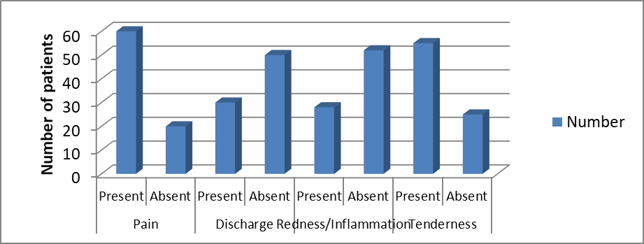
Figure 1: Distribution of patients according to clinical presentation
Demographic and Clinical Profile of Patients
Results:
The study included 80 patients with infected sebaceous cysts. The majority of patients belonged to the 21–40 years age group (35 patients, 43.75%), followed by 41–60 years (25 patients, 31.25%), >60 years (12 patients, 15.00%), and <20 years (8 patients, 10.00%). Males constituted 50 patients (62.50%), while females accounted for 30 patients (37.50%). The scalp was the most common site of involvement (25 patients, 31.25%), followed by the back and trunk (20 patients each, 25.00%), whereas face/neck involvement was observed in 15 patients (18.75%). Regarding cyst size, 2–4 cm lesions were the most common (38 patients, 47.50%), followed by <2 cm (30 patients, 37.50%) and >4 cm (12 patients, 15.00%).
Interpretation:
The findings demonstrate that infected sebaceous cysts were more common among young and middle-aged adults with a clear male predominance. The scalp was the most frequently affected anatomical site, and most patients presented with cysts measuring 2–4 cm, indicating that moderate-sized lesions were the most common at the time of surgical intervention.
Incidence of Postoperative Complications
Results:
Postoperative complications were observed in 12 patients (15.00%), while 68 patients (85.00%) had no complications. The most common postoperative complication was surgical site infection, occurring in 4 patients (5.00%), followed by hematoma/seroma in 3 patients (3.75%). Wound dehiscence and recurrence were each reported in 2 patients (2.50%), whereas hypertrophic scar formation occurred in 1 patient (1.25%).
Interpretation:
The overall postoperative complication rate was low, indicating that surgical treatment of infected sebaceous cysts is generally safe and effective. Surgical site infection remained the most frequently encountered postoperative complication, while other adverse events were relatively uncommon.
Association of Risk Factors with Postoperative Complications
Results:
Patients with preoperative infected or inflamed cysts showed a postoperative complication rate of 40.0% (4/10) compared with 18.1% (13/70) among those without infection. Similarly, patients with diabetes mellitus had a complication rate of 50.0% (3/6) compared with 20.3% (15/74) among non-diabetic patients. Those with cysts larger than 4 cm demonstrated a complication rate of 40.0% (2/5), whereas patients with smaller cysts had a rate of 21.3% (16/75). Patients with ruptured cysts had the highest complication rate of 50.0% (2/4) compared with 21.1% (16/76) among patients without rupture.
Interpretation:
The presence of diabetes mellitus, ruptured cysts, large cyst size, and preoperative infection was associated with a substantially higher rate of postoperative complications. These findings suggest that these clinical factors are important predictors of adverse surgical outcomes and should be considered during preoperative assessment and postoperative management.
Distribution of Patients According to Clinical Presentation
Results:
Pain was the most common presenting symptom, reported by 60 patients (75.00%), followed by tenderness in 55 patients (68.75%). Discharge was present in 30 patients (37.50%), while redness or inflammation was observed in 28 patients (35.00%).
Interpretation:
The majority of patients presented with symptomatic disease characterized by pain and tenderness, reflecting the inflammatory nature of infected sebaceous cysts. Discharge and local redness were also common clinical manifestations, although they were less frequent than pain and tenderness.
Type of Surgical Procedure Performed
Results:
The majority of patients underwent excision under local anesthesia (65 patients, 81.25%). Excision under spinal anesthesia was performed in 10 patients (12.50%), while incision and drainage followed by excision was required in 5 patients (6.25%).
Interpretation:
Excision under local anesthesia was the preferred surgical approach for most patients, indicating that the majority of infected sebaceous cysts could be managed using a simple and minimally invasive procedure. Only a small proportion required spinal anesthesia or staged surgical management.
Duration of Hospital Stay and Outcome
Results:
The majority of patients had a hospital stay of 2 days (50 patients, 62.50%), followed by 3–5 days (24 patients, 30.00%) and more than 5 days (6 patients, 7.50%). Regarding wound healing, 70 patients (87.50%) achieved primary healing, whereas 10 patients (12.50%) experienced delayed healing. The final clinical outcome showed that 72 patients (90.00%) had an uneventful recovery, while 8 patients (10.00%) developed postoperative complications.
Interpretation:
Most patients experienced a short hospital stay and achieved primary wound healing, reflecting effective surgical treatment and postoperative care. The high proportion of uneventful recoveries indicates favorable overall outcomes, with only a small percentage of patients developing complications requiring additional management.
DISCUSSION:
In the present study, infected sebaceous cysts were predominantly observed in the age group of 21–40 years (43.75%), followed by 41–60 years (31.25%), with a male preponderance (62.5%). This demographic pattern is consistent with previous studies describing epidermoid cysts as common benign lesions of young and middle-aged adults, with a slight male predominance and frequent involvement of hair-bearing areas such as the scalp, trunk, and face [11].
The higher incidence in males in our study may be attributed to increased exposure to minor trauma, follicular occlusion, and delayed healthcare-seeking behavior compared to females. Scalp was the most common site of involvement (31.25%), followed by the back and trunk (25% each). This is in agreement with established literature, which reports that epidermoid cysts most frequently occur in areas rich in pilosebaceous units [12,13]. The predominance of scalp lesions may be explained by higher follicular density and an increased likelihood of follicular obstruction leading to cyst formation and secondary infection.Most patients presented with cysts measuring 2–4 cm (47.5%), followed by lesions smaller than 2 cm (37.5%), while only 15% had cysts larger than 4 cm.
This distribution suggests that many patients seek medical attention only after gradual enlargement of the lesion or following the onset of infection, pain, or cosmetic concerns. In the present study, pain (75%) and tenderness (68.75%) were the most common presenting symptoms, followed by discharge (37.5%) and redness or local inflammation (35%). These findings are consistent with previous reports that epidermoid cysts generally remain asymptomatic until rupture or secondary infection occurs, after which they become painful, erythematous, tender, and fluctuant [14,15].
The high frequency of symptomatic presentation in this study indicates that most patients presented during the inflammatory phase rather than when the lesions were quiescent. The overall postoperative complication rate in our study was 15%, with 85% of patients experiencing no postoperative complications. Surgical site infection was the most common complication (5%), followed by hematoma/seroma (3.75%), wound dehiscence (2.5%), recurrence (2.5%), and hypertrophic scar formation (1.25%). These findings are comparable to published surgical series where postoperative complication rates following epidermoid cyst excision generally range between 5% and 15%, with surgical site infection being the most frequently reported complication [16,17].
The relatively low overall complication rate observed in our study highlights the effectiveness and safety of standard surgical excision when performed using meticulous surgical technique and appropriate aseptic precautions. A significant finding of this study was the association of increased postoperative complications with specific clinical risk factors. Patients with preoperative infected or inflamed cysts and those with cysts larger than 4 cm demonstrated complication rates of 40%, while diabetes mellitus and ruptured cysts were associated with the highest complication rates of 50%. These findings are consistent with established evidence that diabetes mellitus adversely affects wound healing through impaired microvascular circulation, reduced immune function, and delayed tissue repair, thereby increasing susceptibility to postoperative infection and delayed recovery [18]. Likewise, ruptured or inflamed cysts are associated with tissue contamination, extensive inflammatory reaction, and difficult surgical dissection, all of which contribute to increased postoperative morbidity [19].
Larger cysts often require wider surgical dissection and create larger dead spaces, predisposing patients to hematoma, seroma, and wound-related complications. These observations are consistent with established surgical principles indicating that larger lesions present greater technical challenges and carry a higher risk of postoperative complications [20]. Therefore, careful patient optimization, appropriate timing of surgery, and meticulous operative technique are essential in patients with these risk factors.Regarding surgical management, the majority of patients underwent complete excision under local anesthesia (81.25%), while 12.5% required excision under spinal anesthesia and 6.25% underwent incision and drainage followed by definitive excision.
These findings are consistent with standard surgical practice, where complete excision under local anesthesia remains the treatment of choice for uncomplicated epidermoid cysts because of its simplicity, reduced cost, shorter hospital stay, and excellent clinical outcomes [16]. Incision and drainage alone is generally discouraged because of the increased risk of recurrence unless it is followed by complete excision after resolution of acute inflammation.In the present study, most patients (62.5%) had a hospital stay of 2 days, while 30% remained hospitalized for 3–5 days and 7.5% required hospitalization for more than 5 days.
Primary wound healing was achieved in 87.5% of patients, whereas delayed wound healing occurred in 12.5%. Overall, 90% of patients experienced an uneventful recovery, while only 10% had adverse postoperative outcomes. These findings are comparable with previous studies demonstrating that surgical excision of epidermoid cysts is associated with excellent postoperative recovery, minimal morbidity, and favorable long-term outcomes when performed appropriately [15,20].The findings of the present study demonstrate that infected sebaceous (epidermoid) cysts are common surgical conditions with predictable demographic characteristics, clinical presentation, and favorable postoperative outcomes.
However, the presence of risk factors such as diabetes mellitus, ruptured cysts, larger cyst size, and preoperative infection significantly increases the likelihood of postoperative complications. Early diagnosis, optimization of associated comorbidities, and meticulous surgical management can substantially improve clinical outcomes and reduce postoperative morbidity in these patients.
CONCLUSION:
We concluded that infected sebaceous cysts predominantly affect young and middle-aged adults, with a clear male preponderance. The scalp was the most frequently involved site, and most lesions measured 2–4 cm at presentation. Pain and tenderness were the leading clinical manifestations, emphasizing the inflammatory nature of the disease. Surgical excision under local anesthesia was the treatment of choice for the majority of patients and was associated with favorable postoperative outcomes. Most patients required only a short hospital stay, achieved primary wound healing, and recovered without significant morbidity.
The overall postoperative complication rate was low, confirming that surgical management of infected sebaceous cysts is generally safe and effective. However, complications were more common in patients with diabetes mellitus, preoperative infection or inflammation, larger cyst size, and ruptured cysts, indicating that these factors significantly influence surgical outcomes. Careful preoperative assessment, adequate control of comorbid conditions, and meticulous postoperative care are therefore essential to minimize complications and improve recovery in high-risk patients.
REFERENCES:
1. Elston D, Ferringer T, Ko CJ, Peckham S, High WA, DiCaudo DJ. Dermatopathology E-book. Elsevier Health Sciences; 2013 Oct 16.
2. Torres-Laboy P, Schmieder SJ. Sebaceous carcinoma. InStatPearls [Internet] 2024 Nov 24. StatPearls Publishing.
3. Yamamoto T, Nishikawa T, Fujii T, Mizuno K. A giant epidermoid cyst demonstrated by magnetic resonance imaging. British Journal of Dermatology. 2001 Jan 1;144(1):217-8.
4. Trinh CT, Nguyen CH, Chansomphou V, Chansomphou V, Tran TT. Overview of epidermoid cyst. European journal of radiology open. 2019 Jan 1;6:291-301.
5. Diven DG, Dozier SE, Meyer DJ, Smith EB. Bacteriology of inflamed and uninflamed epidermal inclusion cysts. Archives of dermatology. 1998 Jan 1;134(1):49-51.
6. Kowalewski C, Kapil A, Shahab Z, Triyono A, Putnam E, Bhosale CR. Cysts in Older Adults: Diagnosis and Care Strategies. InGeriatric Dermatology 2025 May 13 (pp. 1-25). Cham: Springer Nature Switzerland.
7. Handa U, Garg S, Mohan H. Fine needle aspiration in the diagnosis of subcutaneous cysticercosis. Diagnostic cytopathology. 2008 Mar;36(3):183-7.
8. Patel A, Pawar O, Bordeaux JS. Factors associated with time to treatment for definitive surgery for sebaceous carcinoma. Journal of the American Academy of Dermatology. 2025 Jul 1;93(1):289-91.
9. Kumar P, Malhotra R, Urs AB, Augustine J, Mohanty S. Epidermoid cysts of the orofacial region: a clinico-pathological study of 13 cases with review of literature. Indian Journal of Pathology and Microbiology. 2023 Jul 1;66(3):568-72.
10. Kwon SH, Kim M, Shin JW, Na JI, Huh CH. Comparison of Surgical Outcomes of Rectangular Lid Excision Versus Elliptical Excision for the Treatment of Epidermal Cysts. Dermatologic Surgery. 2019 Sep 1;45(9):1141-6.
11. Frank E, Macias D, Hondorp B, Kerstetter J, Inman JC. Incidental squamous cell carcinoma in an epidermal inclusion cyst: a case report and review of the literature. Case Reports in Dermatology. 2018 Jun 18;10(1):61-8.
12. Song A, Carter KD, Syed NA, Song J, Nerad JA. Sebaceous cell carcinoma of the ocular adnexa: clinical presentations, histopathology, and outcomes. Ophthalmic Plastic & Reconstructive Surgery. 2008 May 1;24(3):194-200.
13. Zuber TJ. Minimal excision technique for epidermoid (sebaceous) cysts. American family physician. 2002 Apr 1;65(7):1409-12.
14. Al-Khateeb TH, Al-Masri NM, Al-Zoubi F. Cutaneous cysts of the head and neck. Journal of oral and maxillofacial surgery. 2009 Jan 1;67(1):52-7.
15. Sinha V, Parmar BD, Chaudhary N, Jha SG, Yadav SK. A study on clinical presentation of pseudocyst, dermoid cyst, and sebaceous cyst of pinna and its management at a tertiary care center. Indian Journal of Otology. 2021 Jul 1;27(3):140-3.
16. Wollina U, Langner D, Tchernev G, França K, Lotti T. Epidermoid cysts–a wide spectrum of clinical presentation and successful treatment by surgery: A retrospective 10-year analysis and literature review. Open access Macedonian journal of medical sciences. 2018 Jan 10;6(1):28.
17. Choi MK, Chung KJ. Factors affecting complications after treatment of epidermal cyst. Yeungnam University Journal of Medicine. 2019 Apr 1;36(2):136-40.
18. Zuber TJ. Minimal excision technique for epidermoid (sebaceous) cysts. American family physician. 2002 Apr 1;65(7):1409-12.
19. Owens PL, Barrett ML, Raetzman S, Maggard-Gibbons M, Steiner CA. Surgical site infections following ambulatory surgery procedures. Jama. 2014 Feb 19;311(7).
20. Apollos JR, Ekatah GE, Ng GS, McFadyen AK, Whitelaw SC. Routine histological examination of epidermoid cysts; to send or not to send?. Annals of Medicine and Surgery. 2017 Jan 1;13:24-8.