Developmental Anatomy and Physiological Significance of the Human Fetal Liver During 17–32 Weeks of Gestation: A Morphological Study.
- Shabana Sultana , Assistant Professor: Department of Anatomy: Government medical college, Kamareddy, Telanagana, India.
- Harish Kumar B , Assistant Professor: Department of Physiology: Government Medical College Kamareddy, Telanagana, India.
- M Rajneesh , Associate Professor: Department of Anatomy: Government Medical College, Kodangal, Telanagana, India.
Article Information:
Abstract:
Background: The fetal liver is a vital organ that undergoes rapid structural and functional maturation during intrauterine life. Besides being the largest abdominal organ in the fetus, it serves as the primary site of hematopoiesis and plays an essential role in metabolism, glycogen storage, protein synthesis, and fetal circulation. Understanding the relationship between anatomical development and physiological maturation of the fetal liver is fundamental for interpreting normal fetal growth and identifying developmental abnormalities. Aim: To evaluate the developmental morphology of the human fetal liver and correlate its anatomical changes with physiological maturation during 17–32 weeks of gestation. Materials and Methods: This descriptive cross-sectional study was conducted on 50 preserved human fetal specimens ranging from 17 to 32 weeks of gestation. Gestational age was determined using crown-rump length (CRL). Fetuses were classified into three gestational groups: Group A (17–20 weeks, n=35), Group B (21–24 weeks, n=11), and Group C (25–32 weeks, n=4). Standard anatomical dissection techniques were employed to study the gross morphology, anatomical position, hepatic lobe development, and external features of the liver. The observed anatomical findings were interpreted in relation to the known physiological functions of the fetal liver during intrauterine development. Results: Progressive developmental changes were observed in the fetal liver with advancing gestational age. The liver occupied a relatively larger portion of the abdominal cavity during early gestation and gradually assumed its adult anatomical position beneath the costal margins by 25–32 weeks. The right hepatic lobe became increasingly prominent with advancing gestation, while the quadrate and caudate lobes showed progressive differentiation. These structural changes correspond to the physiological maturation of the liver, including its role in fetal hematopoiesis, hepatic metabolism, glycogen storage, protein synthesis, and adaptation of fetal circulation during intrauterine life. Conclusion: The present study demonstrates that the human fetal liver undergoes coordinated anatomical and physiological maturation during the second and early third trimesters of gestation. The progressive structural changes observed reflect the increasing functional demands placed on the fetal liver before birth. These findings provide baseline developmental data that may be useful in developmental physiology, fetal medicine, prenatal imaging, and the evaluation of congenital and growth-related disorders.
Keywords:
Article :
Introduction:
The liver is the largest internal organ and the largest gland in the human body. It performs numerous vital metabolic, synthetic, storage, detoxification, immunological, and hematopoietic functions that are essential for normal growth and survival. During fetal life, the liver is proportionately much larger than in adults and serves as the principal site of hematopoiesis until the bone marrow becomes functionally mature. Its rapid growth and changing morphology reflect the dynamic developmental processes occurring throughout gestation.
Embryologically, the liver develops during the fourth week of intrauterine life from the hepatic diverticulum, an endodermal outgrowth of the ventral foregut. The cranial portion of this diverticulum (pars hepatica) gives rise to the liver parenchyma and intrahepatic biliary system, whereas the caudal portion (pars cystica) develops into the gallbladder and cystic duct. Hepatic cords proliferate into the septum transversum and are invaded by vitelline and umbilical veins, leading to the formation of hepatic sinusoids and the primitive vascular network. During fetal development, the liver undergoes continuous changes in size, weight, position, lobar proportions, and external morphology before attaining the adult anatomical configuration.[1,2]
The fetal liver occupies a relatively larger proportion of the abdominal cavity compared with the adult liver. In early gestation, it extends well below the costal margin and occupies much of the epigastric region. As gestation advances, differential growth of the thoracic cage and abdominal viscera results in progressive upward migration of the liver, with gradual attainment of its definitive anatomical position beneath the diaphragm. Simultaneously, the relative proportions of the right and left lobes change, producing the characteristic adult configuration. Knowledge of normal fetal liver morphology is important for anatomists, embryologists, radiologists, fetal medicine specialists, pediatric surgeons, and pathologists. Accurate information regarding fetal liver dimensions and developmental anatomy is valuable in prenatal ultrasonography, fetal magnetic resonance imaging, assessment of congenital anomalies, intrauterine growth disorders, and interpretation of autopsy findings. Morphometric standards also aid in distinguishing normal developmental variations from pathological conditions affecting fetal growth.[3,4]
Although numerous studies have described adult liver anatomy, comparatively fewer investigations have documented the sequential anatomical changes occurring in the fetal liver across different gestational ages. Comprehensive morphometric data regarding liver position, dimensions, weight, and lobar development remain limited, particularly in the Indian population.
Therefore, the present study was undertaken to evaluate the gross morphology of the fetal liver in human fetuses of different gestational ages. The study aimed to document developmental changes in the position, extent, weight, lobes, and morphometric measurements of the liver from 17 to 32 weeks of gestation. The findings are expected to provide baseline anatomical data that may contribute to fetal anatomical research, prenatal diagnostic imaging, developmental biology, and clinical practice.
Aim of the Study
· To evaluate the developmental morphology of the human fetal liver and correlate its anatomical changes with physiological maturation during 17–32 weeks of gestation.
Materials and Methods:
Study Design
This descriptive cross-sectional study was conducted to evaluate the gross anatomical development of the human fetal liver and to correlate the observed morphological changes with its known physiological functions during fetal life. The study focused on developmental changes occurring between 17 and 32 weeks of gestation, a period during which the fetal liver undergoes rapid structural and functional maturation.
Study Setting
The study was carried out in the Department of Anatomy in collaboration with the Department of Physiology.
Study Material
The study comprised 50 preserved human fetuses of gestational age ranging from 17 to 32 weeks. Fetuses were obtained from spontaneous abortions, stillbirths, and preterm deliveries after obtaining institutional approval and maintaining complete anonymity.
Specimens showing congenital anomalies involving the liver, abdominal wall defects, or marked autolysis were excluded.
Preservation of Specimens
The fetal specimens were collected within 10–15 hours after delivery and preserved in 10% neutral buffered formalin. Depending on fetal size, formalin was injected into the cranial, thoracic, and abdominal cavities to ensure uniform fixation of internal organs. The specimens remained immersed until adequate fixation was achieved for anatomical dissection.
Determination of Gestational Age
Gestational age was estimated by measuring the crown-rump length (CRL) using a flexible measuring tape. Based on CRL, the specimens were classified into three groups:
|
Group |
Gestational age |
Number of fetuses |
|
A |
17–20 weeks |
35 |
|
B |
21–24 weeks |
11 |
|
C |
25–32 weeks |
4 |
Anatomical Dissection
Each fetus was placed in the supine position, and a standard midline thoraco-abdominal incision was performed. The anterior abdominal wall was carefully reflected to expose the abdominal cavity.
The liver was examined in situ before removal to document:
· Anatomical position
· Extent in relation to the diaphragm and costal margins
· Surface morphology
· Development of hepatic lobes
· Relationship with adjacent organs
· External appearance of the porta hepatis
The liver was then carefully dissected free from its ligamentous attachments and vascular connections to preserve its normal morphology.
Morphometric Analysis
After removal, the following anatomical parameters were recorded:
· Liver weight (g)
· Maximum length
· Maximum breadth
· Maximum thickness
· Morphology of right, left, caudate, and quadrate lobes
· Position of the inferior border relative to anatomical landmarks
Measurements were obtained using a digital weighing balance, Vernier calipers, measuring tape, and a stainless-steel scale. Each measurement was recorded three times, and the average value was used for analysis to minimize observer error.
Physiological Correlation
The observed anatomical changes were interpreted in relation to the physiological development of the fetal liver based on established embryological and physiological principles.
The following functional aspects were considered during interpretation:
· Progressive hepatic growth associated with fetal hematopoiesis
· Development of hepatic lobes in relation to fetal circulation
· Positional changes associated with enlargement of the thoracic cavity
· Morphological maturation corresponding to increasing metabolic activity
· Structural development supporting glycogen storage and protein synthesis during fetal life
These correlations were made using standard embryological and physiological references and were not based on experimental functional measurements.
Instruments Used
· Scalpel with disposable blades
· Dissecting scissors
· Forceps
· Vernier calipers
· Measuring tape
· Stainless steel scale
· Digital analytical balance
· Syringe with 21-gauge needle
· Dissection tray
· Plastic specimen containers
Data Collection
All observations were recorded in a structured proforma. The following variables were documented:
· Gestational age
· Crown-rump length
· Liver position
· Liver morphology
· Hepatic lobe development
· Liver weight
· Morphometric measurements
Statistical Analysis
Data were entered into Microsoft Excel and analyzed using SPSS version 25.0 (or the software you used). Continuous variables were expressed as mean ± standard deviation (SD), while categorical variables were presented as frequencies and percentages. Differences between gestational age groups were assessed using one-way analysis of variance (ANOVA), with p < 0.05 considered statistically significant. Correlation between gestational age, crown-rump length, and liver morphometric parameters was evaluated using Pearson's correlation coefficient.
RESULTS:
A total of 50 human fetal specimens between 17 and 32 weeks of gestation were included in the present study. Gestational age was estimated using crown-rump length (CRL), and the specimens were grouped into three categories according to gestational age.
Table 1. Distribution of fetal specimens according to gestational age
| Group | Gestational age (Weeks) | Number of fetuses | Percentage (%) |
| A | 17–20 | 35 | 70.0 |
| B | 21–24 | 11 | 22.0 |
| C | 25–32 | 4 | 8.0 |
| Total | 17–32 | 50 | 100 |
The majority of specimens (70%) belonged to the 17–20 weeks gestational age group, whereas only four specimens (8%) were available in the 25–32 weeks group.
Crown-Rump Length
The CRL of every fetus was measured before dissection to estimate gestational age. Group A fetuses had CRL values ranging from 15.0 to 19.0 cm, whereas Group B specimens measured 20.0–23.0 cm.
Table 2. Crown-rump length of fetuses in different gestational groups
| Group | Gestational age (Weeks) | Crown-rump length (cm) |
| A | 17 | 15.0–15.5 |
| 18 | 16.0–16.5 | |
| 19 | 17.0–17.5 | |
| 20 | 18.0–19.0 | |
| B | 21 | 20.0–20.5 |
| 22 | 21.0–21.5 | |
| 23 | 22.0–22.5 | |
| 24 | 23.0 | |
| C | 25–32 | >23.0 |
A gradual increase in crown-rump length was observed with advancing gestational age.
Gross Morphology of the Fetal Liver
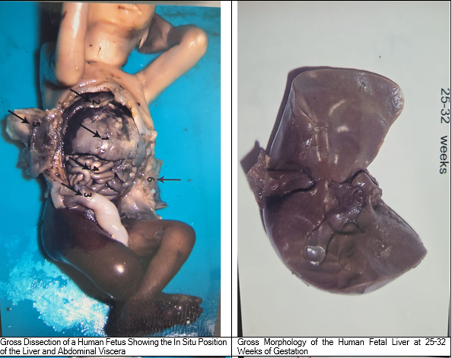
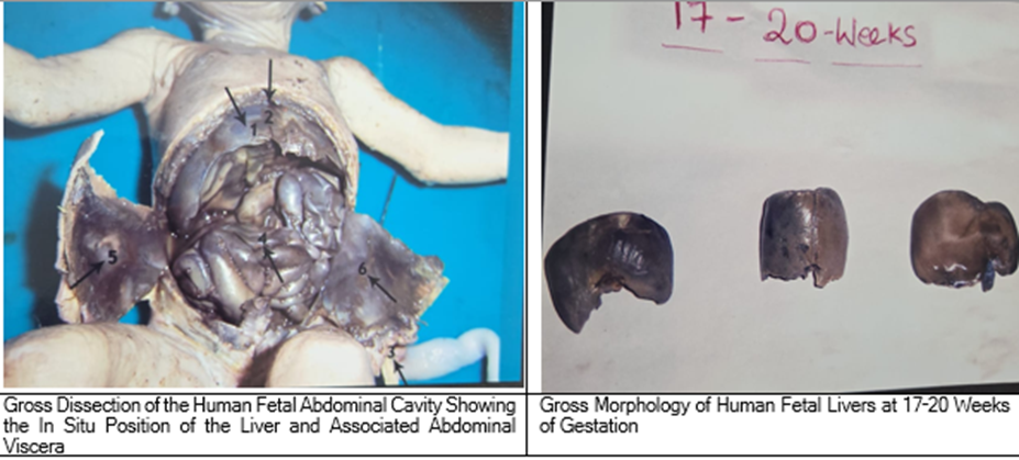
The liver was identified in its normal anatomical position in all specimens. No gross congenital malformations of the liver were observed in the examined fetuses. The liver appeared proportionately large in younger fetuses and occupied a considerable part of the upper abdominal cavity. Progressive changes in its anatomical position and extent were observed with advancing gestational age.
Position of the Liver
The position of the liver was studied in relation to the costal margin, xiphisternum, and umbilicus.
Group A (17–20 weeks)
The liver occupied almost the entire epigastric region. The inferior border projected below the left costal margin. In the youngest fetuses (17 weeks), the inferior margin extended approximately to the midpoint between the xiphisternum and umbilicus. The left lobe extended into the left hypochondrium.
Group B (21–24 weeks)
The inferior border gradually shifted upwards toward the left costal margin. Although the liver continued to occupy the epigastric region, a larger portion became situated beneath the left costal margin. The relative prominence of the left lobe decreased compared with younger fetuses.
Group C (25–32 weeks)
The inferior border of the liver was situated behind both costal margins and was located just posterior to the xiphisternal joint. The position of the liver closely resembled that observed in adults.
Table 3. Position of the liver in different gestational age groups
| Observation | Group A (17–20 weeks) | Group B (21–24 weeks) | Group C (25–32 weeks) |
| Liver occupies epigastrium | Completely | Predominantly | Minimal |
| Inferior border | Below left costal margin | Near left costal margin | Behind both costal margins |
| Left lobe | Prominent | Moderately prominent | Adult configuration |
| Adult anatomical position | Absent | Partial | Present |
A progressive superior migration of the liver was evident with increasing gestational age.
Development of Hepatic Lobes
The right and left hepatic lobes were identified in all specimens.In the younger fetuses, the left lobe appeared relatively larger in proportion to the right lobe. With increasing gestational age, the right lobe exhibited comparatively greater enlargement, resulting in gradual attainment of the adult hepatic configuration.The quadrate and caudate lobes became progressively more distinct in the later gestational groups.
Liver Weight
The liver weight was recorded in all specimens after dissection using an analytical balance. A progressive increase in liver weight was observed with advancing gestational age. The lowest liver weights were recorded in Group A, whereas the highest values were observed in Group C, demonstrating continuous hepatic growth during fetal development.
Morphometric Measurements
The morphometric parameters including liver length, breadth, and thickness were measured after dissection. All morphometric parameters showed a progressive increase with advancing gestational age. Older fetuses demonstrated larger liver dimensions than younger fetuses, indicating normal developmental growth of the organ.
DISCUSSION:
The fetal liver is one of the earliest organs to develop during embryogenesis and serves as the largest abdominal organ throughout intrauterine life. Besides its structural development, it plays a central role in fetal physiology by functioning as the primary hematopoietic organ, participating in carbohydrate metabolism, protein synthesis, glycogen storage, bile formation, and regulation of fetal circulation. The present study evaluated the developmental morphology of the human fetal liver between 17 and 32 weeks of gestation and interpreted the observed anatomical changes in relation to physiological maturation.
Developmental Changes in the Fetal Liver
The present study demonstrated progressive changes in liver morphology with advancing gestational age. The liver occupied a relatively larger proportion of the abdominal cavity in younger fetuses and gradually attained an adult-like anatomical position beneath the costal margins in later gestation. These observations are consistent with Moore, Persaud and Torchia,[1] who described that the liver develops from the hepatic diverticulum during the fourth week of gestation and undergoes rapid enlargement due to proliferation of hepatic cords and extensive hematopoietic activity. Similarly, Sadler (Langman's Medical Embryology) reported that the fetal liver becomes proportionately larger than the adult liver because it serves as the principal hematopoietic organ until the bone marrow assumes this function.[2] The findings of the present study also agree with Standring (Gray's Anatomy), [3]who described progressive cranial migration of the liver during fetal development as a consequence of differential growth of the diaphragm, thoracic cage, and abdominal organs.
Anatomical Position and Physiological Adaptation
One of the important observations of the present study was the gradual superior migration of the liver during gestation. In the early fetal period (17–20 weeks), the inferior border extended below the costal margin, whereas by 25–32 weeks the liver assumed an adult-like position. From a physiological perspective, this positional change reflects coordinated growth of the thoracic cavity and diaphragm. During mid-gestation, the liver occupies a larger proportion of the abdominal cavity because of its intense hematopoietic activity and increasing metabolic requirements. As fetal lung development accelerates during the third trimester, enlargement of the thoracic cavity and descent of the diaphragm facilitate repositioning of the liver. Similar observations have been reported by Hamilton and Mossman, Gray's Anatomy, and Keith L. Moore,[1,5] who attributed these positional changes to normal fetal growth rather than to intrinsic hepatic movement.
Development of Hepatic Lobes
The present study demonstrated progressive enlargement of the right hepatic lobe, whereas the relative prominence of the left lobe gradually decreased with increasing gestational age. These findings correlate well with fetal physiology. During early gestation, the left lobe receives a greater proportion of oxygenated blood through the umbilical vein and ductus venosus, explaining its relatively larger size. As gestation advances and hepatic circulation matures, the right lobe becomes progressively dominant. Similar findings have been described by Arey, Moore and Persaud, and Standring, [1,3,6]who emphasized that fetal hepatic blood flow plays a major role in determining differential growth of the hepatic lobes.
Functional Significance of the Fetal Liver
The structural changes observed in the present study closely correspond to the physiological maturation of the fetal liver. Between the 6th week and approximately the 30th week of gestation, the liver functions as the major hematopoietic organ. Rapid enlargement of the liver during the second trimester reflects intense erythropoiesis, granulopoiesis, and megakaryopoiesis occurring within the hepatic sinusoids. These observations support the descriptions of Sadler, Larsen, and Ross & Pawlina[2,6,8] who identified the fetal liver as the principal site of blood cell formation before bone marrow maturation.
In addition to hematopoiesis, hepatocytes undergo progressive differentiation and begin performing metabolic functions including:
· Glycogen storage
· Albumin synthesis
· Clotting factor synthesis
· Lipid metabolism
· Cholesterol synthesis
· Detoxification of endogenous metabolites
These physiological adaptations prepare the fetus for extrauterine life.
Growth of the Liver
Although detailed morphometric analysis is presented separately, the present study demonstrated a progressive increase in liver size with gestational age. Similar observations have been reported by Szpinda et al[9], who demonstrated a strong positive correlation between gestational age and liver volume using morphometric analysis of human fetuses. Likewise, Ghimire et al[10] reported significant increases in liver dimensions and weight throughout fetal development. The increasing liver size observed in the present study reflects progressive hepatocyte proliferation together with increasing metabolic and hematopoietic activity.
Clinical Significance
Understanding normal developmental anatomy of the fetal liver has considerable physiological and clinical importance.
Knowledge of normal liver development assists in:
· Interpretation of prenatal ultrasonography
· Fetal magnetic resonance imaging
· Diagnosis of congenital hepatobiliary anomalies
· Assessment of fetal growth restriction
· Evaluation of hepatomegaly
· Perinatal autopsy
Accurate anatomical knowledge also facilitates differentiation between physiological developmental variations and pathological abnormalities. These observations support recommendations made by the Federative International Programme for Anatomical Terminology (FIPAT) and the Anatomical Society of India, which emphasize the importance of developmental anatomy in clinical education and prenatal diagnosis.[11]
Table-4: Comparison with Previous Studies
| Author | Observation | Present Study |
| Moore KL, Persaud TVN[1] | Rapid fetal liver growth during mid-gestation | Similar findings observed |
| Sadler TW[2] | Liver is principal hematopoietic organ | Comparable physiological interpretation |
| Standring (Gray's Anatomy)[3] | Progressive superior migration of liver | Confirmed |
| Hamilton & Mossman[5] | Liver occupies large abdominal cavity during fetal life | Similar observations |
| Arey LB[6] | Right hepatic lobe enlarges with gestation | Confirmed |
| Szpinda et al.[9] | Liver dimensions increase with gestational age | Similar trend observed |
| Ghimire et al.[10] | Progressive increase in fetal liver size | Comparable findings |
Strengths of the Study
The study provides direct anatomical observations on fetal liver development over a broad gestational period. Correlating structural changes with physiological maturation enhances understanding of fetal hepatic development and provides baseline reference data for anatomists, physiologists, radiologists, and fetal medicine specialists.
Limitations
The relatively small number of specimens in the late gestational age group limits statistical comparison across all stages of fetal development. The study focused on gross morphology; histological evaluation, vascular casting, and molecular analyses were not performed. Future studies integrating morphometry, histology, Doppler imaging, and immunohistochemistry would provide a more comprehensive understanding of fetal liver development.
Limitations of the Study
The principal limitation of the study was the relatively small number of fetuses in the late gestational age group (25–32 weeks), which may limit detailed comparison across all developmental stages. In addition, histological evaluation and advanced imaging techniques were not included. Future studies with larger sample sizes, comprehensive morphometric analysis, and radiological correlation would further improve understanding of fetal liver development.
CONCLUSION:
The present study demonstrates that the fetal liver undergoes coordinated anatomical growth and physiological maturation between 17 and 32 weeks of gestation. Progressive cranial migration, increasing right lobe dominance, and maturation of hepatic morphology parallel the evolving physiological functions of the liver, including hematopoiesis, metabolism, glycogen storage, protein synthesis, and fetal circulatory adaptation. These findings agree with the observations of Moore, Persaud, Sadler, Standring, Hamilton and Mossman, Arey, Szpinda, and Ghimire, and contribute valuable baseline data for developmental anatomy, physiology, prenatal diagnosis, and perinatal pathology.
References:
1. Moore KL, Persaud TVN, Torchia MG. The Developing Human: Clinically Oriented Embryology. 12th ed. Philadelphia: Elsevier; 2023.
2. Sadler TW. Langman's Medical Embryology. 15th ed. Philadelphia: Wolters Kluwer; 2024.
3. Standring S, editor. Gray's Anatomy: The Anatomical Basis of Clinical Practice. 42nd ed. London: Elsevier; 2020.
4. Sinnatamby CS. Last's Anatomy: Regional and Applied. 13th ed. Edinburgh: Elsevier; 2018.
5. Hamilton WJ, Boyd JD, Mossman HW. Human Embryology. 4th ed. London: Macmillan Press; 1972.
6. Arey LB. Developmental Anatomy. 7th ed. Philadelphia: WB Saunders; 1974.
7. Larsen WJ. Human Embryology. 5th ed. Philadelphia: Churchill Livingstone; 2015.
8. Ross MH, Pawlina W. Histology: A Text and Atlas. 8th ed. Philadelphia: Wolters Kluwer; 2020.
9. Szpinda M, Paruszewska-Achtel M, Woźniak A, et al. Volumetric growth of the liver in the human fetus: an anatomical, hydrostatic and statistical study. Biomed Res Int. 2015;2015:858162.
10. Ghimire P, Bhattarai C, Sharma S, et al. Morphometry of fetal liver from human fetuses between 12–36 weeks gestational age. Cureus. 2022;14(4):e24469.
11. Patten BM. Human Embryology. 3rd ed. New York: McGraw-Hill; 1968.