PROSPECTIVE STUDY OF SURIGCAL MANAGEMENT OF INTRA-ARTICULAR CALCANEUM FRACTURE WITH LOCKING PLATE AND BONE GRAFTING.
- Pradeep Hullatti , Assistant Professor, Department of Orthopaedics, Chikkamagalur Institute of Medical Sciences, Chikkamagalur, Karnataka, India.
- Kavya K B , Assistant Professor, Department of Dermatology, Chikkamagalur Institute of Medical Sciences, Chikkamagalur, Karnataka, India.
- Vinutha Ganesh , MD Anaesthesia Consultant.
- Ganesh P S , Professor, Department of Orthopaedics, Chikkamagalur Institute of Medical Sciences, Chikkamagalur, Karnataka, India.
Article Information:
Abstract:
Background: Displaced intra-articular calcaneal fractures are among the most challenging fractures encountered in orthopaedic practice because of their complex anatomy, significant soft tissue involvement, and potential for long-term functional disability. Surgical management aims to achieve anatomical reduction of the subtalar joint, restore calcaneal morphology, and improve functional outcomes. Open reduction and internal fixation (ORIF) using locking compression plates has emerged as a preferred treatment option for selected fracture patterns. Objectives: 1. To evaluate the functional outcome of intra-articular calcaneal fractures treated with locking compression plate fixation and bone grafting. 2. To assess the incidence of postoperative complications following surgical management. Materials and Methods: A prospective study was conducted among 20 patients with displaced intra-articular calcaneal fractures (Sanders Type II–IV) treated with ORIF using a variable-angle locking compression plate and autologous bone grafting between May 2023 and April 2025. Patients were followed clinically and radiologically at 1, 3, 6, and 12 months postoperatively. Functional outcomes were assessed using the Maryland Foot Score, while radiological outcomes were evaluated using Böhler's and Gissane angles and fracture union. Results: The mean age of the patients was 36.5 years, with males accounting for 85% of cases. Falls from height were the predominant mechanism of injury (90%). Sanders Type III fractures constituted the majority (55%). The mean Böhler's angle improved from 15.4° preoperatively to 29.8° postoperatively, whereas the mean Gissane angle improved from 155.4° to 128.5°. Radiological union was achieved within 12 weeks in 75% of patients and within 20 weeks in the remaining 25%. The mean Maryland Foot Score was 81.5, with 70% of patients demonstrating good functional outcomes. Superficial surgical site infection occurred in 10% of patients, hardware irritation in 15%, and ankle or subtalar stiffness in 10%. No cases of deep infection, implant failure, wound dehiscence, or non-union were observed. Conclusion: Open reduction and internal fixation with locking compression plate and bone grafting provides excellent anatomical restoration, satisfactory fracture union, favorable functional outcomes, and an acceptable complication profile in displaced intra-articular calcaneal fractures. Careful patient selection, optimal timing of surgery, and meticulous surgical technique are essential for achieving successful clinical outcomes.
Keywords:
Article :
INTRODUCTION:
Calcaneal fracture is comparatively the most common fracture of tarsal bone representing 60% of all tarsal fractures in adults, with a reported occurrence of 2% of all fractures. (1) The calcaneus is central to load transmission from the tibia to the forefoot and is essential for normal gait mechanics, balance, and shock absorption. Disruption of its articular congruity, particularly in intra-articular fractures, often leads to altered biomechanics, chronic pain, stiffness, and long-term functional impairment if not adequately addressed.(2) Axial loading is most common mode of injury, fall from height causing bilateral calcaneal fractures.Talus is driven down into calcaneum by axial load which results in primary fracture line which runs across the posterior facet forming anteromedial and posterolateral fragments.(3,4) Sustantacular fragment stays associated with the talus due to strong ligaments. Posterior fragment is important as it contains posterior facet. Essex Lopresti described secondary fracture lines, which can produce tongue type and joint depression type calcaneal fractures. Secondary fracture line extending through tuberosity of the calcaneum produces tongue type fracture and if it extends through dorsal aspect of calcaneum joint, depression type fracture results. (5,6) The annual incidence of calcaneal fractures is 11.5 per 100,000 people, with a male to female ratio of 2.4:1, according to a retrospective study of 752 cases during a 10-year period. Falls are the cause of 72% of these fractures. About 10% cases are associated with spine fractures and 26% associated with other trauma. (7) The calcaneus is the biggest tarsal bone and is responsible for supporting the body. As a result of the fracture, there is made abnormality of calcaneal height and varus and a wider heel. A radiological examination is needed to evaluate the fracture line and compare it with the AO classification or sander's classification. (8) The Bohler's and Gissane's angles of the calcaneum are important parameter characteristics in the diagnosis, treatment, and prognosis of calcaneal fractures (9) . Bohler angle is measured by the intersection angle between a line from the apex of posterior tuberosity to the apex of posterior facet and a line from apex of posterior facet to apex of anterior process on the lateral ankle X-ray (10) . Bohler's angle indicates fracture of the calcaneus with displacement changes. The angle of 20 degrees was strongly predictive of calcaneal fracture that normally ranges from 20◦ to 40◦ (9,10) . However, Gissane's angles measurement made directly inferior to the lateral process of the talus, with the angle normal ranging between 120◦ to 140 . Bohler and Gissene angle also can use as a significant marker of postoperative clinical outcome.
Historically, the management of intra-articular calcaneal fractures has been controversial, with debates surrounding operative versus non-operative treatment and the optimal surgical technique. Advances in imaging, surgical exposure, and implant design have progressively shifted treatment paradigms toward anatomical reduction and stable fixation using plates, aiming to restore calcaneal morphology and subtalar joint congruity. Over the past decades, there has been a marked shift toward surgical intervention. The extended lateral approach has become the preferred route for achieving accurate visualization and reduction, especially for complex intra-articular patterns.(11)The development of low-profile, anatomically contoured locking plates has further improved fixation stability and minimized implant-related complications. Early surgical techniques, including Essex-Lopresti’s percutaneous pinning, offered limited access and often resulted in secondary collapse or pin-tract infection. These shortcomings have since been addressed with open reduction and internal fixation (ORIF) using plating systems, which allow correction in multiple planes and better load distribution across the subtalar joint. This study was undertaken to evaluate the function and radiological outcome of intra-articular calcaneal fracture treated with open reduction and internal fixation with LCP and bone grafting for socio-culturally specific population like Indian which presents with unique challenges.
OBJECTIVES: -
1. To evaluate the functional outcome of patients treated with open reduction and internal fixation with plate and bone grafting for intra-articular fracture of calcaneum for Sander’s Type II, III, IV calcaneal fractures.
2. To assess the incidence of infection and complications following open reduction and internal fixation of calcaneal fractures.
MATERIALS AND METHODS:
It’s a Prospective study of surgical management of 20 patients with Intra articular Calcaneal fracture operated with locking compression plate and bone grafting. This study was conducted in Aralaguppe Mallegowda District Hospital attached to Chikkamagalur Institute of Medical Science from May 2023 to April 2025 with all inclusion and exclusion criteria. Informed written consent was taken from all patients and were followed for minimum of 1 year regularly 1month, 3-month, 6 month and 12months.
Inclusion criteria:-
1. Age > 20 and < 60 years
2. Non-diabetic patients or patients under strict glycaemic control
3. Closed fractures and compound fracture grade I (Sanders type II–IV fractures)
4. Acute Calcaneal fracture
5. Patients with informed consent.
Exclusion criteria:-
1. Age <20 years or >60 years
2. Severe uncontrolled diabetes mellitus.
3. Patient medically not fit for surgery.
4. Severe soft-tissue injury
5. Sanders type I fractures
6. Grade II and III compound fractures
7. Associated vertebral fracture with neurological deficit
8. Non-reducible dislocation of the calcaneum requiring subtalar arthrodesis
9. Injury duration >21 days
10. Patient not willing for surgery.
Surgical technique:-
Patients with clinical suspicion of intra-articular fracture of the calcaneum presenting to the Casualty/OPD of the Department of Orthopaedics were evaluated. After Radiological confirmation of Intra-articular Calcaneum fracture, the patients satisfying the inclusion and exclusion criteria were included in the study after taking the informed consent. The patients’ details were collected, and preoperative radiological imaging (X-ray and CT scan) was performed and pre-operative planning was done.
Patients were initially managed with Splints, oral medications, ice packing and limb elevation till the swelling was reduced. After the appearance of wrinkle sign surgery was done by standard lateral approach. The patient was made to lie in lateral position on a radiolucent table after the patient was anaesthetized. Parts were scrubbed, painted and draped. The lateral skin incision (L shape EXTENDED LATERAL APPROCH) extending from the calcaneal tuberosity to the calcaneo-cuboid joint was made extending upwards between posterior border of distal fibula and the lateral aspect of the Achilles tendon. Thick skin flap was raised and 2 to 3 k wires was put in proximal end of wound at cuboid, talus and metatarsal and bent at 90 degrees to prevent rough handling of soft tissue. Lateral wall of calcaneum was lifted subperiosteally for access. Depressed articular fragments were elevated and the gap was filled with Bone graft (autologous) and fixed temporarily with Multiple k wire. The lateral wall was then closed and appropriate size variable angle LCP was placed over lateral wall and was fixed with appropriate size screw. The whole procedure was done under the guidance of C-arm. After the procedure the skin flaps that were kept elevated by k wire was sutured without any tension. Sterile dressing was done and POP below knee slab was applied for immobilization. Regular dressing of wound was done on post-op day 2,5,10 and 15. Suture removal was done after 3 weeks. Patient was advised for non-weight bear mobilization and ankle pump exercise. Patient was followed up regularly 1,3,6 12 months clinically and radiologically. Weight bearing was advised after fracture union in regular follow up.
RESULTS:
A total of 22 patients with calcaneal fractures were enrolled in this study and underwent open reduction and internal fixation (ORIF) using a variable-angle locking compression plate with bone grafting. All patients were scheduled for regular follow-up; however, 2 patients were lost to follow-up. Therefore, the final analysis included 20 patients. Among the 20 patients who completed follow-up, 17 (85%) were male and 3 (15%) were female. The mean age of the study population was 36.5 years. The majority of patients (12 patients, 60%) belonged to the 30–40 years age group. The youngest patient was 20 years old, while the oldest was 55 years.
Table 1. Demographic Characteristics (n = 20)
|
Variable |
Number (n) |
Percentage (%) |
|
Gender |
||
|
Male |
17 |
85 |
|
Female |
3 |
15 |
|
Age Group (years) |
||
|
20–29 |
3 |
15 |
|
30–40 |
12 |
60 |
|
41–55 |
5 |
25 |
Mean age: 36.5 years.
Age range: 20–55 years
The most common mechanism of injury was a fall from height, accounting for 18 patients (90%), whereas 2 patients (10%) sustained injuries due to motor vehicle accidents (MVAs). There was a slight predominance of right-sided fractures. Patients with bilateral calcaneal fractures and open (compound) fractures were excluded from the study.
Table 2. Mechanism of Injury
|
Mechanism of Injury |
Number (n) |
Percentage (%) |
|
Fall from height |
18 |
90 |
|
Motor vehicle accident |
2 |
10 |
According to the Sanders classification, 6 patients (30%) had Type II fractures, 11 patients (55%) had Type III fractures, and 3 patients (15%) had Type IV fractures. The interval between injury and surgery ranged from 5 to 11 days. Four patients (20%) underwent surgery on the 5th day after injury, 10 patients (50%) on the 6th day, 3 patients (15%) on the 8th day, 2 patients (10%) on the 9th day, and 1 patient (5%) on the 11th day. The majority of patients (50%) underwent surgical intervention on the 6th day following injury.
Table 3. Sanders Classification of Fractures
|
Sanders Type |
Number (n) |
Percentage (%) |
|
Type II |
6 |
30 |
|
Type III |
11 |
55 |
|
Type IV |
3 |
15 |
Table 4. Timing of Surgery After Injury
|
Interval (Days) |
Number (n) |
Percentage (%) |
|
5 |
4 |
20 |
|
6 |
10 |
50 |
|
8 |
3 |
15 |
|
9 |
2 |
10 |
|
11 |
1 |
5 |
Range: 5–11 days
Following surgery, there was a significant improvement in the radiological parameters. The mean Böhler's angle increased from 15.40° preoperatively to 29.80° postoperatively, indicating satisfactory restoration of calcaneal height. Similarly, the mean Gissane angle decreased from 155.40° preoperatively to 128.50° postoperatively, reflecting successful restoration of the calcaneal articular anatomy.
Table 5. Time to Radiological Union
|
Time to Union |
Number (n) |
Percentage (%) |
|
≤12 weeks |
15 |
75 |
|
13–20 weeks |
5 |
25 |
Table 6. Radiological Parameters Before and After Surgery
|
Radiological Parameter |
Preoperative Mean (°) |
Postoperative Mean (°) |
|
Böhler's angle |
15.4 |
29.8 |
|
Gissane angle |
155.4 |
128.5 |
All patients were followed up regularly for clinical and radiological evaluation. Radiological union was achieved in 15 patients (75%) within 12 weeks, while the remaining 5 patients (25%) achieved union by 20 weeks. Functional outcome was assessed using the Maryland Foot Score. Fourteen patients (70%) had good outcomes (score 75–89), 4 patients (20%) had fair/moderate outcomes (score 54–74), and 2 patients (10%) had poor outcomes (score <54). The mean Maryland Foot Score was 81.50, indicating an overall good functional outcome in the study population.
Table 7. Functional Outcome According to Maryland Foot Score
|
Functional Outcome |
Maryland Foot Score |
Number (n) |
Percentage (%) |
|
Good |
75–89 |
14 |
70 |
|
Fair |
54–74 |
4 |
20 |
|
Poor |
<54 |
2 |
10 |
Mean Maryland Foot Score: 81.5
Table 8. Postoperative Complications
|
Complication |
Number (n) |
Percentage (%) |
|
Superficial surgical site infection |
2 |
10 |
|
Hardware irritation |
3 |
15 |
|
Ankle/subtalar stiffness |
2 |
10 |
|
Deep infection |
0 |
0 |
|
Wound dehiscence |
0 |
0 |
|
Implant failure |
0 |
0 |
|
Non-union |
0 |
0 |
No major complications were observed in this study. Two patients (10%) developed superficial surgical site infections, which were successfully treated with intravenous antibiotics, resulting in complete resolution of the infection. Hardware-related irritation was reported in 3 patients (15%). Two patients (10%) developed ankle and/or subtalar joint stiffness during follow-up and were managed with physiotherapy. No cases of deep infection, wound dehiscence, implant failure, or non-union were observed.
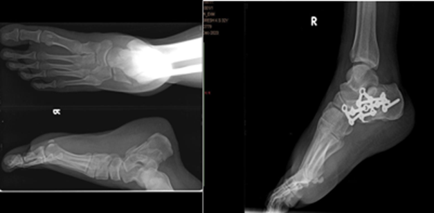
PREOP-XRAY IMMEDIATE POSTOP XRAY
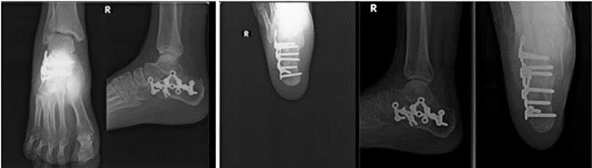
3 MONTHS POST OP X-RAY 1 YEAR POSTOP XRAY
DISCUSSION:
The present prospective study demonstrated that intra-articular calcaneal fractures treated with open reduction and internal fixation (ORIF) using a locking compression plate and autologous bone grafting resulted in favourable radiological and functional outcomes with a low complication rate. The majority of patients were young adult males who sustained injuries following falls from height, reflecting the occupational and high-energy trauma profile commonly reported in the literature. Similar demographic patterns have been described by Rak et al., who reported a predominance of male patients in the economically productive age group sustaining calcaneal fractures due to axial loading injuries.12 The findings of the present study are also consistent with those of Steelman et al., who observed that displaced intra-articular calcaneal fractures predominantly affect young males and are associated with significant functional morbidity if inadequately treated.13
The distribution of Sanders classification in the present study showed that Type III fractures constituted the majority (55%), followed by Type II (30%) and Type IV (15%). Sanders Type III fractures are generally associated with greater articular comminution and pose considerable challenges in achieving anatomical reduction. Popelka et al. reported that ORIF with locking plate fixation remains the preferred treatment modality for Sanders Type III fractures because it facilitates restoration of subtalar joint congruity and improves postoperative function.14 Their prospective study involving displaced intra-articular calcaneal fractures demonstrated satisfactory outcomes when fracture morphology was appropriately matched with surgical intervention. The predominance of Sanders Type III fractures in our study further supports the role of plate osteosynthesis in managing complex intra-articular fracture patterns.
The timing of surgical intervention is an important determinant of postoperative outcomes. In the present study, surgery was performed between 5 and 11 days after injury, with half of the patients undergoing fixation on the sixth day following trauma. Delaying surgery until soft tissue swelling subsides and the wrinkle sign appears is widely recommended to minimize wound-related complications. Rammelt and Zwipp emphasized that waiting for optimal soft tissue conditions significantly reduces postoperative wound complications without adversely affecting fracture reduction or functional outcomes.15 The timing protocol adopted in the present study likely contributed to the absence of wound dehiscence and deep infections.
Radiological restoration of calcaneal morphology remains one of the principal goals of operative management. In our study, the mean Böhler's angle improved significantly from 15.4° preoperatively to 29.8° postoperatively, while the mean Gissane angle decreased from 155.4° to 128.5°. These values fall within the normal radiological range and indicate successful anatomical reduction. Gusic et al. reported postoperative restoration of Böhler's angle to approximately 28°–30° following operative treatment of displaced intra-articular calcaneal fractures and demonstrated a strong correlation between radiological correction and improved clinical outcomes.16 Similarly, Fascione et al. reported that restoration of calcaneal height and subtalar congruity significantly contributed to superior functional scores following operative intervention.17 The radiological findings of the present study therefore support the effectiveness of locking compression plate fixation combined with bone grafting in restoring calcaneal anatomy.
Radiological union was achieved within 12 weeks in 75% of patients, while the remaining 25% achieved union by 20 weeks. No cases of delayed union progressing to non-union were encountered. Comparable findings were reported by da Silva et al., who observed successful fracture union in the majority of surgically managed intra-articular calcaneal fractures within three to five months of surgery.18 Their study concluded that stable fixation provided by plate osteosynthesis facilitates early fracture healing and allows satisfactory restoration of function. The present findings reinforce the biological advantage of achieving rigid internal fixation and maintaining anatomical reduction during the healing process.
Functional outcomes in the present study were favourable, with a mean Maryland Foot Score of 81.5. Seventy percent of patients achieved good functional outcomes, while only two patients had poor outcomes. Steelman et al. reported postoperative functional scores ranging between 80 and 90 following operative management of displaced intra-articular calcaneal fractures, emphasizing that accurate restoration of the posterior facet is associated with improved mobility and reduced pain. Likewise, Rak et al. demonstrated superior functional outcomes in patients treated with locking compression plates compared with conventional non-locking implants. The mean functional score observed in our study is therefore comparable with contemporary literature and highlights the effectiveness of ORIF in achieving satisfactory postoperative rehabilitation.
Postoperative complications were relatively uncommon in the present study. Superficial surgical site infections occurred in two patients (10%), hardware irritation in three patients (15%), and ankle or subtalar stiffness in two patients (10%). No cases of implant failure, non-union, wound dehiscence, or deep infection were observed. Benirschke and Kramer have reported wound complication rates ranging from 10% to 25% following operative treatment of calcaneal fractures, particularly when soft tissue conditions are unfavorable.19 The relatively low incidence of complications in our study may be attributed to appropriate patient selection, delayed surgery until wrinkle sign appearance, meticulous soft tissue handling, and postoperative rehabilitation protocols.
Recent evidence also supports the role of locking plate fixation in maintaining reduction while minimizing implant-related complications. Usami et al. demonstrated that locking plate fixation offers excellent mechanical stability and favourable postoperative outcomes when compared with alternative fixation techniques in intra-articular calcaneal fractures.20 Their findings further substantiate the advantages observed in the present study regarding fracture union, radiological restoration, and functional recovery.
Overall, the findings of the present study suggest that ORIF using locking compression plates with autologous bone grafting provides excellent radiological correction, satisfactory fracture union, favourable functional outcomes, and an acceptable complication profile in displaced intra-articular calcaneal fractures. Careful surgical timing and meticulous soft tissue management remain critical determinants of successful outcomes. These results are comparable with contemporary international literature and support the continued use of locking plate fixation as an effective surgical option for Sanders Type II–IV calcaneal fractures.
CONCLUSION:
The present prospective study demonstrates that open reduction and internal fixation using a locking compression plate with autologous bone grafting is an effective surgical modality for the management of displaced intra-articular calcaneal fractures (Sanders Type II–IV). The procedure provided satisfactory restoration of calcaneal anatomy, as evidenced by significant improvement in Böhler's and Gissane angles, timely fracture union, and favourable functional outcomes based on the Maryland Foot Score. The majority of patients achieved good postoperative functional recovery with minimal complications. Appropriate surgical timing following soft tissue recovery, meticulous operative technique, and structured postoperative rehabilitation contributed significantly to the successful outcomes. The low incidence of wound-related complications and absence of implant failure or non-union further support the safety and reliability of this treatment approach. Overall, locking plate fixation with bone grafting offers excellent radiological and functional results and remains a valuable option in the surgical management of displaced intra-articular calcaneal fractures.
REFERENCES:
1. H.G. Kulkarni, V.S. Mane, K.L. Gaonkar, et al., Plating for intra-articular calcaneal fractures... Is it an overkill? J. Clin. Orthop.Trauma 6 (3) (2015) 153–159, https:// doi.org/10.1016/j.jcot.2015.03.011.
2. Mahale YJ, Chinchole SS, Mishra S. Study of Outcome of Intra-articular Fractures of Calcaneum Treated Surgically by Plating. Journal of Trauma & Orthopaedic Surgery. 2017 Apr;12(2):13-7.
3. Ray makers JTFJ, Dekkers GHG, Brink PRG. Results after operative treatment of intra-articular calcaneal fractures with a minimum follow-up of 2 years. Injury;1998:29(8):593-99.
4. Pendse A, Daveshwar RN, Bhatt J, Shivkumar. Outcome after open reduction and internal fixation of intraarticular fractures of the calcaneum without the useof bone grafts. Indian J orthop. 2006;40:111-14.
5. Mostafa MF, El-Adl G, Hassanin EY, Abdellatif MS. Surgical treatment of displaced intra-articular calcaneal fracture using a single small lateral approach. Strategies Trauma Limb Reconstr. 2010;5(2):87–95.
6. Varela CD, Vaughan TK, Carr JB, Slemmons BK. Fracture blisters: clinical and pathological aspects. J Orthop Trauma. 1993. 7(5):417-27. [Medline].
7. Mitchell MJ, McKinley JC, Robinson CM. The epidemiology of calcaneal fractures. Foot (Edinb) 2009; 19:197.
8. N. Gusic, I. Fedel, N. Darabos, et al., Operative treatment of intraarticular calcaneal fractures: anatomical and functional outcome of three different operative techniques, Injury 46 (2015) S130–S133, https://doi.org/10.1016/j. injury.2015.10.061.
9. R. Ramachandran, S. Shetty, Assessment of Bohler' S and Gissane' S angles of the calcaneum in a group of South Indian population – a radiological study, J. Curr. Res. Rev. 7(15)(2015)17-20, , http://www.ijcrr.com/uploads/479_pdf.pdf.
10. J.D. Isaacs, M. Baba, P. Huang, et al., The diagnostic accuracy of B¨ohler's angle in fractures of the calcaneus, J. Emerg. Med. 45 (6) (2013) 879–884, https://doi.org/ 10.1016/j.jemermed.2013.04.055.
11. Rammelt S, Zwipp H. Calcaneus fractures: facts, controversies and recent developments. Injury. 2004;35(5):443–461.
12. Rak V, Ira D, Masek M. Operative treatment of intra-articular calcaneal fractures with calcaneal plates and its complications. Indian J Orthop. 2009;43(3):271–280. doi:10.4103/0019-5413.53458.
13. Steelman K, Wyrsch B, McBryde A, et al. Evaluation of patient outcomes after operative treatment of intra-articular calcaneus fractures. SICOT J. 2021;7:31. doi:10.1051/sicotj/2021030.
14. Popelka V, Hromádka R, Vavřík P, et al. Operative treatment algorithm for intra-articular calcaneal fractures. Acta Chir Orthop Traumatol Cech. 2018;85(1):18–27. PMID: 30879452.
15. Rammelt S, Zwipp H. Calcaneus fractures: facts, controversies and recent developments. Injury. 2004;35(5):443–461. doi:10.1016/S0020-1383(03)00151-6.
16. Gusic N, Fedel I, Darabos N, et al. Operative treatment of intra-articular calcaneal fractures: anatomical and functional outcome of three different operative techniques. Injury. 2015;46(Suppl 6):S130–S133. doi:10.1016/j.injury.2015.10.061.
17. Fascione F, Rammelt S, et al. Surgical treatment of displaced intra-articular calcaneal fractures using Calcanail®. Injury. 2019;50(Suppl 4):S86–S92. doi:10.1016/j.injury.2018.10.012.
18. da Silva LCA, Pingel A, et al. Surgical treatment of intra-articular fractures of the calcaneus: comparison between calcaneal plate and flat plate fixation. Rev Bras Ortop. 2016;52(4):383–389. doi:10.1016/j.rboe.2016.08.003.
19. Benirschke SK, Kramer PA. Wound healing complications in closed and open calcaneal fractures. J Orthop Trauma. 2004;18(1):1–6. doi:10.1097/00005131-200401000-00001.
20. Usami T, Yamanaka Y, et al. Fixation of intra-articular calcaneal fractures: a comparative study of locking plate fixation. J Orthop Surg Res. 2023;18:164. doi:10.1186/s13018-023-03657-5.