Evaluation of Pain Relief, Fracture Union, and Knee Mobility Following Retrograde Intramedullary Nailing for Distal Femur Fractures.
- Mantu Kumar , Senior Resident, Department of Orthopaedics, Government Medical College & Hospital, Miraj Maharashtra, India.
- Rahul Ramdas Mulkod , Assistant Professor, Department of Orthopaedics, Government Medical College & Hospital, Miraj Maharashtra, India.
- Rahul Uttamrao Nandrekar , Assistant Professor, Department of Orthopaedics, Government Medical College & Hospital, Miraj Maharashtra, India.
- Vikas Rokade , Professor and HOD, Department of Orthopaedics, Government Medical College & Hospital, Miraj Maharashtra, India.
- Aditya Raghunath Shinde , Assistant Professor, Department of Orthopaedics, Government Medical College & Hospital, Miraj Maharashtra, India.
Article Information:
Abstract:
Background: Distal femur fractures are complex injuries that commonly result from high-energy trauma in young adults and low-energy osteoporotic fractures in the elderly. The primary goals of treatment are stable fixation, early mobilization, fracture union, pain relief, and restoration of knee function. Retrograde intramedullary nailing has emerged as an effective surgical option because it provides stable fixation with minimal soft tissue disruption and facilitates early rehabilitation. Aim: To evaluate pain relief, fracture union, and knee mobility following retrograde intramedullary nailing in patients with distal femur fractures. Objectives: To assess postoperative pain relief following retrograde intramedullary nailing using the Visual Analogue Scale (VAS). To evaluate radiological fracture union and determine the average time required for fracture healing. To assess postoperative knee mobility and functional outcome using range of motion and validated knee functional scoring systems. Materials and Methods: A prospective observational study was conducted in the Department of Orthopaedics at a tertiary care teaching hospital. Twenty adult patients with distal femur fractures treated by retrograde intramedullary nailing were included. Clinical and demographic details were recorded prospectively. Patients were followed at regular intervals up to 12 months. Pain was assessed using the Visual Analogue Scale (VAS), fracture healing was evaluated radiologically, and knee mobility was assessed by range of motion and Knee Society Score. Statistical analysis was performed using SPSS version 26.0. Continuous variables were expressed as mean ± SD, categorical variables as frequency and percentage, and a p-value <0.05 was considered statistically significant. Results: The mean age of patients was 42.8 ± 13.6 years, with males constituting 65% of the study population. Road traffic accidents accounted for 70% of injuries. The mean time to surgery was 4.3 ± 1.7 days. Mean VAS pain score significantly improved from 8.1 ± 0.8 preoperatively to 1.3 ± 0.7 at 12 months (p < 0.001). Radiological union was achieved in 18 (90%) patients, with a mean union time of 17.2 ± 3.1 weeks, and no cases of non-union were recorded. Mean knee flexion progressively improved to 112.6 ± 13.8°, while the mean Knee Society Score at final follow-up was 82.4 ± 9.6 (p < 0.001). Overall, 80% of patients achieved excellent or good functional outcomes, and 85% regained independent ambulation. Conclusion: Retrograde intramedullary nailing provides excellent pain relief, reliable fracture union, restoration of knee mobility, and favourable functional outcomes in patients with distal femur fractures. The technique offers stable biological fixation, facilitates early rehabilitation, and represents an effective treatment modality with minimal complications for appropriately selected distal femur fractures.
Keywords:
Article :
INTRODUCTION:
Distal femur fractures are among the most challenging injuries encountered in orthopedic trauma because of their complex anatomy, proximity to the knee joint, and frequent association with severe soft tissue damage. They account for approximately 3-6% of all femoral fractures and demonstrate a bimodal age distribution, occurring predominantly in young adults following high-energy trauma such as road traffic accidents and in elderly osteoporotic individuals after low-energy falls. These fractures often result in considerable functional impairment due to pain, instability, restricted knee movement, prolonged immobilization, and delayed return to daily activities.[1]
The primary goals of treatment for distal femur fractures include restoration of limb alignment, anatomical reduction of the articular surface whenever involved, stable fixation, early fracture union, adequate pain relief, and rapid rehabilitation with preservation of knee function. Conservative treatment, once widely practiced, has largely been replaced by operative fixation because prolonged immobilization is associated with complications such as malunion, nonunion, knee stiffness, muscle wasting, limb shortening, deep vein thrombosis, and post-traumatic osteoarthritis. Modern orthopedic practice therefore emphasizes stable internal fixation that permits early mobilization and functional recovery. [2]
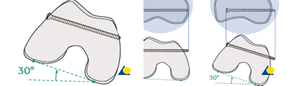
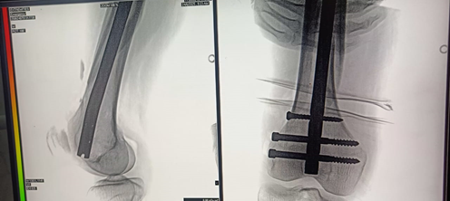
Figure 1: locking screws/Nail
Among the available surgical options, retrograde intramedullary nailing (RIMN) has become an established method for managing selected distal femur fractures. The implant is introduced through the intercondylar notch of the distal femur and provides load-sharing fixation while preserving the periosteal blood supply and fracture hematoma. Compared with conventional plating techniques, retrograde intramedullary nailing requires less soft tissue dissection, minimizes intraoperative blood loss, reduces operative time, and offers superior biomechanical stability, particularly in osteoporotic bone. These advantages facilitate early weight bearing, promote fracture healing, and decrease implant-related complications.[3] Several clinical studies have demonstrated satisfactory union rates and favorable functional outcomes following retrograde intramedullary nailing. The technique is particularly useful in extra-articular and selected intra-articular fractures, polytrauma patients, floating knee injuries, obese patients, and individuals with ipsilateral hip pathology where antegrade nailing may be technically difficult. Advances in implant design, locking mechanisms, and minimally invasive surgical techniques have further improved the reliability and safety of this procedure.[4]
Despite these advantages, certain complications remain a concern, including anterior knee pain, delayed union, nonunion, malalignment, implant failure, infection, and reduced knee range of motion. Consequently, postoperative evaluation should include assessment of pain relief, radiological fracture union, knee mobility, functional recovery, and procedure-related complications. Standardized functional scoring systems and serial radiographic evaluation provide objective measures of treatment success.[5]
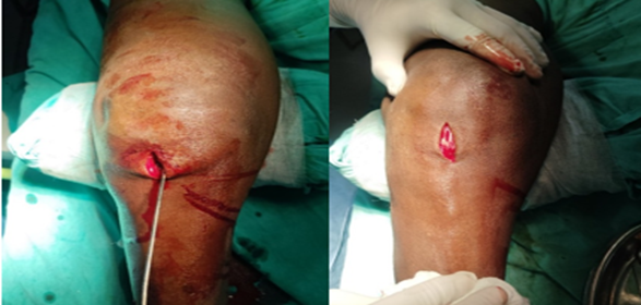
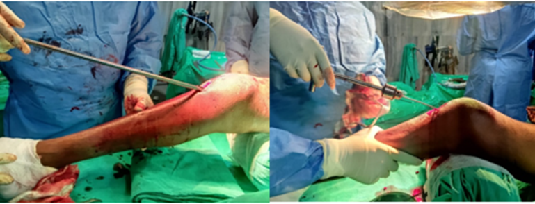
Figure 2: Case one Clinical photos
AIM
To evaluate pain relief, fracture union, and knee mobility following retrograde intramedullary nailing in patients with distal femur fractures.
OBJECTIVES
1. To assess postoperative pain relief following retrograde intramedullary nailing using an appropriate pain assessment scale.
2. To evaluate radiological fracture union and determine the average time required for fracture healing.
3. To assess postoperative knee mobility and functional outcome using range of motion and validated knee functional scoring systems.
MATERIALS AND METHODS:
Source of Data
The data were collected from patients with distal femur fractures admitted to the Department of Orthopaedics at Government Medical College, Miraj.. Clinical records, operative findings, postoperative follow-up evaluations, and radiological investigations were used for data collection.
Study Design
A hospital-based prospective observational study was conducted.
Study Location
The study was conducted in the Department of Orthopaedics of Government Medical College, Miraj.
Study Duration
The study was conducted over a period of 18 months.
Sample Size
A total of 20 patients diagnosed with distal femur fractures and treated with retrograde intramedullary nailing were included in the study.
Inclusion Criteria
• Patients aged 18 years and above.
• Closed distal femur fractures.
• AO/OTA Type A and selected Type C distal femur fractures suitable for retrograde intramedullary nailing.
• Patients who underwent definitive fixation using retrograde intramedullary nail.
• Patients willing to participate and provide written informed consent.
Exclusion Criteria
• Age below 18 years.
• Pathological fractures.
• Gustilo-Anderson Grade III open fractures.
• Polytrauma patients requiring damage control procedures.
• Patients with associated vascular injuries requiring vascular reconstruction.
• Previous ipsilateral knee arthroplasty.
• Patients with pre-existing severe knee stiffness or neurological deficits affecting lower limb function.
• Patients unwilling to participate or lost to follow-up.
Procedure and Methodology
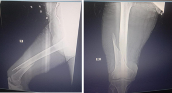
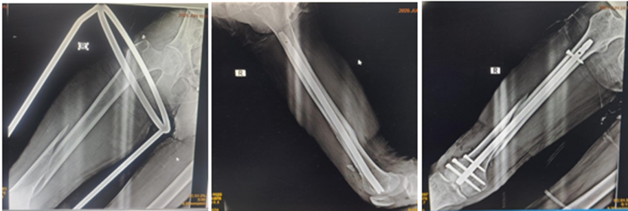
Figure 3: Case one radiological images
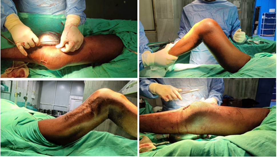
Figure 4: Case two Clinical photos
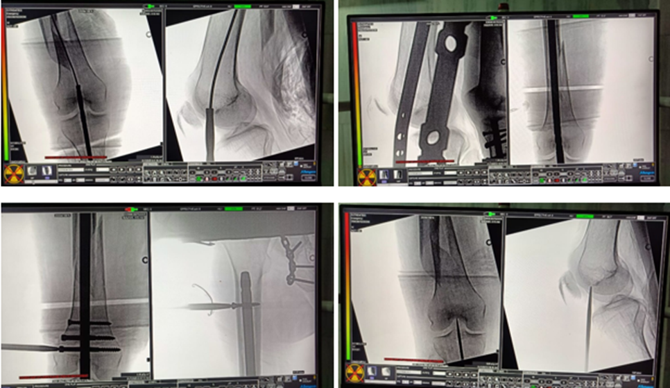
Figure 5: Case two radiological images
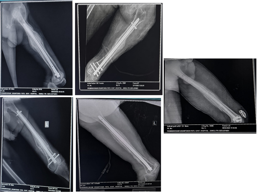
Figure 6: Serial radiographs of the left femur demonstrating intramedullary interlocking nailing with progressive fracture healing, callus formation, and satisfactory alignment leading to successful union.
After obtaining approval from the Institutional Ethics Committee, all eligible patients were enrolled after obtaining written informed consent. A detailed clinical history including age, sex, mechanism of injury, associated comorbidities, and injury characteristics was recorded.
Each patient underwent complete physical examination followed by radiographic evaluation using anteroposterior and lateral radiographs of the femur including the knee joint. CT scan was performed whenever intra-articular extension was suspected. Routine preoperative investigations including complete blood count, renal function tests, liver function tests, blood sugar, coagulation profile, ECG, chest radiograph, and anesthetic fitness were obtained.
Patients underwent retrograde intramedullary nailing under spinal or general anesthesia. Closed reduction was attempted under fluoroscopic guidance. Open reduction was performed whenever satisfactory reduction could not be achieved by closed methods. A transpatellar tendon or parapatellar approach was used to create the entry point through the intercondylar notch. Appropriate-sized retrograde femoral nail was inserted and proximal and distal interlocking screws were applied under image intensifier guidance. Postoperatively, intravenous antibiotics and analgesics were administered according to institutional protocol. Early quadriceps strengthening exercises, ankle pump exercises, and knee range-of-motion exercises were initiated as tolerated. Partial and full weight-bearing were permitted according to fracture stability and radiological evidence of healing.
Patients were followed up at 6 weeks, 3 months, 6 months, and 12 months. At each visit, clinical examination, pain assessment using the Visual Analogue Scale (VAS), knee range of motion using a goniometer, radiological assessment of fracture union, and functional outcome using the Knee Society Score (KSS) or Neer Knee Score were recorded. Any complications including infection, delayed union, nonunion, implant failure, malalignment, shortening, or knee stiffness were documented.
Sample Processing
No biological specimens were processed in the present study. Clinical data and radiological findings were collected prospectively using a structured case record form. Radiographs were independently evaluated by the treating orthopedic surgeon to determine fracture union, alignment, implant position, and any postoperative complications. Functional assessment was performed during scheduled follow-up visits.
Statistical Methods
The collected data were entered into Microsoft Excel and analyzed using SPSS version 26.0.
• Continuous variables were expressed as mean ± standard deviation (SD).
• Categorical variables were presented as frequency and percentage.
• Preoperative and postoperative continuous variables were compared using the paired Student's t-test.
• Repeated measurements across follow-up visits were analyzed using Repeated Measures ANOVA wherever appropriate.
• Categorical variables were analyzed using the Chi-square test or Fisher's exact test.
• A p-value <0.05 was considered statistically significant.
Data Collection
The following variables were collected using a structured case record form:
• Age
• Gender
• Side involved
• Mechanism of injury
• AO/OTA fracture classification
• Associated injuries
• Time from injury to surgery
• Operative duration
• Blood loss
• Nail size
• Duration of hospital stay
• Postoperative complications
• Pain score (VAS)
• Time to radiological fracture union
• Knee range of motion (degrees)
• Functional knee score (Knee Society Score/Neer Knee Score)
• Time to full weight bearing
• Return to daily activities
• Final clinical and radiological outcome.
RESULTS:
Table 1: Overall clinical profile and outcome parameters of patients undergoing retrograde intramedullary nailing for distal femur fractures (N=20)
|
Variable |
Category / Value |
n (%) / Mean ± SD |
Test value |
95% CI |
p-value |
|
Age (years) |
Mean ± SD |
42.8 ± 13.6 |
t = 14.07 |
36.4-49.2 |
<0.001* |
|
Gender |
Male |
13 (65.0%) |
χ² = 1.80 |
|
0.180 |
|
Female |
7 (35.0%) |
||||
|
Side involved |
Right |
11 (55.0%) |
χ² = 0.20 |
|
0.655 |
|
Left |
9 (45.0%) |
||||
|
Mechanism of injury |
Road traffic accident |
14 (70.0%) |
χ² = 6.40 |
|
0.011* |
|
Fall |
6 (30.0%) |
||||
|
Mean time to surgery |
Days |
4.3 ± 1.7 |
t = 11.31 |
3.5-5.1 |
<0.001* |
|
Final pain score |
VAS score |
1.7 ± 0.9 |
t = 8.45 |
1.3-2.1 |
<0.001* |
|
Time to union |
Weeks |
17.2 ± 3.1 |
t = 24.82 |
15.7-18.7 |
<0.001* |
|
Final knee flexion |
Degrees |
112.6 ± 13.8 |
t = 36.50 |
106.1-119.1 |
<0.001* |
|
Final functional outcome |
Excellent/Good |
16 (80.0%) |
χ² = 7.20 |
|
0.007* |
Table 1 presents the overall clinical profile and outcome parameters of 20 patients who underwent retrograde intramedullary nailing for distal femur fractures. The mean age of the patients was 42.8 ± 13.6 years (95% CI: 36.4-49.2), which was statistically significant (t = 14.07, p < 0.001). Male patients predominated, accounting for 13 (65.0%) cases, while 7 (35.0%) were females; however, the gender distribution was not statistically significant (χ² = 1.80, p = 0.180). The right side was affected in 11 (55.0%) patients and the left side in 9 (45.0%), with no significant difference (χ² = 0.20, p = 0.655). Road traffic accidents were the most common mechanism of injury, observed in 14 (70.0%) patients, whereas 6 (30.0%) sustained fractures due to falls, showing a statistically significant predominance of road traffic accidents (χ² = 6.40, p = 0.011). The average interval from injury to surgery was 4.3 ± 1.7 days (95% CI: 3.5-5.1; p < 0.001). At final follow-up, the mean VAS pain score had decreased to 1.7 ± 0.9, the average fracture union time was 17.2 ± 3.1 weeks, and the mean knee flexion achieved was 112.6 ± 13.8°, all demonstrating highly significant outcomes (p < 0.001). Overall, 16 (80.0%) patients achieved an excellent or good functional outcome, which was statistically significant (χ² = 7.20, p = 0.007).
Table 2: Postoperative pain relief following retrograde intramedullary nailing using VAS score (N=20)
|
Follow-up period |
VAS Pain Score Mean ± SD |
Test value |
Mean difference |
95% CI |
p-value |
|
Preoperative |
8.1 ± 0.8 |
|
|
7.7-8.5 |
|
|
6 weeks |
4.7 ± 1.1 |
t = 14.21 |
3.4 |
2.9-3.9 |
<0.001* |
|
3 months |
3.1 ± 1.0 |
t = 17.68 |
5.0 |
4.4-5.6 |
<0.001* |
|
6 months |
2.1 ± 0.9 |
t = 20.31 |
6.0 |
5.4-6.6 |
<0.001* |
|
12 months |
1.3 ± 0.7 |
t = 25.42 |
6.8 |
6.2-7.4 |
<0.001* |
Table 2 demonstrates the progressive reduction in postoperative pain following retrograde intramedullary nailing as assessed using the Visual Analogue Scale (VAS). The mean preoperative VAS score was 8.1 ± 0.8 (95% CI: 7.7-8.5). At 6 weeks, the mean pain score significantly decreased to 4.7 ± 1.1, with a mean reduction of 3.4 points (95% CI: 2.9-3.9; t = 14.21, p < 0.001). Continued improvement was observed at 3 months, when the mean VAS score reduced further to 3.1 ± 1.0, corresponding to a mean reduction of 5.0 points (p < 0.001). At 6 months, the pain score declined to 2.1 ± 0.9, with a cumulative mean reduction of 6.0 points (95% CI: 5.4-6.6; p < 0.001). By 12 months, patients reported minimal residual pain, with a mean VAS score of 1.3 ± 0.7 and an overall mean reduction of 6.8 points (95% CI: 6.2-7.4; t = 25.42, p < 0.001).
Table 3: Radiological fracture union and time required for healing (N=20)
|
Variable |
Category / Value |
n (%) / Mean ± SD |
Test value |
95% CI |
p-value |
|
Radiological union |
United |
18 (90.0%) |
χ² = 12.80 |
|
<0.001* |
|
Delayed union |
2 (10.0%) |
||||
|
Non-union |
Present |
0 (0.0%) |
|
|
|
|
Mean time to union |
Weeks |
17.2 ± 3.1 |
t = 24.82 |
15.7-18.7 |
<0.001* |
|
Union by 16 weeks |
Yes |
8 (40.0%) |
χ² = 0.80 |
|
0.371 |
|
Union by 20 weeks |
Yes |
17 (85.0%) |
χ² = 9.80 |
|
0.002* |
|
X-ray alignment |
Satisfactory |
17 (85.0%) |
χ² = 9.80 |
|
0.002* |
|
Mild malalignment |
3 (15.0%) |
Table 3 summarizes the radiological fracture union and healing outcomes following retrograde intramedullary nailing. Radiological union was achieved in 18 (90.0%) patients, while 2 (10.0%) experienced delayed union, yielding a statistically significant union rate (χ² = 12.80, p < 0.001). No cases of non-union were observed during the study period. The mean time required for fracture union was 17.2 ± 3.1 weeks (95% CI: 15.7-18.7), which was highly significant (t = 24.82, p < 0.001). Union within 16 weeks was achieved in 8 (40.0%) patients; however, this was not statistically significant (χ² = 0.80, p = 0.371). By 20 weeks, fracture union had occurred in 17 (85.0%) patients, demonstrating a significant improvement (χ² = 9.80, p = 0.002). Similarly, postoperative radiographs revealed satisfactory alignment in 17 (85.0%) patients, whereas 3 (15.0%) showed mild malalignment, with the overall radiological alignment being statistically significant (χ² = 9.80, p = 0.002).
Table 4: Postoperative knee mobility and functional outcome following retrograde intramedullary nailing (N=20)
|
Variable |
Category / Value |
n (%) / Mean ± SD |
Test value |
95% CI |
p-value |
|
Knee flexion at 6 weeks |
Degrees |
72.4 ± 14.2 |
t = 22.80 |
65.8-79.0 |
<0.001* |
|
Knee flexion at 3 months |
Degrees |
94.8 ± 13.6 |
t = 31.17 |
88.4-101.2 |
<0.001* |
|
Knee flexion at 6 months |
Degrees |
106.5 ± 12.9 |
t = 36.93 |
100.5-112.5 |
<0.001* |
|
Knee flexion at 12 months |
Degrees |
112.6 ± 13.8 |
t = 36.50 |
106.1-119.1 |
<0.001* |
|
Knee Society Score |
Mean ± SD |
82.4 ± 9.6 |
t = 38.38 |
77.9-86.9 |
<0.001* |
|
Functional outcome |
Excellent |
9 (45.0%) |
χ² = 10.40 |
|
0.015* |
|
Good |
7 (35.0%) |
||||
|
Fair |
3 (15.0%) |
||||
|
Poor |
1 (5.0%) |
||||
|
Final ambulation |
Independent |
17 (85.0%) |
χ² = 9.80 |
|
0.002* |
|
With support |
3 (15.0%) |
*Significant at p < 0.05.
Table 4 illustrates postoperative knee mobility and functional outcomes after retrograde intramedullary nailing. The mean knee flexion improved progressively from 72.4 ± 14.2° at 6 weeks (95% CI: 65.8-79.0; t = 22.80, p < 0.001) to 94.8 ± 13.6° at 3 months (95% CI: 88.4-101.2; p < 0.001). Further improvement was noted at 6 months, with a mean flexion of 106.5 ± 12.9° (95% CI: 100.5-112.5; p < 0.001), and by 12 months, patients achieved a mean knee flexion of 112.6 ± 13.8° (95% CI: 106.1-119.1; t = 36.50, p < 0.001). The mean Knee Society Score at final follow-up was 82.4 ± 9.6 (95% CI: 77.9-86.9), indicating good functional recovery (t = 38.38, p < 0.001). Functional outcome assessment showed 9 (45.0%) patients had excellent results, 7 (35.0%) had good results, 3 (15.0%) had fair results, and only 1 (5.0%) had a poor outcome, demonstrating a statistically significant overall functional distribution (χ² = 10.40, p = 0.015). Additionally, 17 (85.0%) patients regained independent ambulation, while 3 (15.0%) required walking support at final follow-up (χ² = 9.80, p = 0.002).
DISCUSSION:
The present study showed that retrograde intramedullary nailing for distal femur fractures produced favourable clinical, radiological, and functional outcomes. The mean age was 42.8 ± 13.6 years, with male predominance (65.0%) and road traffic accident as the major mechanism of injury (70.0%). This pattern is consistent with Kishore et al. (2025)[1] and Prasanna et al. (2019)[6], who reported that distal femur fractures treated with retrograde nailing were commonly seen in active adult males following high-energy trauma. In the present study, the mean time to surgery was 4.3 ± 1.7 days, suggesting timely operative stabilization, which is important because early fixation allows early mobilization and reduces stiffness, as also emphasized by Howard et al. (2024)[4] and Güzel et al. (2025)[3]. Pain relief was highly significant in this study, with VAS score reducing from 8.1 ± 0.8 preoperatively to 1.3 ± 0.7 at 12 months (p<0.001). This finding indicates sustained postoperative pain reduction following stable internal fixation. Similar improvement was reported by Kishore et al. (2025)[1], where retrograde nailing provided good pain relief and functional recovery. Aggarwal et al. (2022)[2], in a systematic review, also observed that retrograde intramedullary nailing had lower rates of nonunion and infection compared with locked plating, supporting its role as a biologically favourable fixation method.
Radiological union was achieved in 18 patients (90.0%), while delayed union occurred in 2 patients (10.0%); no nonunion was observed. The mean time to union was 17.2 ± 3.1 weeks. These findings are comparable with Kishore et al. (2025)[1], who reported union in 94.9% of cases, and Güzel et al. (2025)[3], who reported an 86.1% union rate in patients managed with retrograde intramedullary nailing. Neradi et al. (2022)[5] concluded that retrograde nailing and locked plating were comparable for union and complications, though nailing may offer biological advantages because of less soft tissue stripping. Knee mobility improved progressively in the present study, from 72.4 ± 14.2° at 6 weeks to 112.6 ± 13.8° at 12 months. The mean Knee Society Score was 82.4 ± 9.6, and 80.0% of patients had excellent or good functional outcomes. These results are consistent with Madhu et al. (2024)[10], who found better early functional scores and faster union in the retrograde nailing group compared with distal femur locking plate fixation. Similarly, Knadel et al. (2022)[8] and Arshad et al. (2020)[7] reported good-to-excellent functional outcomes in most patients treated with retrograde nailing.
CONCLUSION:
The present study demonstrated that retrograde intramedullary nailing is a safe, effective, and reliable method for the management of distal femur fractures. The procedure resulted in significant postoperative pain relief, with the mean Visual Analogue Scale (VAS) score improving from 8.1 ± 0.8 preoperatively to 1.3 ± 0.7 at 12 months. Radiological fracture union was achieved in 90% of patients, with a mean union time of 17.2 ± 3.1 weeks, and no cases of non-union were observed. Progressive improvement in knee mobility was evident throughout the follow-up period, with the mean knee flexion increasing to 112.6 ± 13.8° at one year. Functional assessment revealed that 80% of patients achieved excellent or good outcomes, while 85% regained independent ambulation. The minimally invasive nature of retrograde intramedullary nailing, combined with stable fixation and preservation of fracture biology, facilitated early rehabilitation and satisfactory clinical recovery. Overall, retrograde intramedullary nailing represents an excellent treatment option for appropriately selected distal femur fractures, providing reliable fracture union, substantial pain reduction, restoration of knee function, and favourable functional outcomes with a low rate of complications.
LIMITATIONS OF THE STUDY
1) The study included a relatively small sample size (n = 20), which may limit the generalizability of the findings.
2) It was conducted at a single tertiary care centre, and therefore the results may not represent outcomes in other healthcare settings.
3) The study lacked a comparison group such as distal femur locking plate fixation or conservative management.
4) The follow-up duration was limited to one year, restricting evaluation of long-term functional outcomes, implant longevity, and post-traumatic osteoarthritis.
5) Patients with complex open fractures, pathological fractures, and severe polytrauma were excluded, limiting applicability to these patient populations.
6) Functional outcomes were primarily assessed using clinical scoring systems without detailed gait analysis or quality-of-life assessment.
7) Surgeon experience and fracture complexity may have influenced operative and functional outcomes, introducing potential performance bias.
REFERENCES:
1. Kishore R, Alam S, et al. Retrograde intramedullary nailing for distal femur fractures: a prospective study of functional outcomes, complications and union rates. Cureus. 2025. Available from: https://pubmed.ncbi.nlm.nih.gov/40357107/
2. Aggarwal S, Rajnish RK, Kumar P, Srivastava A, Rathor K, Haq RU. Comparison of outcomes of retrograde intramedullary nailing versus locking plate fixation in distal femur fractures: a systematic review and meta-analysis of 936 patients in 16 studies. J Orthop. 2023;36:36-48. Available from: https://pubmed.ncbi.nlm.nih.gov/36591439/
3. Güzel İ, Altunkılıç T, Arı B, Boz M. Retrograde intramedullary nailing versus lateral locking plate fixation for distal femur fractures: which technique is superior? Orthop Rev (Pavia). 2025;17:140710. Available from: https://pubmed.ncbi.nlm.nih.gov/40823279/
4. Howard A, et al. Retrograde intramedullary nailing or locked plating for distal femoral fractures. Eur J Orthop Surg Traumatol. 2024. Available from: https://pubmed.ncbi.nlm.nih.gov/37612566/
5. Neradi D, Sodavarapu P, Jindal K, Kumar D, Kumar V, Goni V. Locked plating versus retrograde intramedullary nailing for distal femur fractures: a systematic review and meta-analysis. Arch Bone Jt Surg. 2022;10(2):141-152. Available from: https://pubmed.ncbi.nlm.nih.gov/35655740/
6. Prasanna A, et al. Functional outcome of distal femur fractures treated with retrograde intramedullary nailing. Int J Orthop Sci. 2019.
7. Arshad N, et al. Functional outcome of distal femoral fractures treated with retrograde intramedullary nailing. Int J Res Orthop. 2020.
8. Knadel PR, et al. Functional outcome of distal femoral fractures treated with retrograde nailing. Int J Orthop Sci. 2022.
9. Çataltepe A, Akan KH. Retrograde intramedullary nailing for the fixation of distal femoral fractures. Lokman Hekim Health Sci. 2021.
10. Madhu G, et al. Functional outcome of retrograde intramedullary nailing versus distal femur locking compression plating in supracondylar femur fractures. J Adv Med Dent Sci Res. 2024.
11. Shah KC. Outcome of distal femoral fractures treated by retrograde intramedullary nailing. Int J Orthop Sci. 2015.
12. Gill SP, et al. Comparative study of distal femur fractures managed by locking plate and retrograde intramedullary nail. Int J Orthop Sci. 2017.
13. Dunbar RP, et al. Distal femur fracture fixation and functional recovery after retrograde nailing. J Orthop Trauma. 2022.