Clinical and Radiological Evaluation of Cervical Spondylosis in Patients with Peripheral Vertigo: A Prospective Study.
- Mohit M.P. , Assistant Professor Department of Orthopaedics Chamarajanagar Institute of Medical Sciences (CIMS), Chamarajanagar, Karnataka, India.
- Greeshma K , Senior Resident Department of Otorhinolaryngology (ENT) Chamarajanagar Institute of Medical Sciences (CIMS), Chamarajanagar, Karnataka, India.
- Maruthi C.V , Professor and Head Department of Orthopaedics Chamarajanagar Institute of Medical Sciences (CIMS), Chamarajanagar, Karnataka, India.
Article Information:
Abstract:
Background: Peripheral vertigo is commonly encountered in ENT practice, yet symptom burden and incomplete response to vestibular manoeuvres often prompt evaluation for non-labyrinthine contributors. Cervicogenic dizziness remains debated, but cervical spondylosis is frequently seen on cervical imaging in patients reporting dizziness. Methods: In this prospective observational study, 100 consecutive adults (18–70 years) with a clinical diagnosis of peripheral vertigo were evaluated using standardized history, otoneurological examination, and bedside vestibular tests. All participants underwent lateral cervical spine radiography. Degenerative changes were recorded and graded (0–4). Dizziness Handicap Inventory (DHI) and Neck Disability Index (NDI) were administered. Results: Mean age was 40.75±12.46 years (range 18–67), and 61% were women. BPPV was the most common diagnosis (59%). Radiographic cervical spondylosis was present in 49 participants (49.0%). Prevalence increased significantly with age group (χ²=11.88, df=4, p=0.018) but did not differ by sex (χ²=0.00, df=1, p=1.000). Neck pain and restricted cervical range of motion were strongly associated with radiographic spondylosis (both p<0.001). Mean DHI and NDI scores were higher among those with cervical spondylosis (both p<0.001). Conclusions: Nearly half of peripheral vertigo patients had radiographic cervical spondylosis. Coexisting neck pain or restricted neck m ovements identified a subgroup with higher dizziness and neck-related disability, supporting targeted cervical screening and integrated management in routine ENT practice.
Keywords:
Article :
INTRODUCTION:
Vertigo is one of the most frequent complaints in ENT outpatient clinics. Clinicians usually start where they should: the vestibular apparatus and the central nervous system. Yet a subset of patients doesn’t fit cleanly into a single inner-ear label, or keeps returning with residual unsteadiness after apparently appropriate management. In that grey zone, the entity often termed cervicogenic dizziness becomes clinically tempting, and clinically tricky. There is no standalone confirmatory test, and the diagnosis is typically entertained only after reasonable exclusion of competing vestibular and neurological explanations.1-3
Across routine peripheral vertigo presentations, benign paroxysmal positional vertigo (BPPV), Ménière’s disease, and vestibular neuritis account for much of the workload. Current guidance repeatedly places weight on bedside pattern recognition—positional testing, nystagmus interpretation, and red-flag screening—before investigations are escalated or imaging is ordered reflexively.4-6 When the goal is to quantify impact rather than merely list symptoms, disability scales help. The Dizziness Handicap Inventory (DHI) captures the functional and psychosocial weight of dizziness, while the Neck Disability Index (NDI) records neck-related functional limitation, which is particularly relevant when dizziness travels with neck pain, stiffness, or posture-linked worsening.7,8
Cervical spondylosis complicates interpretation because it is common and often incidental. Degenerative changes and osteophytes increase with age, and population imaging studies have repeatedly shown that “abnormal” cervical scans can appear in people without symptoms.9,10 That mismatch between imaging and illness is exactly why a cautious clinical frame is needed: radiographs should support reasoning, not replace it. A reproducible radiographic grading framework helps standardise severity description, improves reporting consistency, and makes comparisons across studies more meaningful.11
Against this background, the present prospective study examined the prevalence and radiological severity of cervical spondylosis in Indian adults presenting with peripheral vertigo, and explored whether spondylotic change aligned with neck pain, range-of-motion limitation, and disability burden measured using DHI and NDI. A reproducible radiographic grading framework supports consistent reporting and comparability across studies.11
AIMS AND OBJECTIVES
Primary objective: To estimate the prevalence of radiographic cervical spondylosis among patients diagnosed with peripheral vertigo.
Secondary objectives: To assess associations between cervical spondylosis and age group, sex, peripheral vertigo subtype, neck pain, cervical range-of-motion limitation, and DHI/NDI scores; and to explore whether radiological grade correlates with dizziness handicap.
MATERIALS AND METHODS:
Study design and setting: Prospective observational study conducted in the Departments of ENT and Orthopaedics at a tertiary care hospital in India.
Participants: Consecutive adults aged 18–70 years presenting with symptoms of vertigo and diagnosed clinically as peripheral vertigo were recruited until a sample size of 100 was achieved.
Inclusion criteria: (i) clinical diagnosis of peripheral vertigo (BPPV, vestibular neuritis, Meniere’s disease, or other peripheral vestibular disorders) based on history and bedside vestibular tests; (ii) willingness to participate with written informed consent.
Exclusion criteria: (i) suspected or confirmed central causes of vertigo (focal neurological deficit, cerebellar signs, stroke, intracranial mass); (ii) prior cervical spine surgery or acute cervical trauma; (iii) known inflammatory arthropathy; (iv) inability to undergo cervical spine radiography.
Clinical assessment: Demographic variables and symptom duration were recorded. Bedside vestibular assessment included Dix–Hallpike manoeuvre and supine roll test where indicated, head impulse testing when feasible, and neurological screening. Neck pain (present/absent) and clinical limitation of cervical range of motion (present/absent) were recorded on orthopaedic examination.
Radiological assessment: Digital lateral cervical spine radiographs were obtained for all participants. Radiographic cervical spondylosis was defined by degenerative features including osteophyte formation, disc space narrowing, end-plate sclerosis, and/or loss of cervical lordosis on the lateral view. Severity was documented using a study-defined ordinal radiographic grade (0–4) derived from lateral radiographs: Grade 0 (no degenerative change), Grade 1 (mild changes at a single level), Grade 2 (moderate changes involving two levels and/or clear disc narrowing), Grade 3 (multilevel degeneration with prominent osteophytes and disc space reduction), and Grade 4 (severe multilevel degeneration with marked disc space narrowing and extensive osteophyte formation). This simplified grading was used to support consistent reporting in a resource-constrained outpatient setting and was not intended to replicate the full Cervical Degenerative Index (CDI) scoring framework.
Outcome measures: Dizziness Handicap Inventory (DHI; 0–100) and Neck Disability Index (NDI; 0–50) were administered.
Statistical analysis: Categorical variables were summarized as frequency and percentage; continuous variables as mean±SD or median (IQR). Associations were tested using Chi-square test (with Yates correction for 2×2 tables where appropriate) and independent-samples t-test. Multivariable logistic regression was used to identify clinical factors independently associated with radiographic cervical spondylosis. A p value <0.05 was considered statistically significant.
RESULTS:
Among 100 peripheral vertigo patients, 49% showed radiographic cervical spondylosis. Prevalence increased with age (Table 2; Figure 1), while peripheral vertigo diagnoses were dominated by BPPV (Figure 2). Cervical spondylosis clustered with neck pain, cervical motion restriction, and higher DHI/NDI scores (Table 4; Figures 3–5). Among 100 peripheral vertigo patients, 49% showed radiographic cervical spondylosis (Table 1).
Table 1: Baseline characteristics of participants (N=100).
|
Characteristic |
Value |
|
Mean age (years) ± SD |
40.75 ± 12.46 |
|
Age range (years) |
18–67 |
|
Median symptom duration, weeks (IQR) |
4.25 (2.80–6.12) |
|
Female, n (%) |
61 (61.0) |
|
Male, n (%) |
39 (39.0) |
|
BPPV, n (%) |
59 (59.0) |
|
Vestibular neuritis, n (%) |
24 (24.0) |
|
Meniere's disease, n (%) |
12 (12.0) |
|
Others, n (%) |
5 (5.0) |
|
Cervical spondylosis on X-ray, n (%) |
49 (49.0) |
Values are n (%) unless otherwise specified. DHI: Dizziness Handicap Inventory; NDI: Neck Disability Index.
Peripheral vertigo diagnoses were most commonly BPPV (59%), followed by vestibular neuritis (24%) and Ménière’s disease (12%) (Figure 2).
Table 2: Prevalence of cervical spondylosis by age group.
|
Age group (years) |
CS present, n |
CS absent, n |
Prevalence (%) |
|
18-29 |
6 |
16 |
27.3 |
|
30-39 |
9 |
14 |
39.1 |
|
40-49 |
15 |
11 |
57.7 |
|
50-59 |
14 |
10 |
58.3 |
|
60-70 |
5 |
0 |
100.0 |
Chi-square = 11.88, df = 4, p = 0.018.
Cervical spondylosis prevalence rose progressively across age strata, reaching 100% in the 60–70 year group (n=5) (p=0.018) (Table 2; Figure 1).
Table 3: Presence of cervical spondylosis by sex.
|
Sex |
CS present, n |
CS absent, n |
Prevalence (%) |
|
Female |
30 |
31 |
49.2 |
|
Male |
19 |
20 |
48.7 |
Chi-square (Yates corrected) = 0.00, df = 1, p = 1.000 (not significant).
Sex-wise prevalence was comparable between females and males (p=1.000) (Table 3).
Table 4: Clinical correlates of cervical spondylosis.
|
Variable |
CS present (n=49) |
CS absent (n=51) |
P value |
|
Neck pain, n (%) |
32 (65.3) |
13 (25.5) |
<0.001 |
|
Cervical ROM limitation, n (%) |
34 (69.4) |
6 (11.8) |
<0.001 |
|
DHI score, mean ± SD |
43.9 ± 9.3 |
33.3 ± 10.1 |
<0.001 |
|
NDI score, mean ± SD |
22.3 ± 8.5 |
13.4 ± 7.9 |
<0.001 |
P values from Chi-square test (categorical variables) and independent-samples t-test (continuous variables).
Table 5: Study-defined radiographic severity grade of cervical spondylosis (0–4) among affected participants (n=49)
|
Radiological grade |
n |
% |
|
Grade 1 |
17 |
34.7 |
|
Grade 2 |
12 |
24.5 |
|
Grade 3 |
17 |
34.7 |
|
Grade 4 |
3 |
6.1 |
Spearman correlation between grade and DHI: rho = 0.28, p = 0.053.
Table 6: Multivariable logistic regression for factors associated with cervical spondylosis (outcome: CS present).
|
Predictor |
Adjusted OR |
95% CI |
P value |
|
Neck pain (present) |
4.49 |
1.61–12.56 |
0.004 |
|
Cervical ROM limitation (present) |
14.93 |
4.98–44.75 |
<0.001 |
Overall, age group showed a significant association with cervical spondylosis prevalence (p=0.018), whereas sex did not (p=1.000), and clinical variables (neck pain and cervical ROM limitation) remained independently associated in multivariable analysis (Table 6).
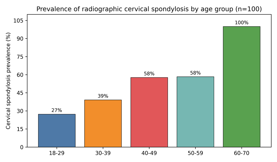
Figure 1: Prevalence of radiographic cervical spondylosis by age group.
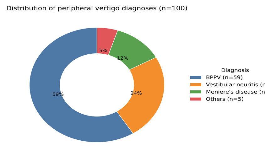
Figure 2: Distribution of peripheral vertigo diagnoses in the study cohort.
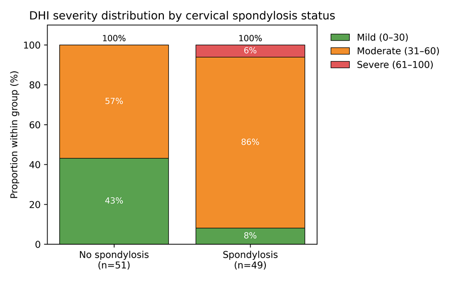
Figure 3: DHI severity distribution by cervical spondylosis status.
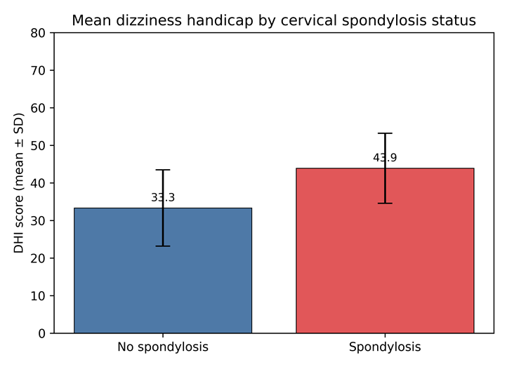
Figure 4: Mean dizziness handicap (DHI) by cervical spondylosis status (mean ± SD).
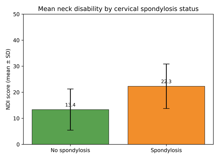
Figure 5: Mean neck disability (NDI) by cervical spondylosis status (mean ± SD).
Neck pain and restricted cervical motion were strongly associated with spondylosis, and affected patients demonstrated higher dizziness‑related and neck‑related disability. In routine ENT practice, especially where advanced imaging is not readily accessible, a lateral cervical spine X‑ray offers a low‑cost screening step when a cervicogenic contribution is clinically suspected.
DISCUSSION:
In this cohort of adults diagnosed with peripheral vertigo, radiographic cervical spondylosis was present in nearly half. At first glance, that looks high. Then you remember the uncomfortable truth: degenerative cervical change is common in the general population and frequently silent. What matters clinically is not the X-ray alone, but the pattern around it. Does the radiograph behave like an incidental background finding, or does it repeatedly cluster with neck symptoms and a heavier dizziness burden?
Two signals were consistent. First, prevalence increased with age, expected for degeneration, but clinically relevant when older patients report “vertigo” that does not fully resolve with vestibular manoeuvres or standard inner-ear–centric management. Second, neck pain and restricted cervical range of motion were strongly associated with radiographic spondylosis, and those patients carried higher DHI and NDI scores. In simpler terms, the neck-symptom subgroup reported more disability, both from dizziness and from neck dysfunction.
Mechanistically, cervicogenic dizziness is often framed as a proprioceptive mismatch problem: altered afferent input from cervical mechanoreceptors interacts with visual and vestibular signalling, producing disequilibrium in susceptible patients.12 This is still debated because boundaries are clinically fuzzy and imaging abnormalities are widespread even in asymptomatic adults. Yet, treatment data (while heterogeneous) repeatedly suggests that cervical-targeted approaches can reduce symptoms in selected patients, particularly when dizziness coexists with neck pain, movement restriction, and provocation by neck motion.13-15
So the pragmatic message for Indian tertiary OPDs is not “X-ray equals cervical vertigo.” It’s more cautious, and more useful: do not label dizziness as cervical on radiology alone. Instead, treat cervical spondylosis on X-ray as a co-finding that gains diagnostic weight when the clinical picture supports it, neck pain, restricted movement, and disproportionate handicap scores. In such settings, a lateral cervical radiograph remains low-cost and widely accessible, and it can help triage patients toward integrated care (vestibular manoeuvres or rehabilitation alongside cervical physiotherapy) rather than serial symptomatic prescriptions.
The grade–DHI relationship in our dataset showed only a borderline trend. Clinically, that is believable. Symptom burden is rarely a neat linear mirror of radiological severity; pain amplification, posture, muscle spasm, sleep debt, anxiety, and vestibular compensation all modulate the final experience. A grading index still has value for standardised reporting and for follow-up comparisons, but it should not be treated as a standalone explanation for dizziness.
Limitations Of Study
First, radiological evaluation relied on a lateral cervical spine radiograph; advanced imaging was not routinely performed, and subtle canal or foraminal compromise may have been missed. Second, while the study screened for central red flags, peripheral vertigo phenotypes can overlap with vestibular migraine or mixed dizziness states, which could dilute associations. Third, the study design cannot prove causality, cervical spondylosis and peripheral vertigo may coexist without a direct causal link in every patient.
CONCLUSON:
Radiographic cervical spondylosis was detected in nearly half of adults presenting with peripheral vertigo, with prevalence rising across age groups. Neck pain and restricted cervical motion identified a subgroup with significantly higher dizziness and neck disability scores. Cervical spine assessment should be considered in peripheral vertigo patients with concurrent neck symptoms, enabling a more comprehensive and rehabilitative management plan.
REFERENCES:
1. Reiley AS, Vickory FM, Funderburg SE, Cesario RA, Clendaniel RA. How to diagnose cervicogenic dizziness. Arch Physiother. 2017;7:12. PMID: 29340206.
2. Wrisley DM, Sparto PJ, Whitney SL, Furman JM. Cervicogenic dizziness: a review of diagnosis and treatment. J Orthop Sports Phys Ther. 2000;30(12):755–766. PMID: 11153554.
3. Li Y, Peng B. Pathogenesis, diagnosis, and treatment of cervical vertigo. Pain Physician. 2015;18(6):E583–E595. PMID: 26218949.
4. Strupp M, Brandt T. Diagnosis and treatment of vertigo and dizziness. Dtsch Arztebl Int. 2008;105(10):173–180. PMID: 19629221.
5. Bhattacharyya N, Gubbels SP, Schwartz SR, et al. Clinical practice guideline: Benign paroxysmal positional vertigo (update). Otolaryngol Head Neck Surg. 2017;156(3_suppl):S1–S47. PMID: 28248609.
6. Basura GJ, Adams ME, Monfared A, et al. Clinical practice guideline: Ménière’s disease. Otolaryngol Head Neck Surg. 2020;162(2_suppl):S1–S55. PMID: 32267799.
7. Jacobson GP, Newman CW. The development of the Dizziness Handicap Inventory. Arch Otolaryngol Head Neck Surg. 1990;116(4):424–427. PMID: 2317323.
8. Vernon H, Mior S. The Neck Disability Index: a study of reliability and validity. J Manipulative Physiol Ther. 1991;14(7):409–415. PMID: 1834753.
9. Gore DR, Sepic SB, Gardner GM. Roentgenographic findings of the cervical spine in asymptomatic people. Spine (Phila Pa 1976). 1986;11(6):521–524. PMID: 3787320.
10. Boden SD, McCowin PR, Davis DO, Dina TS, Mark AS, Wiesel SW. Abnormal magnetic-resonance scans of the cervical spine in asymptomatic subjects. A prospective investigation. J Bone Joint Surg Am. 1990;72(8):1178–1184. PMID: 2398088.
11. Ofiram E, Garvey TA, Schwender JD, Wroblewski JM. Cervical degenerative index: a new quantitative radiographic scoring system for cervical spondylosis with interobserver and intraobserver reliability testing. J Orthop Traumatol. 2009;10(1):21–26. PMID: 19384631.
12. Li Y, Yang L, Dai Y, Peng B. Proprioceptive cervicogenic dizziness: a narrative review of pathogenesis, diagnosis, and treatment. J Clin Med. 2022;11(21):6293. PMID: 36362521.
13. Karlberg M, Magnusson M, Malmström EM, Melander A, Moritz U. Postural and symptomatic improvement after physiotherapy in patients with dizziness of suspected cervical origin. Arch Phys Med Rehabil. 1996;77(9):874–882. PMID: 8822677.
14. Reid SA, Rivett DA. Manual therapy treatment of cervicogenic dizziness: a systematic review. Man Ther. 2005;10(1):4–13. PMID: 15681263.
15. De Vestel C, Vereeck L, Reid SA, Van Rompaey V, Lemmens J, De Hertogh W. Systematic review and meta-analysis of the therapeutic management of patients with cervicogenic dizziness. J Man Manip Ther. 2022;30(5):273–283. PMID: 35383538.