Ultrasonographic Morphology of the Asymptomatic Achilles Tendon in Individuals With and Without Diabetes: A Comparative Cross-Sectional Study.
- Amit Shankhwar , Associate Professor, Department of Radiodiagnosis, M.G.M. Medical College and M.Y. Hospital, Indore, Madhya Pradesh, India.
- Alka Agrawal , Professor and Head , Department of Radiodiagnosis, M.G.M. Medical College and M.Y. Hospital, Indore, Madhya Pradesh, India.
- Chandrajeet Yadav , Associate Professor, Department of Radiodiagnosis, M.G.M. Medical College and M.Y. Hospital, Indore, Madhya Pradesh, India.
- Deepesh Choudhary , PG Resident, Department of Radiodiagnosis, M.G.M. Medical College and M.Y. Hospital, Indore, Madhya Pradesh, India.
Article Information:
Abstract:
Background: Diabetes mellitus can produce subclinical degenerative changes in tendons through collagen glycation, oxidative stress, and microvascular impairment. The Achilles tendon is particularly vulnerable because of its repetitive mechanical loading and limited vascularity. This study compared the ultrasonographic morphology of asymptomatic Achilles tendons in individuals with and without diabetes. Methods: This hospital-based comparative cross-sectional study included 240 participants, comprising 120 patients with diabetes mellitus and 120 age- and sex-matched non-diabetic controls. Both Achilles tendons were examined using high-resolution ultrasonography with a 7–16 MHz linear transducer. Tendon thickness, midportion and enthesis abnormalities, echotexture, vascularity, and calcification were assessed. Group comparisons were performed using appropriate statistical tests, with p<0.05 considered significant. Results: The mean age was comparable between diabetic and non-diabetic participants (59.8±11.49 vs. 59.6±10.81 years). Irrespective of side, diabetic participants had significantly higher frequencies of increased tendon thickness (65.8% vs. 51.7%; p=0.026), midportion abnormalities (84.2% vs. 46.7%; p<0.001), enthesis abnormalities (76.7% vs. 56.7%; p=0.001), increased vascularity (78.3% vs. 47.5%; p<0.001), abnormal echotexture (75.8% vs. 50.8%; p<0.001), and calcification (80.0% vs. 47.5%; p<0.001). Overall asymptomatic sonographic abnormalities were more common in diabetics than controls (68.3% vs. 41.7%; p<0.001). Conclusion: Diabetes mellitus is strongly associated with subclinical structural and vascular abnormalities of the Achilles tendon. High-resolution ultrasonography may serve as a useful, non- invasive tool for early detection and preventive management.
Keywords:
Article :
INTRODUCTION:
Diabetes mellitus is a chronic metabolic disorder characterized by persistent hyperglycemia resulting from impaired insulin secretion, insulin action, or both. Beyond its metabolic effects, diabetes is associated with widespread structural and functional alterations involving the vascular, neural, and connective tissues, leading to significant long-term morbidity [1]. With the global prevalence of diabetes continuing to rise, recognition of both classical and less frequently appreciated complications has become increasingly important. According to recent estimates, more than 537 million adults were living with diabetes in 2021, and this number is projected to increase substantially over the coming decades [2]. India accounts for a considerable proportion of this burden, with over 101 million individuals affected, making diabetes a major public health concern [3].
Although diabetic retinopathy, nephropathy, neuropathy, and cardiovascular disease are well- recognized complications, musculoskeletal manifestations remain relatively underdiagnosed despite their substantial impact on physical function and quality of life. Reported prevalence of musculoskeletal disorders in individuals with diabetes ranges from 24% to 58% worldwide and from 42% to 52.9% in Indian populations, with higher rates observed among patients with prolonged disease duration and inadequate glycemic control [4]. Chronic hyperglycemia promotes non-enzymatic collagen glycation, resulting in the accumulation of advanced glycation end products, oxidative stress, and microvascular impairment, all of which adversely affect the biomechanical properties of connective tissues by increasing stiffness and reducing elasticity [5].
Among the various connective tissue structures, tendons are particularly susceptible to these metabolic alterations because of their collagen-rich composition and relatively low metabolic turnover [6]. The Achilles tendon, the largest and strongest tendon in the human body, plays a critical role in gait and weight-bearing activities and is exposed to considerable mechanical stress throughout daily life [7]. Previous studies have demonstrated increased tendon thickness, altered echotexture, and vascular changes in the Achilles tendon of individuals with diabetes, even in the absence of clinical symptoms, suggesting that tendon involvement may precede overt clinical disease [8].
Musculoskeletal ultrasonography provides a rapid, non-invasive, and cost-effective method for evaluating tendon morphology, allowing assessment of tendon thickness, echogenicity, structural integrity, and vascularity in real time [9]. Despite its clinical utility, evidence regarding ultrasonographic changes in asymptomatic Achilles tendons among diabetic individuals remains limited. Therefore, the present study was undertaken to compare the ultrasonographic morphology of the asymptomatic Achilles tendon in patients with and without diabetes mellitus and to identify early subclinical tendon changes that may facilitate timely intervention and preventive management.
MATERIALS AND METHODS:
This hospital-based cross-sectional observational study was conducted in the Department of Radiodiagnosis, M.G.M. Medical College and M.Y. Hospital, Indore, Madhya Pradesh, India, over a period of one year after obtaining approval from the Institutional Scientific Review Board and Institutional Ethics Committee. A total of 240 participants were enrolled, comprising 120 patients with diabetes mellitus and 120 age- and sex-matched non-diabetic controls. Participants were recruited from the Department of Medicine and referred to the Department of Radiodiagnosis for ultrasonographic evaluation of the Achilles tendon.Adults aged more than 40 years with a confirmed diagnosis of type 1 or type 2 diabetes mellitus who provided written informed consent were included in the diabetic group. The control group consisted of healthy age- and sex-matched individuals with fasting blood glucose levels below 110 mg/dL and no history of diabetes. Participants with HbA1c values between 6.0% and 6.4% were excluded to avoid overlap between diabetic and non-diabetic groups. Diabetes was defined as HbA1c ≥6.5%, while HbA1c <6.0% was considered normal according to the World Health Organization criteria.
Individuals with Achilles tendon pain or tenderness, previous tendon injury or trauma, congenital ankle deformities, known lower limb musculoskeletal disorders, systemic metabolic disorders other than diabetes, or neurological disorders unrelated to diabetes were excluded from both groups. Control participants with diabetes or any condition likely to influence Achilles tendon morphology were also excluded.
After obtaining written informed consent, demographic details and relevant clinical history were recorded. Height and weight were measured using standard methods, and body mass index (BMI) was calculated for all participants. Ultrasonographic examination was performed using a high-resolution linear-array transducer with a frequency range of 7–16 MHz. All examinations were carried out with participants in the prone position, with the feet hanging freely beyond the edge of the examination table to maintain the ankle in approximately 90° of dorsiflexion.
Both Achilles tendons were examined in longitudinal and transverse planes along their entire course, including the musculotendinous junction, midportion, and calcaneal insertion. Tendon thickness was measured at the midportion, and a thickness greater than 6 mm was considered abnormal. Qualitative assessment included evaluation of echotexture, fibrillar architecture, hypoechoic areas, intratendinous defects, calcifications, ossifications, enthesopathy, cortical irregularity, and degenerative changes. Midportion abnormalities were defined by the presence of hypoechogenicity, loss of normal fibrillar pattern, focal intratendinous defects, or Doppler signal within the tendon, whereas insertional abnormalities included tendon thickening, hypoechogenicity, enthesophytes, and cortical irregularity at the calcaneal attachment. All findings were documented in a predesigned data collection form (Figure 1).
Data were entered into Microsoft Excel and analyzed using SPSS statistical software. Continuous variables were expressed as mean ± standard deviation, while categorical variables were presented as frequencies and percentages. Comparisons between groups were performed using the Student's t-test or Mann-Whitney U test for continuous variables and the Chi-square test for categorical variables. A p- value of <0.05 was considered statistically significant.
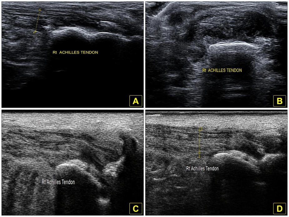 Figure 1. (a) Right Achilles tendon demonstrating increased tendon thickness measuring 0.70 cm with intratendinous calcification. (b) Right Achilles tendon showing a midportion abnormality in the form of an intratendinous hypoechoic area. (c) Right Achilles tendon demonstrating an enthesis abnormality in the form of an enthesophyte (bony spur). (d) Right Achilles tendon showing focal abnormal echotexture
RESULTS:
A total of 240 participants were included in the study, comprising 120 patients with diabetes mellitus and 120 age- and sex-matched non-diabetic controls. The mean age of the diabetic and non-diabetic groups was 59.8 ± 11.49 years and 59.6 ± 10.81 years, respectively. Males constituted a slightly higher proportion of participants in both groups (61.7% vs. 59.2%). The mean body mass index was comparable between the two groups (27.1 ± 2.77 kg/m² vs. 26.9 ± 2.60 kg/m²), with obesity being the most common BMI category. Among diabetic participants, 45.8% had a disease duration of more than 10 years (Table 1).
Side-specific ultrasonographic evaluation demonstrated that all assessed abnormalities were significantly more frequent among diabetic participants than non-diabetic controls. Increased tendon thickness, midportion abnormalities, enthesis abnormalities, increased vascularity, abnormal echotexture, and calcification were observed more commonly on both the right and left sides in the diabetic group. Most comparisons showed highly significant differences (p<0.001), whereas right-sided increased tendon thickness (p=0.016), left-sided enthesis abnormality (p=0.033), left-sided abnormal echotexture (p=0.013), and right-sided calcification (p=0.001) also reached statistical significance (Table 2).
Assessment of bilateral Achilles tendon involvement demonstrated significantly greater abnormalities among diabetic participants. Bilateral increased vascularity (35.8% vs. 7.5%), midportion abnormalities (33.3% vs. 10.0%), abnormal echotexture (25.8% vs. 10.8%), calcification (25.8% vs. 7.5%), enthesis abnormalities (22.5% vs. 6.7%), and increased tendon thickness (18.3% vs. 7.5%) were all significantly more frequent in the diabetic group (Table 3).
When ultrasonographic findings were analyzed irrespective of side, diabetic participants consistently showed a higher prevalence of tendon abnormalities than non-diabetic controls. Midportion abnormalities (84.2%), calcification (80.0%), increased vascularity (78.3%), enthesis abnormalities (76.7%), abnormal echotexture (75.8%), and increased tendon thickness (65.8%) were significantly more prevalent among diabetic participants (all p≤0.026) (Table 4).
Overall, asymptomatic sonographic abnormalities of the Achilles tendon were detected in 132 (55.0%) participants. The prevalence of these abnormalities was significantly higher in diabetic participants than in non-diabetic controls (68.3% vs. 41.7%; χ² = 17.239, p<0.001), indicating a strong association between diabetes mellitus and subclinical Achilles tendon changes (Table 5).
Table 1. Baseline demographic and clinical characteristics of the study participants
|
Characteristic |
Diabetic group (n=120) |
Non-diabetic group (n=120) |
|
|
Age, years, mean ± SD |
59.8 ± 11.49 |
59.6 ± 10.81 |
|
|
Age group, n (%) |
41–50 years |
33 (27.5) |
28 (23.3) |
|
51–60 years |
27 (22.5) |
38 (31.7) |
|
|
61–70 years |
33 (27.5) |
31 (25.8) |
|
|
71–80 years |
27 (22.5) |
23 (19.2) |
|
|
Sex, n (%) |
Male |
74 (61.7) |
71 (59.2) |
|
Female |
46 (38.3) |
49 (40.8) |
|
|
BMI, kg/m², mean ± SD |
27.1 ± 2.77 |
26.9 ± 2.60 |
|
|
BMI category, n (%) |
Normal (18.5–24.9 kg/m²) |
8 (6.7) |
6(5.0) |
|
Overweight (25.0–29.9 kg/m²) |
28 (23.3) |
29 (24.2) |
|
|
Obese (≥30 kg/m²) |
84 (70.0) |
85 (70.8) |
|
|
Duration of diabetes, n (%) |
≤5 years |
30 (25.0) |
- |
|
6–10 years |
35 (29.2) |
- |
|
|
|
>10 years |
55 (45.8) |
- |
Table 2. Side-specific ultrasonographic abnormalities of the Achilles tendon in diabetic and non- diabetic participants
|
Ultrasonographic finding |
Side |
Diabetic group (n=120), n (%) |
Non-diabetic group (n=120), n (%) |
Total (N=240), n (%) |
χ² |
p-value |
|
Increased tendon thickness |
Right |
53 (44.2) |
35 (29.2) |
88 (36.7) |
5.813 |
0.016 |
|
Left |
66 (55.0) |
31 (25.8) |
97 (40.4) |
21.195 |
<0.001 |
|
|
Midportion abnormality |
Right |
79 (65.8) |
34 (28.3) |
113 (47.1) |
33.865 |
<0.001 |
|
Left |
62 (51.7) |
34 (28.3) |
96 (40.0) |
13.611 |
<0.001 |
|
|
Enthesis abnormality |
Right |
66 (55.0) |
39 (32.5) |
105 (43.8) |
12.343 |
<0.001 |
|
Left |
53 (44.2) |
37 (30.8) |
90 (37.5) |
4.551 |
0.033 |
|
|
Increased vascularity |
Right |
66 (55.0) |
31 (25.8) |
97 (40.4) |
21.195 |
<0.001 |
|
Left |
71 (59.2) |
35 (29.2) |
106 (44.2) |
21.898 |
<0.001 |
|
|
Abnormal echotexture |
Right |
61 (50.8) |
32 (26.7) |
93 (38.8) |
14.764 |
<0.001 |
|
Left |
61 (50.8) |
42 (35.0) |
103 (42.9) |
6.140 |
0.013 |
|
|
Calcification |
Right |
62 (51.7) |
37 (30.8) |
99 (41.3) |
10.746 |
0.001 |
|
Left |
65 (54.2) |
29 (24.2) |
94 (39.2) |
22.664 |
<0.001 |
Table 3. Comparison of bilateral ultrasonographic abnormalities of the Achilles tendon between diabetic and non-diabetic participants
|
Bilateral ultrasonographic abnormality |
Diabetic group (n=120), n (%) |
Non-diabetic group (n=120), n (%) |
Total (N=240), n (%) |
χ² |
p-value |
|
Increased tendon thickness |
22 (18.3) |
9 (7.5) |
31 (12.9) |
6.260 |
0.012 |
|
Midportion abnormality |
40 (33.3) |
12 (10.0) |
52 (21.7) |
19.247 |
<0.001 |
|
Enthesis abnormality |
27 (22.5) |
8 (6.7) |
35 (14.6) |
12.075 |
0.001 |
|
Increased vascularity |
43 (35.8) |
9 (7.5) |
52 (21.7) |
28.380 |
<0.001 |
|
Abnormal echotexture |
31 (25.8) |
13 (10.8) |
44 (18.3) |
9.017 |
0.003 |
|
Calcification |
31 (25.8) |
9 (7.5) |
40 (16.7) |
14.520 |
<0.001 |
Table 4. Participant-level prevalence of Achilles tendon ultrasonographic abnormalities irrespective of side
|
Ultrasonographic abnormality |
Diabetic group (n=120), n (%) |
Non-diabetic group (n=120), n (%) |
Total (N=240), n (%) |
χ² |
p-value |
|
Increased tendon thickness |
79 (65.8) |
62 (51.7) |
141 (58.8) |
4.969 |
0.026 |
|
Midportion abnormality |
101 (84.2) |
56 (46.7) |
157 (65.4) |
37.296 |
<0.001 |
|
Enthesis abnormality |
92 (76.7) |
68 (56.7) |
160 (66.7) |
10.800 |
0.001 |
|
Increased vascularity |
94 (78.3) |
57 (47.5) |
151 (62.9) |
24.448 |
<0.001 |
|
Abnormal echotexture |
91 (75.8) |
61 (50.8) |
152 (63.3) |
16.148 |
<0.001 |
|
Calcification |
96 (80.0) |
57 (47.5) |
153 (63.7) |
27.424 |
<0.001 |
Table 5. Overall prevalence of asymptomatic sonographic abnormalities (ASA) of the Achilles tendon
|
Variable |
Diabetic group (n=120),n (%) |
Non-diabetic group (n=120),n (%) |
Total (N=240), n (%) |
χ² |
p-value |
|
ASA present |
82 (68.3) |
50 (41.7) |
132 (55.0) |
17.239 |
<0.001 |
|
ASA absent |
38 (31.7) |
70 (58.3) |
108 (45.0) |
DISCUSSION:
Diabetes mellitus is associated with progressive changes in connective tissues that extend beyond the well-recognized vascular and neurological complications. Persistent hyperglycemia promotes the formation of advanced glycation end products, oxidative stress, and microvascular dysfunction, leading to collagen cross-linking, impaired tendon remodeling, and reduced tissue elasticity. The Achilles tendon is particularly susceptible to these changes because of its high collagen content, continuous mechanical loading, and relatively poor vascular supply [10]. As a result, structural alterations may develop long before the onset of clinical symptoms, making imaging an important tool for early detection [11].
In the present study, the diabetic and non-diabetic groups were comparable with respect to age, sex, and body mass index, minimizing the influence of potential confounding factors. Nearly half of the diabetic participants had a disease duration of more than 10 years, suggesting prolonged exposure to hyperglycemia, which has been associated with progressive tendon degeneration [4]. Similar baseline
characteristics have been reported by Afolabi et al., supporting the validity of the present comparison [12].
Ultrasonographic evaluation demonstrated significantly greater structural abnormalities in the Achilles tendon among diabetic participants. Side-specific analysis showed a higher prevalence of increased tendon thickness, midportion abnormalities, enthesis abnormalities, increased vascularity, abnormal echotexture, and calcification in diabetics than in controls. These findings are consistent with previous studies by Prajwal et al., Afolabi et al., and Roesch et al., who also reported increased tendon thickness and structural alterations in diabetic individuals [8], [12], [13]. The observed increase in tendon thickness is likely related to collagen glycation and extracellular matrix disorganization caused by chronic hyperglycemia [4].
Bilateral tendon involvement was also significantly more frequent in diabetic participants, indicating that diabetes produces diffuse rather than localized tendon changes. Likewise, when abnormalities were analyzed irrespective of side, all ultrasonographic parameters remained significantly more prevalent in the diabetic group, with midportion abnormalities, calcification, and increased vascularity showing the highest frequencies. The predominance of midportion abnormalities is biologically plausible because the Achilles tendon midportion represents a relatively hypovascular region that is particularly vulnerable to degeneration [11]. Increased Doppler vascularity observed in the present study probably reflects pathological neovascularization associated with tendon remodeling rather than acute inflammation, as previously described by Liu et al. [14]. Similarly, abnormal echotexture and calcification indicate chronic collagen disorganization and long-standing degenerative changes, findings that agree with those reported by Ursini et al. and Page et al. [9], [15].
Overall, asymptomatic sonographic abnormalities were significantly more common in diabetic participants than in non-diabetic controls (68.3% vs. 41.7%). This supports earlier reports that substantial tendon pathology may remain clinically silent in diabetes [8], [16]. The present findings therefore highlight the value of high-resolution ultrasonography as a simple, non-invasive, and accessible method for identifying subclinical Achilles tendon involvement. Early recognition of these changes may facilitate timely interventions, including improved glycemic control, weight management, and preventive rehabilitation strategies, thereby reducing the risk of symptomatic tendinopathy, tendon rupture, and long-term functional impairment.
CONCLUSION:
Diabetes mellitus is associated with a significantly higher prevalence of subclinical Achilles tendon abnormalities compared with non-diabetic individuals, even in the absence of symptoms. In the present study, diabetic participants demonstrated increased tendon thickness, midportion and enthesis abnormalities, increased vascularity, abnormal echotexture, calcification, and overall asymptomatic sonographic abnormalities more frequently than controls. These findings suggest that structural and
vascular tendon changes occur early in the course of diabetes and may precede clinical manifestations. High-resolution ultrasonography is a simple, non-invasive, and reliable imaging modality for detecting these early changes and may be valuable for routine assessment of diabetic patients to facilitate timely intervention and prevent progression to symptomatic tendon disease and related complications.
REFERENCES:
1. Harding JL, Pavkov ME, Magliano DJ, Shaw JE, Gregg EW. Global trends in diabetes complications. Diabetologia. 2019;62(1):3–16.
2. Sun H, Saeedi P, Karuranga S, et al. IDF Diabetes Atlas: Global, regional and country-level diabetes prevalence estimates for 2021 and projections for 2045. Diabetes Res Clin Pract. 2022;183:109119.
3. Anjana RM, Deepa M, Pradeepa R, et al. Prevalence of diabetes and prediabetes in 15 states of India: results from the ICMR-INDIAB population-based cross-sectional study. Lancet Diabetes Endocrinol. 2017;5(8):585-596.
4. Kaka B, Maharaj SS, Fatoye F. Prevalence of musculoskeletal disorders in patients with diabetes mellitus: A systematic review and meta-analysis. J Back Musculoskelet Rehabil. 2019;32(2):223- 235.
5. Xu J, Wang J, Ji Y, et al. The impact of diabetes mellitus on tendon pathology: a review. Front Pharmacol. 2024;15:1491633.
6. Ward H, Jawad AS. Musculoskeletal manifestations of diabetes mellitus - an update. Clin Med (Lond). 2026;26(1):100498.
7. Yammine K, Daccache E, Assi C. Morphological changes and pathological findings in the Achilles tendons of diabetic patients: a meta-analysis of comparative clinical studies. Muscles Ligaments Tendons J. 2023;13(2):201-210.
8. Prajwal R, Sakalecha AK, Dudekula A, Varma N. Ultrasound Evaluation of Achilles Tendon Thickness in Diabetic Patients With and Without Foot Complications. Cureus. 2024;16(6):e62831.
9. Page P, Manske RC, Voight M, Wolfe C. Diagnostic Musculoskeletal Ultrasound of the Achilles Tendon. Int J Sports Phys Ther. 2025;20(2):296-304.
10. Winnicki K, Ochała-Kłos A, Rutowicz B, Pękala PA, Tomaszewski KA. Functional anatomy, histology and biomechanics of the human Achilles tendon - A comprehensive review. Ann Anat. 2020;229:151461.
11. Elsheikh AM, Fouad SEM, Ibrahim SA, Alsowey AM. Ultrasound evaluation of Achilles tendon among diabetic patients (type 2 diabetes mellitus). J Neonatal Surg. 2025;14(9 Suppl):781-789.
12. Afolabi BI, Idowu BM, Onigbinde SO. Achilles tendon degeneration on ultrasound in type 2 diabetic patients. J Ultrason. 2021;20(83):e291-e299.
13. Roesch HJ, Banwell H, Jones M, Saunders S, Milanese S. An exploration of the acute response of the asymptomatic Achilles tendon to exercise using diagnostic ultrasound. A systematic review of cohort studies. J Sci Med Sport. 2025;28(4):313-323.
14. Cannata F, Vadalà G, Ambrosio L, et al. Diabetes and tendinopathy. Diabetes Metab Res Rev. 2021;37(6):e3417.
15. Ursini F, Arturi F, D’Angelo S, et al. High Prevalence of Achilles Tendon Enthesopathic Changes in Patients with Type 2 Diabetes Without Peripheral Neuropathy. J Am Podiatr Med Assoc. 2017;107(2):99-105.
16. Bakkegaard M, Johannsen FE, Højgaard B, Langberg H. Ultrasonography as a prognostic parameter in Achilles tendinopathy. Eur J Radiol. 2015;84(3):458–462.