Ultrasonographic Assessment of First-Trimester Nuchal Translucency and Population-specific Reference Values in Central India.
- Alka Agrawal , Professor and Head of Department, Department of Radiodiagnosis, MGMMC MYH Indore (M.P).
- Archana Bhatnagar , Associate Professor, Department of Radiodiagnosis, MGMMC MYH Indore (M.P).
- Faraz Khan , Assistant Professor, Department of Radiodiagnosis, MGMMC MYH Indore (M.P).
- Raj Bahor Patel , Junior Resident, Department of Radiodiagnosis, MGMMC MYH Indore (M.P).
- Anjana Kumari , Junior Resident, Department of Radiodiagnosis, MGMMC MYH Indore (M.P).
Article Information:
Abstract:
Background: Nuchal translucency (NT) measurement is an established first-trimester ultrasonographic marker for screening fetal chromosomal and structural abnormalities. As NT values vary across populations, locally derived reference values are essential for accurate prenatal risk assessment. Methods: This hospital-based cross-sectional observational study included 1,000 singleton pregnant women undergoing routine first-trimester ultrasonography between 11 and 14 weeks of gestation at a tertiary care centre in Central India. NT was measured using standardized ultrasonographic techniques. Gestational age- and crown–rump length (CRL)-specific reference values were generated, and correlations of NT with gestational age, CRL, maternal age, and gravidity/parity were analysed. Results: The mean NT thickness was 1.455 ± 0.279 mm, with a median of 1.50 mm and a 95th percentile of 1.90 mm. NT demonstrated an overall increasing trend with advancing gestational age, with mean values rising from 1.326 ± 0.260 mm at 11 weeks to 1.638 ± 0.289 mm at 14 weeks. A similar trend was observed with increasing CRL. NT showed significant positive correlations with gestational age (r = 0.281, p <0.001) and CRL (r = 0.270, p <0.001), while maternal age showed a weak positive correlation (r = 0.068, p = 0.031). Conclusion: The study establishes gestational age- and CRL-specific NT reference values for a Central Indian population. These population-specific normative data may improve the interpretation of first-trimester NT measurements and support more accurate prenatal risk assessment.
Keywords:
Article :
INTRODUCTION:
First-trimester prenatal screening plays a pivotal role in the early identification of fetuses at increased risk of chromosomal abnormalities and structural anomalies, allowing appropriate counselling and timely diagnostic evaluation. Among the available screening modalities, ultrasonography remains the most widely accepted, non-invasive, and cost-effective investigation in routine obstetric practice. Nuchal translucency (NT), defined as the sonographic appearance of the subcutaneous fluid-filled space at the posterior aspect of the fetal neck, is measured between 11 and 14 weeks of gestation and constitutes an essential component of first-trimester screening [1], [2].
An increased NT measurement has been associated with chromosomal abnormalities, particularly trisomy 21, as well as congenital heart defects, genetic syndromes, and adverse pregnancy outcomes, even in fetuses with a normal karyotype [3], [4].Large prospective studies have demonstrated that NT measurement provides effective risk stratification for fetal aneuploidy and has therefore become an integral part of prenatal screening programmes worldwide. The interpretation of NT values is based on gestational age and crown–rump length (CRL), with measurements above the 95th or 99th percentile indicating an increased likelihood of fetal abnormalities [5], [6].
Despite standardized measurement techniques, NT values may vary across populations because of differences in ethnicity, genetic background, maternal characteristics, and environmental influences. Most reference charts currently used in clinical practice are derived from Western populations and may not accurately reflect NT distribution in Indian pregnancies [7], [8]. The use of population-specific reference values is therefore important to improve the accuracy of prenatal risk assessment, reduce unnecessary invasive investigations, and facilitate appropriate counselling.
Given the high burden of congenital anomalies and the continued reliance on ultrasonography as the primary prenatal screening tool in India, establishing normative NT values in the local population is clinically relevant [9].The present study was undertaken to evaluate ultrasonographic NT measurements in first-trimester pregnancies, establish gestational age-specific normative data, assess their relationship with CRL, maternal age, and gravidity, and compare the findings with published reference values from other populations.
MATERIALS AND METHODS:
This hospital-based, cross-sectional observational study was conducted in the Department of Radiodiagnosis, M.G.M. Medical College and M.Y. Hospital, Indore, Madhya Pradesh, India, over a period of one year following approval from the Institutional Scientific Review Board (ISRB) and the Institutional Ethics Committee. Written informed consent was obtained from all participants before enrolment.
A total of 1,000 consecutive pregnant women presenting for routine first-trimester antenatal ultrasonography between 11 and 14 weeks of gestation were included in the study. Gestational age was determined based on the last menstrual period and confirmed by ultrasonographic crown–rump length (CRL) measurements.
Pregnant women with singleton pregnancies between 11 and 14 weeks of gestation who were registered under the Pre-Conception and Pre-Natal Diagnostic Techniques (PCPNDT) Act and provided written informed consent were eligible for inclusion. Only Indian pregnant women with Indian partners were enrolled to establish population-specific normative data. Women with maternal systemic illnesses, multiple gestations, intrauterine fetal demise, known fetal structural abnormalities, or CRL measurements outside the recommended range of 45–84 mm were excluded. Cases in which optimal fetal positioning did not permit accurate NT measurement or where consent was declined were also excluded.
All ultrasonographic examinations were performed using a high-resolution ultrasound system equipped with a 3–5 MHz transabdominal transducer. The examination was performed with the participant in the supine position following completion of all PCPNDT documentation. Fetal viability, gestational age, and the presence of major structural abnormalities were assessed before NT measurement.
Nuchal translucency was measured according to established first-trimester ultrasonographic guidelines. A true mid-sagittal view of the fetus was obtained with the fetus in a neutral position and separated from the uterine wall by a visible layer of amniotic fluid. The image was magnified so that the fetal head and upper thorax occupied most of the screen. The calipers were placed on the inner borders of the echogenic lines defining the translucency, and the maximum vertical thickness of the subcutaneous fluid at the posterior aspect of the fetal neck was recorded. Crown–rump length was measured during the same examination using standard techniques (Figure 1). Demographic and obstetric details, including maternal age, gravidity, gestational age, CRL, and NT measurements, were recorded in a structured data collection proforma.
Statistical analysis was performed using IBM SPSS Statistics for Windows, Version 23.0 (IBM Corp., Armonk, NY, USA), while data entry and preliminary data management were carried out using Microsoft Excel. Continuous variables were expressed as mean ± standard deviation (SD), whereas categorical variables were presented as frequencies and percentages. Pearson's correlation coefficient was used to evaluate the relationships between NT thickness and gestational age, crown–rump length (CRL), and maternal age. Differences in mean NT thickness across gravidity/parity groups were assessed using one-way analysis of variance (ANOVA). Gestational age- and CRL-specific NT percentile charts (5th, 10th, 25th, 50th, 75th, 90th, and 95th percentiles) were generated. A p-value <0.05 was considered statistically significant.
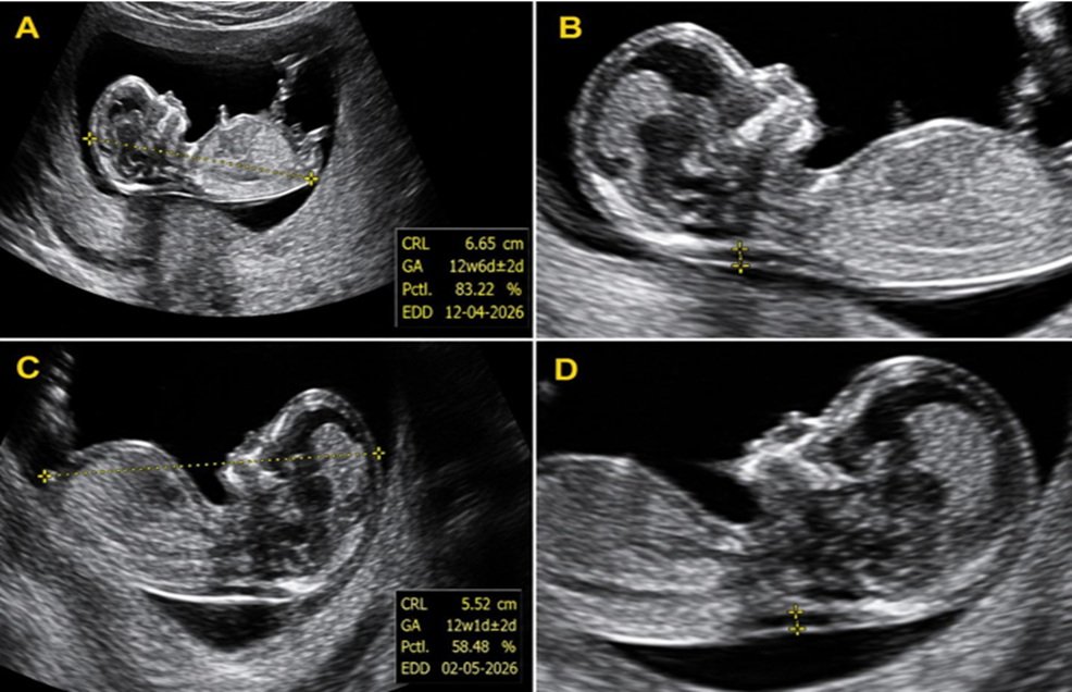
Figure 1. Representative first-trimester ultrasonographic images demonstrating crown–rump length (CRL) and nuchal translucency (NT) measurements. (A) Mid-sagittal fetal image showing CRL measurement of approximately 66 mm, corresponding to a gestational age of 12 weeks 6 days. (B) Mid-sagittal fetal image demonstrating NT measurement of approximately 1.51 mm. (C) Mid-sagittal fetal image showing CRL measurement of approximately 55 mm, corresponding to a gestational age of 12 weeks 1 day. (D) Mid-sagittal fetal image demonstrating NT measurement of approximately 1.69 mm.
RESULTS:
A total of 1,000 pregnant women with singleton pregnancies between 11 and 14 weeks of gestation were included in the study. The majority of participants were aged 21–25 years (39.7%), followed by 26–30 years (30.1%), while only 2.9% were older than 35 years. Most examinations were performed at 12 weeks of gestation (35.9%), followed by 13 weeks (30.0%), 11 weeks (27.6%), and 14 weeks (6.5%). Regarding obstetric status, G2P1 women constituted the largest subgroup (31.8%), closely followed by primigravidae (30.0%) (Table 1).
Table 1. Baseline Maternal and Obstetric Characteristics of the Study Population (N = 1000)
|
Characteristic |
Category |
n |
% |
|
Maternal age (years) |
18–20 |
150 |
15.0 |
|
21–25 |
397 |
39.7 |
|
|
26–30 |
301 |
30.1 |
|
|
31–35 |
123 |
12.3 |
|
|
>35 |
29 |
2.9 |
|
|
Gestational age (weeks) |
11 |
276 |
27.6 |
|
12 |
359 |
35.9 |
|
|
13 |
300 |
30.0 |
|
|
14 |
65 |
6.5 |
|
|
Gravidity/Parity |
G1P0 |
300 |
30.0 |
|
G2P1 |
318 |
31.8 |
|
|
G3P2 |
230 |
23.0 |
|
|
G4P3 |
104 |
10.4 |
|
|
G5P4 |
48 |
4.8 |
The overall mean nuchal translucency (NT) thickness was 1.455 ± 0.279 mm, with values ranging from 0.70 to 2.20 mm. The median NT was 1.50 mm (interquartile range: 1.30–1.69 mm). The 5th and 95th percentile values were 1.00 mm and 1.90 mm, respectively. Most fetuses (90.9%) had NT measurements between 1.01 and 2.00 mm, whereas only 1.4% had NT values exceeding 2.00 mm (Table 2).
Table 2. Distribution and Descriptive Statistics of Nuchal Translucency Thickness (N = 1000)
|
A. NT thickness categories |
n |
% |
|
≤1.00 mm |
77 |
7.7 |
|
1.01–1.50 mm |
554 |
55.4 |
|
1.51–2.00 mm |
355 |
35.5 |
|
>2.00 mm |
14 |
1.4 |
|
Total |
1000 |
100.0 |
|
B. Descriptive statistics |
Value |
|
|
Mean ± SD (mm) |
1.455 ± 0.279 |
|
|
Median (IQR), mm |
1.50 (1.30–1.69) |
|
|
Minimum–Maximum (mm) |
0.70–2.20 |
|
|
5th Percentile (mm) |
1.00 |
|
|
95th Percentile (mm) |
1.90 |
|
Gestational age-specific NT reference values demonstrated a gradual increase in NT thickness with advancing gestation. The mean NT increased from 1.326 ± 0.260 mm at 11 weeks to 1.638 ± 0.289 mm at 14 weeks. Similarly, the median NT increased from 1.30 mm to 1.60 mm, while the 95th percentile increased from 1.71 mm to 2.00 mm across the study period (Table 3).
Table 3. Gestational Age-specific Reference Values for Fetal Nuchal Translucency
|
Gestational age (weeks) |
n |
Mean ± SD (mm) |
5th |
10th |
25th |
Median |
75th |
90th |
95th |
|
11 |
276 |
1.326 ± 0.260 |
1.00 |
1.00 |
1.10 |
1.30 |
1.50 |
1.70 |
1.71 |
|
12 |
359 |
1.495 ± 0.269 |
1.00 |
1.10 |
1.30 |
1.50 |
1.70 |
1.80 |
1.90 |
|
13 |
300 |
1.487 ± 0.261 |
1.00 |
1.16 |
1.30 |
1.50 |
1.70 |
1.80 |
1.90 |
|
14 |
65 |
1.638 ± 0.289 |
1.20 |
1.30 |
1.50 |
1.60 |
1.90 |
2.00 |
2.00 |
A comparable trend was observed with increasing crown–rump length (CRL). Mean NT increased from 1.308 mm in fetuses with a CRL of 45–50 mm to 1.611 mm in those with a CRL of 81-84 mm. The corresponding 95th percentile values ranged from 1.70 mm to 2.03 mm across the evaluated CRL groups (Table 4).
Table 4. Crown–Rump Length-specific Reference Values for Fetal Nuchal Translucency
|
CRL (mm) |
n |
Mean (mm) |
5th |
25th |
Median |
75th |
95th |
|
45–50 |
231 |
1.308 |
1.00 |
1.10 |
1.30 |
1.50 |
1.70 |
|
51–55 |
123 |
1.433 |
1.00 |
1.25 |
1.40 |
1.60 |
1.90 |
|
56–60 |
153 |
1.492 |
1.00 |
1.30 |
1.50 |
1.70 |
1.86 |
|
61–65 |
113 |
1.552 |
1.20 |
1.40 |
1.50 |
1.70 |
1.93 |
|
66–70 |
150 |
1.494 |
1.00 |
1.30 |
1.50 |
1.70 |
1.97 |
|
71–75 |
112 |
1.484 |
1.10 |
1.30 |
1.50 |
1.70 |
1.84 |
|
76–80 |
100 |
1.540 |
1.20 |
1.30 |
1.50 |
1.70 |
2.00 |
|
81-84 |
18 |
1.611 |
1.19 |
1.40 |
1.60 |
1.80 |
2.03 |
NT thickness showed minimal variation among women aged 18–35 years, whereas participants aged ≥36 years demonstrated a higher mean NT (1.721 ± 0.204 mm) and a higher 95th percentile (2.06 mm). Mean NT values were comparable across G1P0 to G4P3 groups, while the G5P4 group had relatively higher NT measurements (Table 5).
Table 5. Nuchal Translucency Thickness According to Maternal Age and Gravidity/Parity
|
Characteristic |
Category |
n |
Mean ± SD (mm) |
Median (mm) |
5th Percentile (mm) |
95th Percentile (mm) |
|
Maternal age (years) |
18–20 |
150 |
1.419 ± 0.291 |
1.40 |
1.00 |
1.90 |
|
21–25 |
397 |
1.468 ± 0.270 |
1.50 |
1.00 |
1.88 |
|
|
26–30 |
301 |
1.437 ± 0.275 |
1.40 |
1.00 |
1.90 |
|
|
31–35 |
123 |
1.441 ± 0.291 |
1.46 |
1.00 |
1.90 |
|
|
≥36 |
29 |
1.721 ± 0.204 |
1.70 |
1.40 |
2.06 |
|
|
Gravidity/Parity |
G1P0 |
300 |
1.454 ± 0.284 |
1.50 |
1.00 |
1.90 |
|
G2P1 |
318 |
1.446 ± 0.268 |
1.50 |
1.00 |
1.85 |
|
|
G3P2 |
230 |
1.444 ± 0.276 |
1.40 |
1.00 |
1.90 |
|
|
G4P3 |
104 |
1.437 ± 0.290 |
1.48 |
1.00 |
1.90 |
|
|
G5P4 |
48 |
1.619 ± 0.277 |
1.65 |
1.17 |
2.06 |
|
|
Statistical analysis: Maternal age: Pearson correlation, r = 0.068, p = 0.031. Gravidity/parity groups: one-way ANOVA, F = 4.464, p = 0.001. |
||||||
Correlation analysis demonstrated significant positive associations between NT thickness and both CRL (r = 0.270, p <0.001) and gestational age (r = 0.281, p <0.001). Maternal age showed a weak but statistically significant positive correlation with NT thickness (r = 0.068, p = 0.031). Comparison of NT measurements across gravidity/parity groups also demonstrated a statistically significant difference (one-way ANOVA, F = 4.464, p = 0.001) (Table 6).
Table 6. Association of Nuchal Translucency Thickness with Clinical Variables
|
Variable |
Test statistic |
p-value |
|
Crown–rump length (CRL) |
r = 0.270 |
<0.001 |
|
Gestational age |
r = 0.281 |
<0.001 |
|
Maternal age |
r = 0.068 |
0.031 |
|
Gravidity/parity groups |
F = 4.464 |
0.001 |
|
Values of r represent Pearson's correlation coefficient. F represents the one-way ANOVA test statistic. A p-value <0.05 was considered statistically significant. |
||
DISCUSSION:
Nuchal translucency (NT) measurement is an established component of first-trimester prenatal screening and plays a central role in identifying pregnancies at increased risk of chromosomal abnormalities and structural anomalies. As NT values are influenced by fetal growth and population characteristics, the availability of population-specific reference ranges is essential for accurate risk assessment. The present study established gestational age- and crown–rump length (CRL)-specific NT reference values in a cohort of 1,000 normal singleton pregnancies from Central India. The overall mean NT thickness in the present study was 1.455 ± 0.279 mm, with a 95th percentile of 1.90 mm. These findings are comparable with reports from Indian populations by Mahale et al. (2013) and are broadly consistent with observations from neighbouring South Asian populations such as the Nepalese study by Karki et al. (2013)[10], [11]. The observed differences in NT values across populations reinforce the need for locally derived reference standards for accurate prenatal risk assessment.
A progressive increase in NT thickness with advancing gestational age and CRL was observed in the present study, consistent with the physiological increase reported in earlier studies [12].Significant positive correlations between NT and both gestational age (r = 0.281, p < 0.001) and CRL (r = 0.270, p < 0.001) reinforce the importance of interpreting NT measurements in relation to fetal size rather than using a fixed threshold. These findings are in accordance with recommendations from the Fetal Medicine Foundation and previous investigations demonstrating that CRL-adjusted NT assessment provides more reliable prenatal risk stratification [2], [6], [12].
Maternal age demonstrated a statistically significant but weak positive correlation with NT thickness, while women aged ≥36 years showed slightly higher mean NT values than younger participants. Similar observations have been reported by Spencer et al., suggesting that maternal age contributes primarily to overall aneuploidy risk rather than substantially influencing NT measurements [13]. Likewise, NT values were largely comparable across gravidity/parity groups, indicating that parity has minimal influence on NT distribution, although grand multiparous women showed relatively higher values. This finding should be interpreted cautiously because of the relatively small number of women in this subgroup.
Compared with published international reference values, the upper reference limits observed in the present study were generally lower than those reported in several Western populations [10], [12]. This suggests that the use of non-local reference standards may not accurately reflect NT distribution in the Central Indian population and could potentially influence prenatal risk estimation. The major strengths of this study include its large sample size, standardized ultrasonographic technique, and the development of both gestational age- and CRL-specific reference values. However, the study was conducted at a single tertiary care centre, and biochemical screening markers and long-term fetal outcome data were not available. Multicentric studies with postnatal follow-up are required to further validate these findings and refine population-specific NT reference standards for routine clinical practice.
CONCLUSION:
The present study established gestational age- and crown–rump length-specific normative reference values for first-trimester nuchal translucency in a Central Indian population. NT thickness demonstrated a significant positive correlation with gestational age and CRL, confirming that fetal growth is the primary determinant of NT measurements. The derived population-specific normogram provides a reliable reference for routine prenatal screening and may improve the accuracy of identifying fetuses requiring further evaluation. Adoption of locally validated NT reference ranges can enhance risk stratification, reduce unnecessary invasive investigations, and support evidence-based clinical decision-making. Further multicentric studies are recommended to validate these findings across diverse Indian populations.
REFERENCES:
1. American College of Obstetricians and Gynecologists’ Committee on Practice Bulletins—Obstetrics; Committee on Genetics; Society for Maternal-Fetal Medicine. Screening for Fetal Chromosomal Abnormalities: ACOG Practice Bulletin, Number 226. Obstet Gynecol. 2020;136(4):e48-e69.
2. Nicolaides KH. Nuchal translucency and other first-trimester sonographic markers of chromosomal abnormalities. Am J Obstet Gynecol. 2004;191(1):45-67.
3. Souka AP, Snijders RJM, Novakov A, Soares W, Nicolaides KH. Defects and syndromes associated with increased nuchal translucency thickness at 10–14 weeks of gestation. Ultrasound Obstet Gynecol. 1998;11(6):391–400.
4. Nicolaides KH. First-trimester screening for chromosomal abnormalities. Semin Perinatol. 2005;29(4):190–194.
5. Souka AP, Von Kaisenberg CS, Hyett JA, Sonek JD, Nicolaides KH. Increased nuchal translucency with normal karyotype. Am J Obstet Gynecol. 2005;192(4):1005–1021.
6. Snijders RJ, Noble P, Sebire N, Souka A, Nicolaides KH. UK multicentre project on assessment of risk of trisomy 21 by maternal age and fetal nuchal-translucency thickness at 10-14 weeks of gestation. Fetal Medicine Foundation First Trimester Screening Group. Lancet. 1998;352(9125):343-346.
7. Kagan KO, Wright D, Spencer K, Molina FS, Nicolaides KH. First-trimester screening for trisomy 21 by free β-hCG and PAPP-A: impact of maternal and pregnancy characteristics. Ultrasound Obstet Gynecol. 2008;31(5):493–502.
8. Sahota DS, Leung TY, Chan LW, Law LW, Fung TY, Lau TK. Fetal nuchal translucency thickness in the Chinese population. Ultrasound Obstet Gynecol. 2009;34(4):376–380.
9. Basu P, Alvi S, Kulkarni R, et al. Epidemiology of birth defects in India: prevalence, patterns and public health implications. Indian J Pediatr. 2019;86(11):1013–1020.
10. Mahale N, Kumar A, Rayapureddi VCM, Mahale A, Krishnapriya. Variaton of nuchal translucency with increasing crown rump length and gestational age in normal singleton pregnancies. IOSR J Dent Med Sci. 2013;6(3):16-19.
11. Karki C, Shrestha S, Sharma S, et al. First-trimester nuchal translucency screening: experience from a Nepalese population. Journal of Nepal Health Research Council. 2013;11(25):1-6.
12. Hasegawa J, Nakamura M, Hamada S, Matsuoka R, Ichizuka K, Sekizawa A, et al. Distribution of nuchal translucency thickness in Japanese fetuses. J Obstet Gynaecol Res. 2013;39(4):766-9.
13. Spencer K, Crossley JA, Aitken DA, et al. Screening for single gene disorders in the first trimester: evaluation of nuchal translucency in pregnancies at risk for Mendelian disorders. Prenat Diagn. 2000;20(3):180–185.